
Teach me endometrial biopsy reporting Cytopathology
endometrial biopsy cytopathology reporting Bethesda categories classification
endometrial biopsy histology normal proliferative secretory endometrium
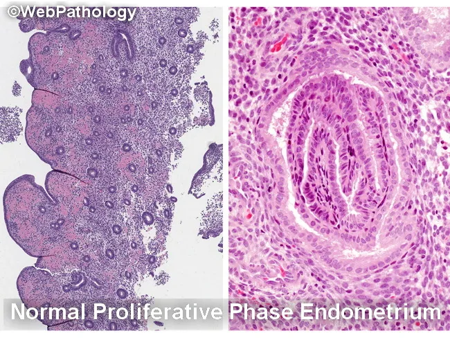
Histology image illustrating the endometrium in two complementary views. The left panel depicts normal proliferative phase endometrium with relatively sparse glands embedded in a cellular stroma, producing a low gland-to-stroma ratio and open gland spacing. The right panel shows characteristic epithelial morphology from proliferative endometrium with elongated, back-to-back glands and prominent, basally oriented nuclei at high power. The specimen emphasizes normal architectural patterns and avoids overcalling hyperplasia when glands are well separated or when fragmentation and telescoping create artifactual crowding. Key differential considerations include endometrial hyperplasia (EH) with or without atypia, disordered proliferative endometrium, reparative or regenerative changes, and endocervical/metaplastic proliferations. Within EH, nonatypical EH must be distinguished from atypical endometrial hyperplasia/endometrioid intraepithelial neoplasia (AEH/EIN), and AEH/EIN must be differentiated from endometrioid carcinoma and from endocervical lesions. Normal proliferative glands show minimal cytologic atypia, whereas AEH/EIN demonstrates architectural complexity (crumpled, crowded glands) and cytologic atypia. Artifacts such as glandular fragmentation, fragmentation- or telescoping-related crowding, and sample artifact can mimic AEH/EIN if the specimen is not intact. This image supports educational differentiation for gynecologic pathology, endometrial biopsy interpretation, and clinical correlation with bleeding patterns and imaging findings. Keywords: endometrium, proliferative phase, hyperplasia, AEH, EIN, histology, biopsy, gland-stroma ratio, artifacts, differential diagnosis teaching.
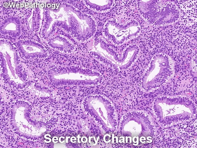
Endometrial histology in secretory phase demonstrated by light microscopy on a hematoxylin and eosin stained endometrial biopsy specimen. Glandular architecture shows irregular serration with gland crowding in focal areas, while glands are lined by tall, columnar secretory epithelium. Nuclei are enlarged and may display mild pleomorphism and loss of polarity, consistent with secretory transformation. The background stroma is edematous and commonly predecidualized, with cytoplasmic vacuolization of stromal cells. Together, these findings reflect physiologic secretory changes rather than overt endometrial hyperplasia. Notably, secretory endometrium can mimic endometrial hyperplasia/endometrioid intraepithelial neoplasia (EIN) in some cases, but the absence of marked architectural crowding, complex glandular patterns, or cytologic atypia argues against a neoplastic process. Differential considerations include secretory change, simple endometrial hyperplasia without atypia, and EIN in a background of secretory endometrium; careful architectural assessment and clinical correlation are essential. Diagnostic significance lies in distinguishing benign secretory transformation from premalignant or malignant entities to avoid overtreatment. This image is educational for pathology training, differential diagnosis exercises, and research on endometrial cycle pathology. Correlate with patient menstrual history, pregnancy status, and cycle timing; repeat sampling or adjunct studies may be warranted when discordance is suspected. Interpret interpretation should integrate histology with clinical data and follow-up.
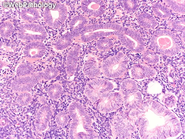
High-resolution hematoxylin and eosin stained endometrial biopsy reveals nonatypical endometrial hyperplasia (NEH). The histology shows proliferative-type glands that are simple to mildly irregular and tightly packed, with a gland to stroma ratio greater than one. Gland architecture is predominantly tubular, with minimal branching and without complex cribriform patterns. The glandular epithelium is lined by pseudostratified columnar cells showing uniform nuclei without marked pleomorphism. Cytologic atypia is absent or very subtle, with no conspicuous nuclear enlargement, hyperchromasia, or prominent nucleoli. The surrounding stroma is fibrous and moderately edematous, enriched with scattered lymphocytes, without desmoplastic reaction. Overall, the glandular proliferation mimics a proliferative endometrium but with increased density and crowding appropriate for NEH. No stromal invasion or myometrial involvement is evident in this section. The image highlights hallmark NEH features: gland crowding, preserved maturation, lack of cytologic atypia, and a relatively benign architectural pattern. Clinically relevant distinctions include differentiation from endometrial hyperplasia with atypia and endometrial carcinoma, which carry higher malignant risk and require different management. This image, attributed to WebPathology, serves as an educational reference for pathologists and trainees to recognize NEH morphology and to support differential diagnosis in gynecologic pathology exams and clinical practice. Used for teaching and diagnostic guidance.
This is a light microscopy image of an endometrial curettage specimen stained with hematoxylin and eosin (H&E), illustrating atypical endometrial hyperplasia within the decidualized endometrium. The biopsy is derived from the uterine cavity lining (endometrium) in a 45-year-old female with abnormal uterine bleeding. Histology shows proliferative glands with marked architectural crowding and back-to-back arrangement, irregular gland outlines, and cribriform-like branching, coupled with cytologic atypia of glandular epithelium. Nuclei are enlarged and hyperchromatic with mild to moderate pleomorphism; nuclear stratification is evident, and mitotic activity may be increased. Stroma remains relatively fibromuscular and less cellular compared to the epithelium. These features define atypical endometrial hyperplasia, a premalignant lesion with substantial risk of progression to endometrioid adenocarcinoma if left untreated. In the clinical context, unopposed estrogen stimulation from estrogen-secreting ovarian neoplasms such as adult granulosa cell tumor may drive endometrial proliferation; therefore, endometrial pathology must be interpreted alongside ovarian findings. Diagnostic significance lies in risk stratification and management decisions, including surveillance, hormonal therapy, or definitive surgical intervention. This image underlines the intersection of gynecologic oncology and endometrial pathology, with educational utility for histology, pathology, gynecology, and medical trainees. Correlation with receptor status and molecular markers may further refine prognosis and therapy.
endometrial hyperplasia atypical EIN cytology adenocarcinoma histopathology
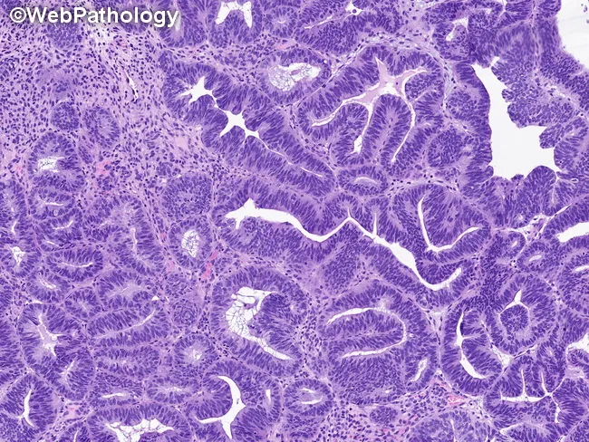
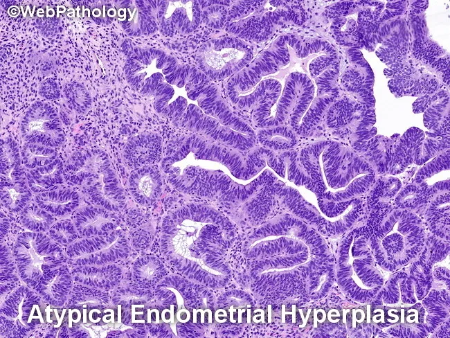
Histopathology of an endometrial biopsy showing atypical hyperplasia/endometrioid intraepithelial neoplasia (AEH/EIN). The section demonstrates endometrial glands arranged in a back-to-back, crowded pattern with minimal intervening stroma, resulting in a gland-to-stroma ratio greater than one. Glandular profiles are round to tubular or exhibit complex, stellate, or irregular contours with branching and occasional outpouchings. Cytologic atypia is evident by enlarged, round to elongated nuclei with increased nuclear-to-cytoplasmic ratio, loss of nuclear polarity, irregular nuclear distribution, and, in some glands, conspicuous nucleoli. Mitotic activity is variably increased, reflecting proliferative potential. Notably, metaplastic changes are present in several glands, including squamous, tubal, secretory, eosinophilic, mucinous, or papillary phenotypes, which can complicate interpretation. The background stroma remains relatively scant, supporting a diagnosis of AEH/EIN rather than benign proliferative processes. Clinically, this lesion is considered a precursor to endometrioid carcinoma and carries an elevated risk of concurrent carcinoma; thus, accurate grading and thorough sampling are essential. This histologic pattern is typically identified on formalin-fixed, paraffin-embedded H&E-stained sections examined under light microscopy at variable magnifications (low-power architecture and high-power cytology). Relevance spans diagnostic pathology, gynecologic oncology, and research on endometrial carcinogenesis; implications include risk stratification and guiding management (surgical or conservative) and surveillance.
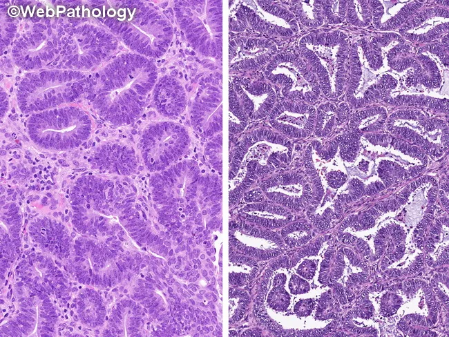
This is a paired histopathology image of endometrial neoplasia stained with hematoxylin and eosin. The left panel highlights atypical endometrial hyperplasia/endometrial intraepithelial neoplasia (AEH/EIN), showing glandular crowding, complex architecture, pseudostratified or stratified nuclei, and prominent cytologic atypia confined to the epithelium with preserved stroma. The right panel shows a well-differentiated endometrioid adenocarcinoma, with confluent back-to-back glands, solid sheets, cribriform and maze-like growth, and more marked cytologic atypia; a desmoplastic stromal response may be evident adjacent to malignant glands. Architectural patterns such as villoglandular, papillary, or microacinar arrangements can appear in either lesion but are more characteristic of carcinoma in this context. Necrosis and foamy macrophages may be present in carcinoma areas. Distinguishing AEH/EIN from carcinoma on limited biopsy can be challenging due to overlapping features and fragmentary material. Diagnostic thresholds cited by experts include gland size and extent (often >2 mm) and the presence of infiltrative or confluent growth with reduced stromal support. When features are intermediate, terminology such as atypical hyperplasia cannot rule out well-differentiated carcinoma or AEH/EIN bordering on well-differentiated carcinoma may be reported. This image emphasizes the spectrum and informs clinical decision making regarding surveillance, medical therapy, and surgical management.
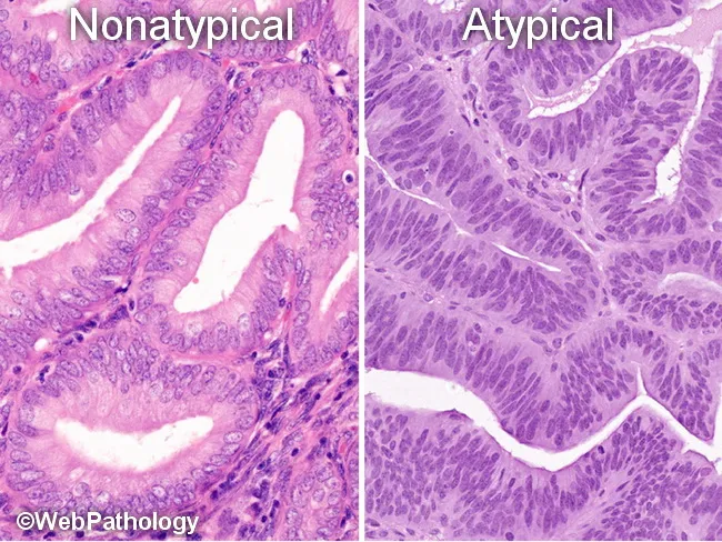
This is a paired histopathology image of endometrium illustrating the 2014 World Health Organization (WHO) revision to a two-tiered classification of endometrial hyperplasia (EH): nonatypical EH (benign hyperplasia) on the left and atypical EH/EIN on the right. The left panel displays simple to moderately complex glandular architecture with mild or absent cytologic atypia, lined by columnar epithelium and relatively orderly nuclear orientation, consistent with benign reactive changes due to unopposed estrogen. The right panel demonstrates marked architectural complexity with back-to-back crowded glands and pronounced cytologic atypia, including nuclear enlargement, loss of polarity, prominent nucleoli, and stratification, compatible with atypical EH and endometrioid intraepithelial neoplasia (EIN). The textural differences reflect a histologic continuum from nonatypical EH to clonal premalignant lesions. This classification emphasizes molecular alterations such as PTEN, PAX2, KRAS mutations, and microsatellite instability associated with EIN, and correlates with clinical risk of progression to endometrial adenocarcinoma (≈<2% for nonatypical EH versus ~25–30% for atypical EH/EIN if untreated). Clinically, this distinction guides management decisions, surveillance, and potential hormonal versus surgical therapy. The image underscores the transition from benign hyperplasia to a premalignant/neoplastic process, highlighting the diagnostic significance of cytologic atypia and glandular architecture in endometrial pathology.
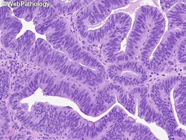
This is a histopathology image of endometrial tissue stained with hematoxylin and eosin. The section shows endometrial glands embedded in scant stroma with a marked gland crowding characteristic of atypical hyperplasia/endometrioid intraepithelial neoplasia (AEH/EIN). The glands are arranged back-to-back, with gland-to-stroma ratio greater than 1, and display rounded, tubular, or complex, stellate profiles with branching and occasional outpouchings. Epithelial cells demonstrate cytologic atypia, including enlarged nuclei, elevated nuclear-to-cytoplasmic ratio, loss of polarity, and irregular nuclear distribution; nucleoli may be distinct in some areas. Mitotic figures are variably increased, reflecting proliferative activity. Some cases show metaplastic changes such as squamous, tubal-type, secretory, eosinophilic, mucinous, or papillary differentiation, which can complicate interpretation. The image highlights the diagnostic challenge of distinguishing AEH/EIN from benign hyperplasia or early endometrial carcinoma. Clinically, AEH/EIN denotes a premalignant lesion with heightened risk of progression to endometrioid carcinoma and potential for synchronous endometrial neoplasia. This morphologic pattern informs risk stratification and management decisions, including conservative progestin therapy in select patients or definitive surgical treatment in others. In educational and research contexts, this image illustrates diagnostic criteria, histologic criteria, and the importance of integrating architecture, cytology, and metaplasia in endometrial neoplasia assessment.
endometrial carcinoma histology grade 1 2 3 FIGO endometrioid
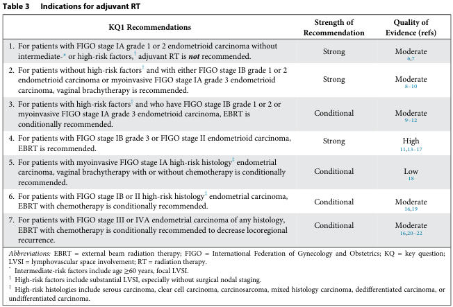
Table 3 Indications for adjuvant RT <table><thead><tr><th>KQ1 Recommendations</th><th>Strength of Recommendation</th><th>Quality of Evidence (refs)</th></tr></thead><tbody><tr><td>1. For patients with FIGO stage IA grade 1 or 2 endometrioid carcinoma without intermediate-* or high-risk factors,† adjuvant RT is not recommended.</td><td>Strong</td><td>Moderate 6,7</td></tr><tr><td>2. For patients without high-risk factors† and with either FIGO stage IB grade 1 or 2 endometrioid carcinoma or myoinvasive FIGO stage IA grade 3 endometrioid carcinoma, vaginal brachytherapy is recommended.</td><td>Strong</td><td>Moderate 8-10</td></tr><tr><td>3. For patients with high-risk factors† and who have FIGO stage IB grade 1 or 2 or myoinvasive FIGO stage IA grade 3 endometrioid carcinoma, EBRT is conditionally recommended.</td><td>Conditional</td><td>Moderate 9-12</td></tr><tr><td>4. For patients with FIGO stage IB grade 3 or FIGO stage II endometrioid carcinoma, EBRT is recommended.</td><td>Strong</td><td>High 11,13-17</td></tr><tr><td>5. For patients with myoinvasive FIGO stage IA high-risk histology‡ endometrial carcinoma, vaginal brachytherapy with or without chemotherapy is conditionally recommended.</td><td>Conditional</td><td>Low 18</td></tr><tr><td>6. For patients with FIGO stage IB or II high-risk histology‡ endometrial carcinoma, EBRT with chemotherapy is conditionally recommended.</td><td>Conditional</td><td>Moderate 16,19</td></tr><tr><td>7. For patients with FIGO stage III or IVA endometrial carcinoma of any histology, EBRT with chemotherapy is conditionally recommended to decrease locoregional recurrence.</td><td>Conditional</td><td>Moderate 16,20-22</td></tr><tr><td colspan="3">Abbreviations: EBRT = external beam radiation therapy; FIGO = International Federation of Gynecology and Obstetrics; KQ = key question; LVSI = lymphovascular space involvement; RT = radiation therapy.</td></tr><tr><td colspan="3">* Intermediate-risk factors include age ≥60 years, focal LVSI.</td></tr><tr><td colspan="3">† High-risk factors include substantial LVSI, especially without surgical nodal staging.</td></tr><tr><td colspan="3">‡ High-risk histologies include serous carcinoma, clear cell carcinoma, carcinosarcoma, mixed histology carcinoma, dedifferentiated carcinoma, or undifferentiated carcinoma.</td></tr></tbody></table>
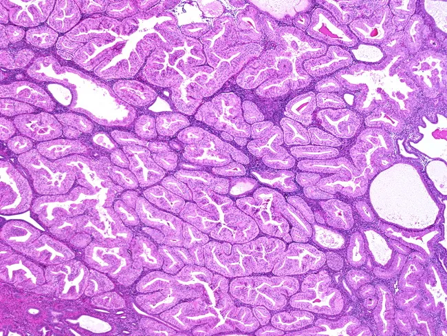
This histology image demonstrates an endometrial glandular neoplasm with well-differentiated, elongated, angulated, and branching glands embedded in a scant stroma. The glandular architecture is predominantly back-to-back, with occasional cribriform-like contours and minimal intervening cytoplasm, consistent with endometrioid histology. Cytologic atypia is mild, featuring uniform nuclei and rare mitotic figures, indicating a low-grade phenotype. The lesion contains a small solid component, described as less than or equal to 5% of tumor volume, which supports a FIGO Grade 1 classification for endometrial adenocarcinoma. The specimen originates from a formalin-fixed paraffin-embedded tissue section, stained with Hematoxylin and Eosin for routine morphologic assessment, and evaluated under bright-field microscopy. Clinically, FIGO Grade 1 endometrioid adenocarcinoma is associated with a relatively favorable prognosis compared with higher-grade counterparts, though invasion depth and myometrial involvement critically influence staging and treatment planning. Diagnostic significance centers on accurate grading, as it guides surgical management and adjuvant therapy decisions. Differential considerations include benign proliferative endometrium, endometrial hyperplasia with atypia, higher-grade endometrioid carcinomas, and serous-endometrial carcinoma. This image is valuable for gynecologic oncology education, pathology training, and research in endometrial carcinogenesis, pattern recognition, and grading criteria. This descriptive caption supports digital teaching libraries, case reviews, and AI-assisted pattern analysis in oncology education.
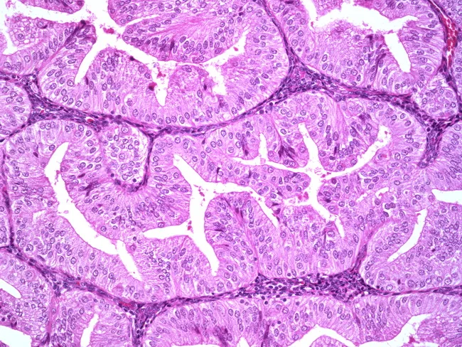
A high-magnification hematoxylin and eosin-stained histopathology image of endometrial tissue showing a well-differentiated endometrioid adenocarcinoma. Glandular architecture dominates with tubule-forming glands that resemble normal endometrium but exhibit mild cytologic atypia, including uniform nuclei and minimal pleomorphism. The tumor displays a low-grade pattern with limited solid growth, consistent with FIGO grade 1 endometrioid carcinoma; however, grading can be influenced by cytologic atypia and the relative proportion of solid components. Notable considerations include ensuring exclusion of high-grade serous carcinoma, which would require different architectural and nuclear features. The finding has significant diagnostic and clinical implications: prognosis is generally favorable for low-grade, well-differentiated lesions when confined to the uterus, but deeper myometrial invasion, lymphovascular invasion, or higher grade would alter management and adjuvant therapy. In practice, pathologists assess the balance between glandular differentiation and cytologic atypia to assign FIGO grade, with grade 2 arising if a tumor with predominant architectural pattern grade 1 shows severe atypia. Differential diagnoses include benign mimics and other endometrial carcinomas; correlation with clinical data and ancillary studies may be necessary for definitive classification. Contextual notes may include imaging findings, hormone receptor status, and molecular profiling if required for treatment planning and prognosis. These data can influence eligibility for targeted therapies and clinical trials.
Endometrial Biopsy Reporting in Cytopathology
Part 1 - Specimen Types and Adequacy
Types of samples
| Sample | Method | Primary use |
|---|---|---|
| Pipelle/Novak biopsy | Office aspiration | First-line evaluation of AUB |
| Dilatation & Curettage (D&C) | OR procedure | Definitive sampling |
| Hysteroscopy-directed biopsy | Direct visualization | Targeted lesion sampling |
| Endometrial cells on Pap smear | Cervical cytology | Incidental / screening |
Adequacy criteria (histological biopsy)
- Adequate: Minimum of 5-6 fragments of endometrial tissue with both glands and stroma; or unequivocal pathology (carcinoma, hyperplasia)
- Inadequate (scant): Fewer than 5 fragments; blood and mucus only; no recognizable endometrial tissue
- Report as: "Insufficient for diagnosis - clinical correlation advised, consider repeat sampling"
Key point: A negative/inadequate EMB does NOT exclude endometrial cancer. Up to 15% false-negative rate exists with office biopsy for focal lesions.
Part 2 - Normal Endometrium: Know What's Normal First
Menstrual cycle phases on biopsy
- Straight, tubular glands lined by pseudostratified columnar epithelium
- Mitoses present in glands and stroma
- Dense, cellular stroma
- Gland-to-stroma ratio: <1
- Subnuclear vacuoles appear first (day 17-18)
- Glands become tortuous/serrated (saw-tooth pattern)
- Stromal edema then predecidua forms
- Spiral arterioles prominent in late secretory phase
- Fragmented, hemorrhagic tissue
- Stromal breakdown and inflammatory cells
- Fragmented glands with degenerative changes
- Thin, scant strips of inactive glands
- Glands lined by low cuboidal epithelium with scant stroma
- Cystic atrophy: dilated glands with flat epithelium ("Swiss cheese" pattern)
Part 3 - WHO 2014 / 2020 Classification of Endometrial Hyperplasia
| Old WHO (4-tier) | New WHO (2-tier) | Risk of progression |
|---|---|---|
| Simple hyperplasia without atypia | Nonatypical (benign) hyperplasia | <2% |
| Complex hyperplasia without atypia | Nonatypical (benign) hyperplasia | <2% |
| Simple hyperplasia with atypia | Atypical hyperplasia / EIN | 25-30% |
| Complex hyperplasia with atypia | Atypical hyperplasia / EIN | 25-30% |
Part 4 - Nonatypical (Benign) Endometrial Hyperplasia
- Increased gland-to-stroma ratio (>1)
- Glands may be simple/tubular to mildly complex or cystically dilated
- No cytologic atypia - nuclei are elongated, pseudostratified, bland
- Mitotic activity may be present
- Stroma is present between glands
Part 5 - Atypical Hyperplasia / EIN
Diagnostic criteria (all three required):
- Architectural: Gland-to-stroma ratio >1 (glands crowd out stroma; >50% of the lesion is glands); complex, branching, or back-to-back glands
- Cytologic atypia: Nuclear enlargement, rounding (loss of elongated shape), prominent nucleoli, loss of polarity, irregular nuclear membranes - nuclei look different from the background endometrium
- Size: Lesion typically >1 mm
Key reporting points:
- State: "Atypical endometrial hyperplasia / Endometrial intraepithelial neoplasia (AEH/EIN)"
- Add a note: "Concurrent endometrial carcinoma cannot be excluded on biopsy material; hysterectomy is recommended for definitive assessment"
- ~25-40% of AEH/EIN cases have concurrent carcinoma at hysterectomy
Part 6 - Endometrial Carcinoma - Histological Classification and Grading
Type I vs. Type II Endometrial Carcinoma
| Feature | Type I | Type II |
|---|---|---|
| Histology | Endometrioid | Serous, clear cell, carcinosarcoma |
| Precursor | AEH/EIN | EIC (endometrial intraepithelial carcinoma) |
| Estrogen-driven | Yes | No |
| Prognosis | Favorable | Unfavorable |
| Mutations | PTEN, KRAS, MLH1 | TP53, HER2 |
FIGO Grading of Endometrioid Carcinoma
| Grade | Solid growth | Description |
|---|---|---|
| Grade 1 | ≤5% | Well-differentiated; almost entirely glandular |
| Grade 2 | 6-50% | Moderately differentiated |
| Grade 3 | >50% | Poorly differentiated; mostly solid |
Upgrade rule: If nuclear atypia is severe (grade 3 nuclei) in an otherwise grade 1 or 2 tumor, upgrade by one FIGO grade.
Squamous differentiation (morules) is not counted in the solid component.
Endometrioid Adenocarcinoma - Histological Features
- Low grade (G1/G2): Back-to-back glands, minimal stroma, villous or cribriform pattern; mild-moderate nuclear atypia; diagnosis of invasion = confluent glandular growth >2.1 mm, or irregular infiltration with stromal reaction
- High grade (G3): Solid sheets, marked nuclear pleomorphism, prominent nucleoli, necrosis, brisk mitoses
- Confluent glandular growth (>2.1 mm) - most common
- Irregular infiltration with desmoplastic stroma
- Extensive papillary pattern
Serous Carcinoma (Type II)
- Papillary/micropapillary architecture with tufting
- Marked nuclear pleomorphism ("hobnail" cells)
- High mitotic index
- Psammoma bodies in ~30%
- TP53 mutation; p53 overexpression on IHC
- Even limited myometrial invasion carries high risk of spread
Clear Cell Carcinoma
- Tubulocystic, papillary, or solid growth
- Clear or hobnail cells
- Hyaline basement membrane deposits
- Excellent marker: Napsin A and HNF1β positive
Part 7 - Endometrial Cells on Cervical Cytology (Pap Smear Reporting)
When to report endometrial cells:
- Benign-appearing endometrial cells: Report if patient is ≥45 years (changed from ≥40 years in TBS 2014)
- Atypical endometrial cells: Always report regardless of age
- Endometrial adenocarcinoma cells: Always report
TBS reporting categories for endometrial findings:
| Finding | Bethesda category | Action |
|---|---|---|
| Normal endometrial cells in ≥45 yr | "Other" category | Endometrial biopsy if symptomatic/postmenopausal |
| Atypical endometrial cells | AGC - endometrial (NOS) | Endometrial biopsy |
| Adenocarcinoma, endometrial | Adenocarcinoma, endometrial | Immediate referral |
Cytological features of endometrial adenocarcinoma cells on Pap:
- Small clusters or single cells
- Nuclei variably enlarged, irregular
- Prominent nucleoli
- Scant, vacuolated ("foamy") cytoplasm
- Intracytoplasmic neutrophils (characteristic)
- Watery/thin tumor diathesis (vs. thick dirty diathesis of SCC)
Part 8 - Direct Endometrial Aspiration Cytology (Cytological Reporting)
Specimen categories:
| Category | Definition |
|---|---|
| Unsatisfactory | No endometrial cells; blood/mucus only |
| Negative for malignancy | Normal endometrial cells, benign findings |
| Endometrial cells, favor benign | Reactive/reparative changes, IUD effect |
| Atypical endometrial cells | Abnormal features, cannot exclude neoplasia |
| Suspicious for malignancy | High suspicion but not definitive |
| Positive for malignancy | Diagnostic of carcinoma |
Part 9 - Special Situations in Endometrial Biopsy Reporting
IUD (Intrauterine Device) Effect
- Stromal decidualization
- Glandular atrophy or inactive appearance
- Arias-Stella-like reaction
- Actinomyces may be seen (a finding to report)
- Report as "Changes consistent with IUD effect"
Disordered Proliferative Endometrium
- Inconsistent glandular development; some cystic, some tubular
- No architectural crowding or cytologic atypia
- Gland-to-stroma ratio remains <1
- Seen in anovulatory cycles
- NOT hyperplasia - important not to over-call
Secretory vs. Arias-Stella Reaction
Endometritis
- Acute: Neutrophils within glands/stroma (excluding menstrual)
- Chronic: Plasma cells in the stroma - plasma cells are the key (normally absent in endometrium); report as "Chronic endometritis - clinical correlation advised"
Synchronous Endometrial and Ovarian Tumors
- ~10-15% of endometrioid carcinomas are associated with concurrent ovarian endometrioid carcinoma
- Report each separately; molecular analysis may help determine if one primary with metastasis or two independent primaries
Part 10 - Immunohistochemistry in Endometrial Biopsies
| Antibody | Use |
|---|---|
| ER/PR | Endometrioid carcinoma (positive); serous (negative) |
| p53 | Wildtype (scattered) = endometrioid; Aberrant (diffuse strong or null) = serous |
| PTEN | Loss in EIN and endometrioid carcinoma |
| PAX2 | Loss in EIN |
| Vimentin | Positive in endometrial; negative in endocervical adenocarcinoma |
| p16 | Diffuse strong in serous carcinoma; patchy in endometrioid |
| Ki-67 | High in high-grade tumors |
| Napsin A, HNF1β | Clear cell carcinoma |
| MLH1, MSH2, MSH6, PMS2 | Mismatch repair (Lynch syndrome screening) |
Endometrial vs. Endocervical Adenocarcinoma (key differential):
| Feature | Endometrial | Endocervical |
|---|---|---|
| Vimentin | + | - |
| CEA | - / focal | + |
| ER/PR | + | - |
| p16 | patchy | diffuse |
| HPV | - | + |
Part 11 - Structured Report Template
SPECIMEN TYPE: Endometrial biopsy / D&C / Hysteroscopy-directed biopsy
ADEQUACY: Satisfactory / Unsatisfactory (explain)
CLINICAL INFORMATION: Age, menopausal status, indication (e.g., postmenopausal bleeding, AUB, abnormal imaging)
GROSS DESCRIPTION: Fragmented tissue X cc, tan-brown, soft
HISTOLOGICAL FINDINGS:
- Endometrial phase / pattern
- Gland-to-stroma ratio
- Presence/absence of atypia
- Specific diagnosis
DIAGNOSIS:
[Category from below]
COMMENT (when needed):
- Risk of concurrent carcinoma
- Recommendation for repeat sampling or hysterectomy
- IHC results
Summary Diagnostic Categories at a Glance
| Diagnosis | Key feature | Action |
|---|---|---|
| Adequate, negative | Normal cycling endometrium | Correlate clinically |
| Disordered proliferative | Inconsistent glands, no atypia | Hormonal evaluation |
| Nonatypical hyperplasia | Gland crowding, NO atypia, G:S >1 | Progestin therapy |
| Atypical hyperplasia/EIN | Crowding + cytologic atypia | Hysterectomy preferred |
| Well-diff endometrioid Ca (G1) | Confluent glands >2.1mm, mild atypia | Surgical staging |
| High-grade Ca (G3/serous/clear cell) | Solid growth, marked atypia, necrosis | Urgent surgery + staging |
| Chronic endometritis | Plasma cells in stroma | Antibiotics; exclude PID |
| Inadequate | No endometrial tissue | Repeat biopsy / D&C |
Quick Reference: "What Atypia Means"
- No atypia = nuclei are elongated, pseudostratified, orderly, cells look like normal proliferative endometrium
- Atypia present = nuclei are round (not elongated), enlarged, prominent nucleoli, irregular membranes, loss of polarity - cells look different from the background endometrium
Create a summary PDF with diagnostic criteria and images for quick reference
mkdir -p /tmp/workspace/endometrial-pdf/images && ls /tmp/workspace/
cd /tmp/workspace/endometrial-pdf/images && \ curl -sL "https://cdn.orris.care/cdss_images/Pathology_1760056794449_5ad5edc0-79e9-4bda-9338-9937d34d522e.jpg" -o normal_proliferative.jpg && \ curl -sL "https://cdn.orris.care/cdss_images/Pathology_1760056795236_14b53763-c598-458d-a616-e6c9002f626c.jpg" -o normal_secretory.jpg && \ curl -sL "https://cdn.orris.care/cdss_images/Pathology_1760056803389_e9f2535d-4396-4983-860f-5ff70181fba4.jpg" -o nonatypical_hyperplasia.jpg && \ curl -sL "https://cdn.orris.care/cdss_images/Pathology_1760056782609_6d75506c-b5a5-44f9-b53c-d12c6cd6595e.jpg" -o who_2tier.jpg && \ curl -sL "https://cdn.orris.care/cdss_images/Pathology_1760056791443_83e88391-9903-42aa-a57f-2460d911479e.jpg" -o aeh_ein.jpg && \ curl -sL "https://cdn.orris.care/cdss_images/Pathology_1760056800808_2c1023e0-52dc-4a09-ba59-cb9ea34164f6.jpg" -o aeh_vs_carcinoma.jpg && \ curl -sL "https://cdn.orris.care/cdss_images/Pathology_1760056807429_088333c0-222b-454c-a4e5-770aa8321ec6.jpg" -o endo_ca_grade1.jpg && \ curl -sL "https://cdn.orris.care/cdss_images/Pathology_1760057415654_8a3859b3-1e0b-4de4-a2de-4f91a47cff3d.jpg" -o atyp_hyperplasia_curettage.jpg && \ ls -lh
curl -sL "https://cdn.orris.care/cdss_images/Pathology_1760056803389_e9f2535d-4396-4983-860f-5ff70181fba4.jpg" -v -o /tmp/workspace/endometrial-pdf/images/nonatypical_hyperplasia.jpg 2>&1 | tail -5; \ # Try correct URL curl -sL "https://cdn.orris.care/cdss_images/Pathology_1760056803389_e9f2535d-4396-4860-9f64-5ff70181fba4.jpg" -o /tmp/workspace/endometrial-pdf/images/nonatypical_hyperplasia2.jpg; \ ls -lh /tmp/workspace/endometrial-pdf/images/nonatypical*.jpg
~/endometrial-pdf/generate_pdf.py
"""
Endometrial Biopsy Reporting - Quick Reference PDF
Generated with ReportLab
"""
import os
from reportlab.lib.pagesizes import A4
from reportlab.lib import colors
from reportlab.lib.units import cm, mm
from reportlab.lib.styles import getSampleStyleSheet, ParagraphStyle
from reportlab.lib.enums import TA_LEFT, TA_CENTER, TA_JUSTIFY
from reportlab.platypus import (
SimpleDocTemplate, Paragraph, Spacer, Table, TableStyle,
Image, HRFlowable, PageBreak, KeepTogether
)
from reportlab.platypus.flowables import BalancedColumns
from reportlab.graphics.shapes import Drawing, Rect, String
from reportlab.pdfgen import canvas
IMG_DIR = "/tmp/workspace/endometrial-pdf/images"
OUT_PATH = "/tmp/workspace/endometrial-pdf/Endometrial_Biopsy_Reporting_Quick_Reference.pdf"
# ── Colour palette ──────────────────────────────────────────────────────────
DARK_BLUE = colors.HexColor("#1B3A6B")
MID_BLUE = colors.HexColor("#2E6DA4")
LIGHT_BLUE = colors.HexColor("#D6E4F0")
ACCENT_TEAL = colors.HexColor("#1A7A6E")
TEAL_LIGHT = colors.HexColor("#D0EDE9")
RED_WARN = colors.HexColor("#C0392B")
RED_LIGHT = colors.HexColor("#FADBD8")
AMBER = colors.HexColor("#E67E22")
AMBER_LIGHT = colors.HexColor("#FDEBD0")
GREEN = colors.HexColor("#1E8449")
GREEN_LIGHT = colors.HexColor("#D5F5E3")
GREY_DARK = colors.HexColor("#2C3E50")
GREY_MID = colors.HexColor("#5D6D7E")
GREY_LIGHT = colors.HexColor("#EAF0F6")
WHITE = colors.white
W, H = A4
MARGIN = 1.8 * cm
INNER_W = W - 2 * MARGIN
# ── Styles ───────────────────────────────────────────────────────────────────
styles = getSampleStyleSheet()
def style(name, **kw):
s = ParagraphStyle(name, **kw)
return s
TITLE_S = style("title_s", fontName="Helvetica-Bold", fontSize=22,
textColor=WHITE, alignment=TA_CENTER, spaceAfter=4)
SUBTITLE_S = style("subtitle_s", fontName="Helvetica", fontSize=11,
textColor=LIGHT_BLUE, alignment=TA_CENTER, spaceAfter=2)
DATE_S = style("date_s", fontName="Helvetica-Oblique", fontSize=9,
textColor=LIGHT_BLUE, alignment=TA_CENTER)
H1 = style("H1", fontName="Helvetica-Bold", fontSize=13,
textColor=WHITE, spaceBefore=6, spaceAfter=4)
H2 = style("H2", fontName="Helvetica-Bold", fontSize=11,
textColor=DARK_BLUE, spaceBefore=8, spaceAfter=3)
H3 = style("H3", fontName="Helvetica-Bold", fontSize=10,
textColor=ACCENT_TEAL, spaceBefore=5, spaceAfter=2)
BODY = style("BODY", fontName="Helvetica", fontSize=9,
textColor=GREY_DARK, leading=14, spaceAfter=4)
BODY_SM = style("BODY_SM", fontName="Helvetica", fontSize=8,
textColor=GREY_MID, leading=12, spaceAfter=2)
BOLD_SM = style("BOLD_SM", fontName="Helvetica-Bold", fontSize=8,
textColor=GREY_DARK, leading=12)
CAPTION = style("CAPTION", fontName="Helvetica-Oblique", fontSize=7.5,
textColor=GREY_MID, alignment=TA_CENTER, spaceAfter=6)
WARN = style("WARN", fontName="Helvetica-Bold", fontSize=8.5,
textColor=RED_WARN, leading=13)
KEY_POINT = style("KEY_POINT", fontName="Helvetica-Bold", fontSize=9,
textColor=ACCENT_TEAL, leading=13)
CELL_HEADER = style("CELL_HEADER", fontName="Helvetica-Bold", fontSize=8.5,
textColor=WHITE, alignment=TA_CENTER)
CELL_BODY = style("CELL_BODY", fontName="Helvetica", fontSize=8,
textColor=GREY_DARK, alignment=TA_LEFT, leading=12)
CELL_BODY_C = style("CELL_BODY_C", fontName="Helvetica", fontSize=8,
textColor=GREY_DARK, alignment=TA_CENTER, leading=12)
# ── Helper: section header banner ────────────────────────────────────────────
def section_banner(title, color=DARK_BLUE, text_color=WHITE):
data = [[Paragraph(title, H1)]]
t = Table(data, colWidths=[INNER_W])
t.setStyle(TableStyle([
("BACKGROUND", (0,0), (-1,-1), color),
("LEFTPADDING", (0,0), (-1,-1), 8),
("RIGHTPADDING", (0,0), (-1,-1), 8),
("TOPPADDING", (0,0), (-1,-1), 5),
("BOTTOMPADDING",(0,0), (-1,-1), 5),
("ROUNDEDCORNERS", [4]),
]))
return t
def teal_banner(title):
return section_banner(title, color=ACCENT_TEAL)
def amber_banner(title):
return section_banner(title, color=AMBER)
def red_banner(title):
return section_banner(title, color=RED_WARN)
# ── Helper: info box ─────────────────────────────────────────────────────────
def info_box(text, bg=LIGHT_BLUE, border=MID_BLUE):
data = [[Paragraph(text, BODY)]]
t = Table(data, colWidths=[INNER_W])
t.setStyle(TableStyle([
("BACKGROUND", (0,0), (-1,-1), bg),
("BOX", (0,0), (-1,-1), 1, border),
("LEFTPADDING", (0,0), (-1,-1), 10),
("RIGHTPADDING", (0,0), (-1,-1), 10),
("TOPPADDING", (0,0), (-1,-1), 6),
("BOTTOMPADDING",(0,0), (-1,-1), 6),
]))
return t
def warn_box(text):
return info_box(f"<font color='#C0392B'><b>! </b></font>{text}", bg=RED_LIGHT, border=RED_WARN)
def key_box(text):
return info_box(f"<font color='#1A7A6E'><b>KEY: </b></font>{text}", bg=TEAL_LIGHT, border=ACCENT_TEAL)
# ── Helper: image with caption ───────────────────────────────────────────────
def img_cap(filename, caption, width=None, height=None):
path = os.path.join(IMG_DIR, filename)
if not os.path.exists(path) or os.path.getsize(path) < 500:
return []
w = width or (INNER_W * 0.92)
try:
im = Image(path, width=w, height=height or (w * 0.55))
im.hAlign = "CENTER"
return [im, Paragraph(caption, CAPTION)]
except Exception:
return []
# ── Helper: two-column image layout ─────────────────────────────────────────
def two_imgs(f1, cap1, f2, cap2, col_w=None):
cw = col_w or (INNER_W / 2 - 0.3*cm)
p1 = os.path.join(IMG_DIR, f1)
p2 = os.path.join(IMG_DIR, f2)
cells = []
for p, cap, ok_size in [(p1, cap1, True), (p2, cap2, True)]:
ok_size = os.path.exists(p) and os.path.getsize(p) > 500
if ok_size:
try:
cells.append([Image(p, width=cw, height=cw*0.65),
Paragraph(cap, CAPTION)])
except Exception:
cells.append([Paragraph(cap, CAPTION)])
else:
cells.append([Paragraph(f"[Image unavailable] {cap}", CAPTION)])
data = [[cells[0], cells[1]]]
t = Table([[cells[0][0], cells[1][0]],
[cells[0][1], cells[1][1]]],
colWidths=[cw + 0.3*cm, cw + 0.3*cm])
t.setStyle(TableStyle([
("VALIGN", (0,0), (-1,-1), "TOP"),
("ALIGN", (0,0), (-1,-1), "CENTER"),
("LEFTPADDING", (0,0), (-1,-1), 2),
("RIGHTPADDING", (0,0), (-1,-1), 2),
("TOPPADDING", (0,0), (-1,-1), 2),
("BOTTOMPADDING",(0,0), (-1,-1), 2),
]))
return t
# ── Helper: generic table ────────────────────────────────────────────────────
def make_table(headers, rows, col_widths=None, header_bg=DARK_BLUE):
data = [[Paragraph(h, CELL_HEADER) for h in headers]]
for row in rows:
data.append([Paragraph(str(c), CELL_BODY) for c in row])
cw = col_widths or [INNER_W / len(headers)] * len(headers)
t = Table(data, colWidths=cw)
style_cmds = [
("BACKGROUND", (0,0), (-1,0), header_bg),
("TEXTCOLOR", (0,0), (-1,0), WHITE),
("ALIGN", (0,0), (-1,0), "CENTER"),
("VALIGN", (0,0), (-1,-1), "MIDDLE"),
("ROWBACKGROUNDS",(0,1),(-1,-1), [WHITE, GREY_LIGHT]),
("BOX", (0,0), (-1,-1), 0.5, colors.HexColor("#B0BEC5")),
("INNERGRID", (0,0), (-1,-1), 0.4, colors.HexColor("#CFD8DC")),
("LEFTPADDING", (0,0), (-1,-1), 6),
("RIGHTPADDING", (0,0), (-1,-1), 6),
("TOPPADDING", (0,0), (-1,-1), 5),
("BOTTOMPADDING",(0,0), (-1,-1), 5),
]
t.setStyle(TableStyle(style_cmds))
return t
# ── Helper: bullet list ──────────────────────────────────────────────────────
def bullets(items, style=BODY_SM):
return [Paragraph(f"<b>\u2022</b> {item}", style) for item in items]
# ── Cover page canvas callback ───────────────────────────────────────────────
def cover_page(canvas_obj, doc):
canvas_obj.saveState()
# Deep blue gradient background via rectangles
canvas_obj.setFillColor(DARK_BLUE)
canvas_obj.rect(0, 0, W, H, fill=1, stroke=0)
# Decorative bar top
canvas_obj.setFillColor(MID_BLUE)
canvas_obj.rect(0, H - 2.5*cm, W, 2.5*cm, fill=1, stroke=0)
# Accent stripe
canvas_obj.setFillColor(ACCENT_TEAL)
canvas_obj.rect(0, H - 2.7*cm, W, 0.25*cm, fill=1, stroke=0)
# Bottom bar
canvas_obj.setFillColor(MID_BLUE)
canvas_obj.rect(0, 0, W, 1.8*cm, fill=1, stroke=0)
canvas_obj.setFillColor(ACCENT_TEAL)
canvas_obj.rect(0, 1.8*cm, W, 0.2*cm, fill=1, stroke=0)
canvas_obj.restoreState()
def normal_page(canvas_obj, doc):
canvas_obj.saveState()
# Header bar
canvas_obj.setFillColor(DARK_BLUE)
canvas_obj.rect(0, H - 1.4*cm, W, 1.4*cm, fill=1, stroke=0)
canvas_obj.setFillColor(WHITE)
canvas_obj.setFont("Helvetica-Bold", 8)
canvas_obj.drawString(MARGIN, H - 0.9*cm, "ENDOMETRIAL BIOPSY REPORTING")
canvas_obj.setFont("Helvetica", 8)
canvas_obj.drawRightString(W - MARGIN, H - 0.9*cm, "Cytopathology Quick Reference")
# Footer
canvas_obj.setFillColor(LIGHT_BLUE)
canvas_obj.rect(0, 0, W, 1.0*cm, fill=1, stroke=0)
canvas_obj.setFillColor(DARK_BLUE)
canvas_obj.setFont("Helvetica", 7.5)
canvas_obj.drawCentredString(W/2, 0.35*cm, f"Page {doc.page}")
canvas_obj.restoreState()
# ═══════════════════════════════════════════════════════════════════════════════
# BUILD STORY
# ═══════════════════════════════════════════════════════════════════════════════
story = []
# ── PAGE 1: COVER ─────────────────────────────────────────────────────────────
story.append(Spacer(1, 4.5*cm))
story.append(Paragraph("ENDOMETRIAL BIOPSY", TITLE_S))
story.append(Paragraph("REPORTING IN CYTOPATHOLOGY", TITLE_S))
story.append(Spacer(1, 0.5*cm))
story.append(Paragraph("Diagnostic Criteria & Histological Quick Reference", SUBTITLE_S))
story.append(Spacer(1, 0.3*cm))
story.append(Paragraph("WHO 2014/2020 Classification | FIGO Grading | Bethesda System", SUBTITLE_S))
story.append(Spacer(1, 0.5*cm))
story.append(HRFlowable(width=INNER_W*0.5, thickness=2, color=ACCENT_TEAL, hAlign="CENTER"))
story.append(Spacer(1, 5.5*cm))
story.append(Paragraph("Orris Medical Education | August 2026", DATE_S))
story.append(Spacer(1, 0.3*cm))
story.append(Paragraph("For educational use only. Correlate with clinical context.", DATE_S))
story.append(PageBreak())
# ── PAGE 2: SPECIMEN ADEQUACY + NORMAL ENDOMETRIUM ───────────────────────────
story.append(section_banner("1. SPECIMEN TYPES & ADEQUACY"))
story.append(Spacer(1, 0.2*cm))
adq_rows = [
["Pipelle / Novak aspiration", "Office procedure; first-line for AUB"],
["Dilatation & Curettage (D&C)", "OR procedure; definitive sampling"],
["Hysteroscopy-directed biopsy", "Targeted sampling of focal lesions"],
["Endometrial cells on Pap smear", "Incidental / cervical cytology screening"],
]
story.append(make_table(["Specimen Type", "Notes"], adq_rows,
col_widths=[INNER_W*0.4, INNER_W*0.6]))
story.append(Spacer(1, 0.3*cm))
story.append(Paragraph("Adequacy Criteria", H2))
adeq_data = [
["Adequate", "≥5 fragments with both glands AND stroma; or unequivocal pathology present"],
["Scant / Insufficient", "Fewer than 5 fragments; blood and mucus only; no recognizable endometrial tissue"],
]
story.append(make_table(["Result", "Criteria"], adeq_data,
col_widths=[INNER_W*0.28, INNER_W*0.72]))
story.append(Spacer(1, 0.2*cm))
story.append(warn_box("A negative or insufficient biopsy does NOT exclude endometrial carcinoma. Office biopsy has ~15% false-negative rate for focal lesions. Consider repeat sampling or hysteroscopy-directed D&C if clinical suspicion persists."))
story.append(Spacer(1, 0.4*cm))
story.append(teal_banner("2. NORMAL ENDOMETRIUM — KNOW BEFORE YOU DIAGNOSE"))
story.append(Spacer(1, 0.25*cm))
story.append(two_imgs(
"normal_proliferative.jpg",
"Normal Proliferative Phase — tubular glands, pseudostratified nuclei, dense stroma; mitoses present",
"normal_secretory.jpg",
"Normal Secretory Phase — tortuous serrated glands, subnuclear vacuoles, stromal edema / predecidua"
))
story.append(Spacer(1, 0.25*cm))
cycle_rows = [
["Proliferative\n(days 5-14)", "Straight tubular glands", "Pseudostratified columnar cells, mitoses", "Dense, cellular", "<1"],
["Secretory\n(days 15-28)", "Tortuous, serrated ('saw-tooth')", "Subnuclear vacuoles → secretions", "Edematous → predecidualized", "<1"],
["Atrophic\n(post-menopausal)", "Thin inactive / cystically dilated", "Low cuboidal, bland", "Scant fibrous", "Variable"],
["Menstrual", "Fragmented, hemorrhagic", "Degenerative", "Breakdown, inflammation", "N/A"],
]
story.append(make_table(
["Phase", "Glands", "Epithelium", "Stroma", "G:S Ratio"],
cycle_rows,
col_widths=[INNER_W*0.16, INNER_W*0.22, INNER_W*0.26, INNER_W*0.22, INNER_W*0.14]
))
story.append(PageBreak())
# ── PAGE 3: HYPERPLASIA CLASSIFICATION ───────────────────────────────────────
story.append(section_banner("3. WHO 2014/2020 HYPERPLASIA CLASSIFICATION (2-TIER)"))
story.append(Spacer(1, 0.25*cm))
story.append(Paragraph("The 4-tier old WHO system was replaced in 2014. The key driver is <b>cytologic atypia</b>.", BODY))
story.append(Spacer(1, 0.15*cm))
who_rows = [
["Simple hyperplasia w/o atypia", "Nonatypical (Benign) Hyperplasia", "<2%", "Progestin therapy"],
["Complex hyperplasia w/o atypia", "Nonatypical (Benign) Hyperplasia", "<2%", "Progestin therapy"],
["Simple hyperplasia WITH atypia", "Atypical Hyperplasia / EIN", "25–30%", "Hysterectomy preferred"],
["Complex hyperplasia WITH atypia", "Atypical Hyperplasia / EIN", "25–30%", "Hysterectomy preferred"],
]
story.append(make_table(
["Old WHO (4-tier)", "New WHO (2-tier)", "Progression Risk", "Management"],
who_rows,
col_widths=[INNER_W*0.3, INNER_W*0.26, INNER_W*0.14, INNER_W*0.3]
))
story.append(Spacer(1, 0.3*cm))
# Side-by-side: WHO 2-tier image
imgs = img_cap("who_2tier.jpg",
"WHO 2-tier classification — Left: Nonatypical EH (well-spaced glands, elongated bland nuclei). "
"Right: Atypical EH/EIN (back-to-back crowded glands, round enlarged nuclei with prominent nucleoli).",
width=INNER_W*0.85)
for fl in imgs:
story.append(fl)
story.append(Spacer(1, 0.3*cm))
story.append(key_box("The single most important question: Is cytologic atypia present? "
"Nuclei in atypia are ROUND (not elongated), enlarged, with prominent nucleoli and loss of polarity."))
story.append(Spacer(1, 0.4*cm))
# ── Nonatypical Hyperplasia ───────────────────────────────────────────────────
story.append(Paragraph("3a. Nonatypical (Benign) Endometrial Hyperplasia", H2))
nh_items = [
"Gland-to-stroma ratio >1 (glands predominate)",
"Glands may be simple, cystic, or mildly complex",
"<b>No cytologic atypia</b> — nuclei are elongated, pseudostratified, orderly",
"Mitotic activity may be present",
"Stroma clearly present between glands",
"Cause: unopposed estrogen (anovulation, PCOS, obesity, exogenous estrogen)",
]
story.extend(bullets(nh_items))
story.append(Spacer(1, 0.3*cm))
# ── AEH/EIN ──────────────────────────────────────────────────────────────────
story.append(Paragraph("3b. Atypical Hyperplasia / EIN — Diagnostic Criteria (ALL 3 required)", H2))
aeh_criteria = [
["1. Architecture",
"Gland-to-stroma ratio >1 (>50% glands); complex branching or back-to-back glands; glands crowd out stroma"],
["2. Cytologic Atypia",
"Nuclei ROUND (not elongated); enlarged; prominent nucleoli; irregular nuclear membranes; loss of polarity; look DIFFERENT from background endometrium"],
["3. Size",
"Lesion >1 mm (to exclude small foci that may be artifactual)"],
]
story.append(make_table(
["Criterion", "Description"],
aeh_criteria,
col_widths=[INNER_W*0.18, INNER_W*0.82],
header_bg=ACCENT_TEAL
))
story.append(Spacer(1, 0.25*cm))
imgs2 = img_cap("aeh_ein.jpg",
"AEH/EIN — Crowded back-to-back glands with minimal stroma, rounded hyperchromatic nuclei, "
"nuclear enlargement, loss of polarity, and prominent nucleoli.",
width=INNER_W*0.82)
for fl in imgs2:
story.append(fl)
story.append(Spacer(1, 0.25*cm))
story.append(warn_box("AEH/EIN: Report must include — 'Concurrent endometrioid carcinoma cannot be excluded "
"on biopsy material; ~25-40% of cases harbor concurrent carcinoma at hysterectomy.' "
"Hysterectomy is the definitive management in eligible patients."))
story.append(PageBreak())
# ── PAGE 4: CARCINOMA ─────────────────────────────────────────────────────────
story.append(red_banner("4. ENDOMETRIAL CARCINOMA"))
story.append(Spacer(1, 0.25*cm))
story.append(Paragraph("Type I vs. Type II Carcinoma", H2))
type_rows = [
["Histology", "Endometrioid (80%)", "Serous, Clear Cell, Carcinosarcoma"],
["Precursor lesion", "AEH / EIN", "EIC (Endometrial Intraepithelial Carcinoma)"],
["Estrogen-driven", "Yes", "No"],
["Prognosis", "Favorable", "Unfavorable"],
["Key mutations", "PTEN, KRAS, MLH1", "TP53, HER2"],
["p53 IHC", "Wildtype (scattered)", "Aberrant (diffuse strong OR null)"],
]
story.append(make_table(
["Feature", "Type I", "Type II"],
type_rows,
col_widths=[INNER_W*0.28, INNER_W*0.36, INNER_W*0.36],
header_bg=RED_WARN
))
story.append(Spacer(1, 0.3*cm))
story.append(Paragraph("FIGO Grading — Endometrioid Carcinoma", H2))
figo_rows = [
["Grade 1 (Well-diff.)", "≤5%", "Almost entirely glandular; back-to-back glands; mild atypia", "Favorable"],
["Grade 2 (Mod. diff.)", "6–50%", "Mixed glandular and solid areas; moderate atypia", "Intermediate"],
["Grade 3 (Poorly diff.)", ">50%", "Mostly solid sheets; marked pleomorphism; necrosis", "Poor"],
]
story.append(make_table(
["Grade", "Solid Component", "Histological Features", "Prognosis"],
figo_rows,
col_widths=[INNER_W*0.18, INNER_W*0.18, INNER_W*0.44, INNER_W*0.2],
header_bg=RED_WARN
))
story.append(Spacer(1, 0.15*cm))
story.append(info_box(
"<b>FIGO Upgrade Rule:</b> If nuclear atypia is Grade 3 severity in an otherwise Grade 1 or 2 tumor, "
"upgrade by one FIGO grade. <b>Note:</b> Squamous differentiation (morules) is NOT counted in the solid component.",
bg=RED_LIGHT, border=RED_WARN))
story.append(Spacer(1, 0.3*cm))
# Images: AEH vs carcinoma + Grade 1
story.append(two_imgs(
"aeh_vs_carcinoma.jpg",
"Left: AEH/EIN — crowded glands, preserved stroma. "
"Right: Well-diff. endometrioid adenocarcinoma — confluent cribriform glands, desmoplastic stroma.",
"endo_ca_grade1.jpg",
"Grade 1 Endometrioid Adenocarcinoma — back-to-back glands with ≤5% solid component; mild cytologic atypia."
))
story.append(Spacer(1, 0.3*cm))
story.append(Paragraph("Stromal Invasion — How to Recognise", H2))
inv_items = [
"<b>Confluent glandular growth</b> >2.1 mm — most common pattern (maze-like, back-to-back glands with no intervening stroma)",
"<b>Irregular infiltration</b> with desmoplastic (fibrotic) stromal reaction around glands",
"<b>Extensive papillary growth</b> — villoglandular pattern",
]
story.extend(bullets(inv_items))
story.append(PageBreak())
# ── PAGE 5: SEROUS + CLEAR CELL + SPECIAL SITUATIONS ─────────────────────────
story.append(amber_banner("5. HIGH-GRADE CARCINOMA SUBTYPES"))
story.append(Spacer(1, 0.25*cm))
story.append(Paragraph("Serous Carcinoma (Type II — High Grade)", H2))
ser_items = [
"Papillary / micropapillary architecture with cellular tufting and detachment",
"Marked nuclear pleomorphism — 'hobnail' cells with macronucleoli",
"High mitotic index; brisk apoptosis",
"Psammoma bodies in ~30%",
"<b>IHC:</b> Aberrant p53 (diffuse strong or null), p16 diffuse, ER/PR negative",
"<b>Warning:</b> Even superficial myometrial invasion carries high risk of peritoneal spread",
]
story.extend(bullets(ser_items))
story.append(Spacer(1, 0.2*cm))
story.append(Paragraph("Clear Cell Carcinoma (Type II)", H2))
cc_items = [
"Growth patterns: tubulocystic, papillary, or solid",
"Cells: clear cytoplasm (glycogen-rich) OR hobnail cells",
"Hyaline basement membrane deposits in tubules",
"<b>IHC:</b> Napsin A+, HNF1β+, ER/PR negative",
]
story.extend(bullets(cc_items))
story.append(Spacer(1, 0.3*cm))
story.append(teal_banner("6. SPECIAL SITUATIONS IN REPORTING"))
story.append(Spacer(1, 0.2*cm))
special_rows = [
["Disordered Proliferative\nEndometrium",
"Inconsistent glandular development; some cystic, some tubular; G:S <1; NO atypia",
"NOT hyperplasia — do not over-call. Seen in anovulatory cycles."],
["Chronic Endometritis",
"<b>Plasma cells in stroma</b> (key finding — normally absent). ± neutrophils, lymphoid aggregates.",
"Report as chronic endometritis. Treat with doxycycline. Exclude PID."],
["Acute Endometritis",
"Neutrophils within glands or stroma (excluding menstrual phase)",
"Correlate with clinical context. Exclude STI."],
["IUD Effect",
"Stromal decidualization; glandular atrophy; Arias-Stella-like changes; Actinomyces may be present",
"Report as 'Changes consistent with IUD effect'"],
["Arias-Stella Reaction",
"Hobnail nuclei, marked nuclear enlargement, clear cytoplasm — mimics clear cell carcinoma",
"Seen in pregnancy / progesterone effect. Clinical context is key."],
["Atrophic Endometrium",
"Thin scant glands, low cuboidal cells, minimal stroma — can be misread as carcinoma",
"'Atrophic endometrium — benign.' No further action if clinically appropriate."],
]
story.append(make_table(
["Condition", "Histological Features", "Reporting Notes"],
special_rows,
col_widths=[INNER_W*0.22, INNER_W*0.40, INNER_W*0.38],
header_bg=ACCENT_TEAL
))
story.append(Spacer(1, 0.3*cm))
story.append(info_box(
"<b>Atypical hyperplasia curettage specimen:</b> The image below shows a classic case from a patient "
"with AUB — note the back-to-back crowded glands, architectural complexity, and cytologic atypia in "
"curettage material.",
bg=AMBER_LIGHT, border=AMBER))
imgs3 = img_cap("atyp_hyperplasia_curettage.jpg",
"Atypical Endometrial Hyperplasia in a curettage specimen — crowded glands, cribriform-like branching, "
"cytologic atypia with nuclear enlargement and stratification. Compare with background stroma.",
width=INNER_W*0.80)
for fl in imgs3:
story.append(fl)
story.append(PageBreak())
# ── PAGE 6: ENDOMETRIAL CELLS ON PAP + IHC ───────────────────────────────────
story.append(section_banner("7. ENDOMETRIAL CELLS ON PAP SMEAR — BETHESDA SYSTEM (TBS 2014)"))
story.append(Spacer(1, 0.2*cm))
story.append(info_box(
"<b>Age threshold (TBS 2014):</b> Report benign-appearing endometrial cells in women aged <b>≥45 years</b> "
"(changed from ≥40 years in TBS 2001). Always report atypical endometrial cells and adenocarcinoma cells "
"regardless of age.",
bg=LIGHT_BLUE, border=MID_BLUE))
story.append(Spacer(1, 0.2*cm))
pap_rows = [
["Normal endometrial cells, woman <45 yr", "No report required", "None (normal finding)"],
["Benign-appearing endometrial cells, woman ≥45 yr", "'Other' category", "EMB if symptomatic or postmenopausal"],
["Atypical endometrial cells (AGC-E, NOS)", "Glandular cell abnormality", "Endometrial biopsy"],
["Atypical endometrial cells, favor neoplastic", "Glandular cell abnormality", "Endometrial biopsy + colposcopy"],
["Adenocarcinoma, endometrial", "Adenocarcinoma", "Urgent referral; surgical evaluation"],
]
story.append(make_table(
["Pap Finding", "TBS Category", "Clinical Action"],
pap_rows,
col_widths=[INNER_W*0.40, INNER_W*0.28, INNER_W*0.32]
))
story.append(Spacer(1, 0.25*cm))
story.append(Paragraph("Cytological Features of Endometrial Adenocarcinoma Cells on Pap", H2))
pap_feat = [
"Small clusters or single dispersed cells",
"Nuclei variably enlarged, irregular, hyperchromatic",
"Prominent nucleoli",
"Scant, vacuolated ('foamy') cytoplasm",
"<b>Intracytoplasmic neutrophils</b> — characteristic feature",
"<b>Watery / thin tumor diathesis</b> (vs. thick dirty diathesis in squamous cell carcinoma)",
]
story.extend(bullets(pap_feat))
story.append(Spacer(1, 0.35*cm))
story.append(section_banner("8. IMMUNOHISTOCHEMISTRY PANEL"))
story.append(Spacer(1, 0.2*cm))
ihc_rows = [
["ER / PR", "Endometrioid (positive); Serous (negative)", "Hormone receptor status; treatment eligibility"],
["p53", "Wildtype (scattered) = endometrioid; Aberrant (diffuse/null) = serous", "Subtype classification; TP53 mutation proxy"],
["PTEN", "Loss in EIN and endometrioid carcinoma", "Precursor detection; molecular subtype"],
["PAX2", "Loss in EIN", "EIN marker; helps identify focal lesions"],
["Vimentin", "+ve in endometrial; -ve in endocervical", "Endometrial vs. endocervical adenoCa differential"],
["p16", "Patchy in endometrioid; Diffuse strong in serous", "Subtype differentiation; NOT HPV-related here"],
["Napsin A, HNF1β", "Positive in clear cell carcinoma", "Clear cell carcinoma marker"],
["MLH1, MSH2,\nMSH6, PMS2", "Loss indicates MMR deficiency", "Lynch syndrome screening (all endometrial Ca)"],
["Ki-67", "High in high-grade tumors", "Proliferative index; grading support"],
]
story.append(make_table(
["Antibody", "Result / Interpretation", "Clinical Use"],
ihc_rows,
col_widths=[INNER_W*0.20, INNER_W*0.46, INNER_W*0.34]
))
story.append(Spacer(1, 0.25*cm))
story.append(Paragraph("Endometrial vs. Endocervical Adenocarcinoma — Key Differentiators", H3))
diff_rows = [
["Vimentin", "+", "-"],
["CEA", "- / focal", "+"],
["ER / PR", "+", "-"],
["p16", "Patchy", "Diffuse strong"],
["HPV (ISH)", "-", "+"],
["PAX8", "+", "+"],
]
story.append(make_table(
["Marker", "Endometrial", "Endocervical"],
diff_rows,
col_widths=[INNER_W*0.25, INNER_W*0.375, INNER_W*0.375],
header_bg=ACCENT_TEAL
))
story.append(PageBreak())
# ── PAGE 7: MASTER QUICK-REFERENCE TABLE + REPORT TEMPLATE ──────────────────
story.append(section_banner("9. MASTER DIAGNOSTIC SUMMARY TABLE"))
story.append(Spacer(1, 0.2*cm))
master_rows = [
["Normal cycling endometrium", "Phase-appropriate glands and stroma", "None", "Correlate clinically"],
["Atrophic endometrium", "Thin inactive glands, minimal stroma", "None", "Benign — no action"],
["Disordered proliferative", "Inconsistent glands, G:S <1, no atypia", "None", "Hormonal evaluation"],
["Nonatypical hyperplasia", "G:S >1, crowding, NO atypia", "None", "Progestin therapy; 6-month follow-up"],
["Atypical hyperplasia / EIN", "G:S >1, crowding + cytologic atypia", "High (~25%)", "Hysterectomy (preferred)"],
["Endometrioid Ca, Grade 1", "Confluent glands >2.1mm, mild atypia, ≤5% solid", "Malignant", "Surgical staging"],
["Endometrioid Ca, Grade 2", "6–50% solid component", "Malignant", "Surgical staging"],
["Endometrioid Ca, Grade 3", ">50% solid, marked pleomorphism", "Malignant", "Urgent surgery + staging"],
["Serous carcinoma", "Papillary tufting, marked atypia, p53 aberrant", "Malignant (aggressive)", "Urgent surgery"],
["Clear cell carcinoma", "Clear/hobnail cells, Napsin A+, HNF1β+", "Malignant (aggressive)", "Urgent surgery"],
["Chronic endometritis", "Plasma cells in stroma (key)", "None", "Antibiotics; exclude PID"],
["Inadequate specimen", "No recognizable endometrial tissue", "N/A", "Repeat biopsy / D&C"],
]
story.append(make_table(
["Diagnosis", "Key Histological Features", "Malignant Risk", "Action"],
master_rows,
col_widths=[INNER_W*0.25, INNER_W*0.38, INNER_W*0.17, INNER_W*0.20]
))
story.append(Spacer(1, 0.4*cm))
story.append(teal_banner("10. STRUCTURED REPORT TEMPLATE"))
story.append(Spacer(1, 0.2*cm))
template_text = """<font name="Courier" size="8.5">
<b>SPECIMEN TYPE:</b> Endometrial biopsy / D&C / Hysteroscopy-directed biopsy<br/><br/>
<b>ADEQUACY:</b> Satisfactory (≥5 fragments, glands and stroma present) /
Unsatisfactory (specify reason)<br/><br/>
<b>CLINICAL INFORMATION:</b> Age: ___ yr | Menopausal status: Pre / Post
Indication: □ Postmenopausal bleeding □ AUB □ Abnormal imaging
□ Follow-up hyperplasia □ Other: _________<br/><br/>
<b>GROSS DESCRIPTION:</b> Fragmented tan-brown tissue measuring ___ cc (aggregate).<br/><br/>
<b>HISTOLOGICAL FINDINGS:</b><br/>
- Gland-to-stroma ratio: _____<br/>
- Endometrial phase / pattern: _____<br/>
- Cytologic atypia: Present / Absent<br/>
- Mitotic activity: _____<br/>
- Specific features: _____<br/><br/>
<b>DIAGNOSIS:</b><br/>
[ ] Adequate, negative for hyperplasia or malignancy — ___ phase endometrium<br/>
[ ] Nonatypical (benign) endometrial hyperplasia<br/>
[ ] Atypical endometrial hyperplasia / Endometrial intraepithelial neoplasia (AEH/EIN)<br/>
[ ] Endometrioid adenocarcinoma, FIGO Grade ___<br/>
[ ] Serous carcinoma<br/>
[ ] Clear cell carcinoma<br/>
[ ] Other: _____<br/>
[ ] Insufficient for diagnosis<br/><br/>
<b>COMMENT (if applicable):</b><br/>
- Concurrent carcinoma risk<br/>
- IHC results (p53, MLH1/MSH2/MSH6/PMS2, ER/PR, Napsin A, etc.)<br/>
- Recommendation for repeat sampling or hysterectomy
</font>"""
story.append(info_box(template_text, bg=GREY_LIGHT, border=GREY_MID))
story.append(Spacer(1, 0.3*cm))
story.append(key_box(
"The KEY diagnostic decision: Is cytologic atypia present? "
"ABSENT → Nonatypical hyperplasia (benign, manage with progestins). "
"PRESENT → AEH/EIN (premalignant; recommend hysterectomy)."))
story.append(PageBreak())
# ── FINAL PAGE: ATYPIA DECISION TREE ─────────────────────────────────────────
story.append(section_banner("11. DIAGNOSTIC DECISION FLOWCHART"))
story.append(Spacer(1, 0.3*cm))
flow_data = [
[Paragraph("<b>ENDOMETRIAL BIOPSY RECEIVED</b>", style("fc0", fontName="Helvetica-Bold",
fontSize=10, textColor=WHITE, alignment=TA_CENTER))],
]
flow_tbl = Table(flow_data, colWidths=[INNER_W])
flow_tbl.setStyle(TableStyle([
("BACKGROUND", (0,0),(-1,-1), DARK_BLUE),
("TOPPADDING", (0,0),(-1,-1), 8),
("BOTTOMPADDING",(0,0),(-1,-1), 8),
("ROUNDEDCORNERS",[4]),
]))
story.append(flow_tbl)
story.append(Spacer(1, 0.2*cm))
def flow_box(text, bg, border, width=INNER_W):
d = [[Paragraph(text, style("fbt", fontName="Helvetica", fontSize=9,
textColor=GREY_DARK, alignment=TA_CENTER))]]
t = Table(d, colWidths=[width])
t.setStyle(TableStyle([
("BACKGROUND",(0,0),(-1,-1),bg),
("BOX",(0,0),(-1,-1),1.5,border),
("TOPPADDING",(0,0),(-1,-1),7),
("BOTTOMPADDING",(0,0),(-1,-1),7),
("ROUNDEDCORNERS",[4]),
]))
return t
def arrow():
return Paragraph("<font size=14>▼</font>",
style("arr", fontName="Helvetica", fontSize=14,
textColor=GREY_MID, alignment=TA_CENTER, spaceAfter=2))
story.append(flow_box("STEP 1: Is the specimen adequate?\n(≥5 fragments with glands + stroma)",
GREY_LIGHT, GREY_MID))
story.append(Spacer(1,0.1*cm))
row2 = Table(
[[flow_box("NO → Inadequate\nReport: Insufficient for diagnosis\nRecommend: Repeat biopsy / D&C",
RED_LIGHT, RED_WARN, (INNER_W/2 - 0.3*cm)),
flow_box("YES → Proceed to Step 2",
GREEN_LIGHT, GREEN, (INNER_W/2 - 0.3*cm))]],
colWidths=[INNER_W/2, INNER_W/2])
row2.setStyle(TableStyle([("VALIGN",(0,0),(-1,-1),"MIDDLE"),
("LEFTPADDING",(0,0),(-1,-1),4),
("RIGHTPADDING",(0,0),(-1,-1),4)]))
story.append(row2)
story.append(Spacer(1,0.15*cm))
story.append(arrow())
story.append(flow_box("STEP 2: Is gland-to-stroma ratio >1?\n(Glands crowd out stroma in >50% of lesion)",
GREY_LIGHT, GREY_MID))
story.append(Spacer(1,0.1*cm))
row3 = Table(
[[flow_box("NO → Normal endometrium or\nDisordered proliferative\n(correlate with cycle phase)",
GREEN_LIGHT, GREEN, (INNER_W/2 - 0.3*cm)),
flow_box("YES → Hyperplasia (some form)\nProceed to Step 3",
AMBER_LIGHT, AMBER, (INNER_W/2 - 0.3*cm))]],
colWidths=[INNER_W/2, INNER_W/2])
row3.setStyle(TableStyle([("VALIGN",(0,0),(-1,-1),"MIDDLE"),
("LEFTPADDING",(0,0),(-1,-1),4),
("RIGHTPADDING",(0,0),(-1,-1),4)]))
story.append(row3)
story.append(Spacer(1,0.15*cm))
story.append(arrow())
story.append(flow_box("STEP 3: Is CYTOLOGIC ATYPIA present?\n(Round nuclei, nuclear enlargement, prominent nucleoli, loss of polarity)",
GREY_LIGHT, GREY_MID))
story.append(Spacer(1,0.1*cm))
row4 = Table(
[[flow_box("NO → NONATYPICAL HYPERPLASIA\n(Benign — <2% progression risk)\nManage: Progestin therapy",
GREEN_LIGHT, GREEN, (INNER_W/2 - 0.3*cm)),
flow_box("YES → ATYPICAL HYPERPLASIA / EIN\n(Premalignant — 25-30% progression risk)\nManage: Hysterectomy preferred",
RED_LIGHT, RED_WARN, (INNER_W/2 - 0.3*cm))]],
colWidths=[INNER_W/2, INNER_W/2])
row4.setStyle(TableStyle([("VALIGN",(0,0),(-1,-1),"MIDDLE"),
("LEFTPADDING",(0,0),(-1,-1),4),
("RIGHTPADDING",(0,0),(-1,-1),4)]))
story.append(row4)
story.append(Spacer(1,0.15*cm))
story.append(arrow())
story.append(flow_box("STEP 4: If architectural crowding is very marked — is there stromal invasion?\n"
"(Confluent glands >2.1mm, desmoplasia, irregular infiltration)",
GREY_LIGHT, GREY_MID))
story.append(Spacer(1,0.1*cm))
row5 = Table(
[[flow_box("NO → AEH/EIN\n(add: cannot exclude concurrent Ca)",
AMBER_LIGHT, AMBER, (INNER_W/2 - 0.3*cm)),
flow_box("YES → ENDOMETRIAL CARCINOMA\nGrade 1/2/3 per FIGO\nUrgent surgical referral",
RED_LIGHT, RED_WARN, (INNER_W/2 - 0.3*cm))]],
colWidths=[INNER_W/2, INNER_W/2])
row5.setStyle(TableStyle([("VALIGN",(0,0),(-1,-1),"MIDDLE"),
("LEFTPADDING",(0,0),(-1,-1),4),
("RIGHTPADDING",(0,0),(-1,-1),4)]))
story.append(row5)
story.append(Spacer(1, 0.3*cm))
story.append(info_box(
"<b>Remember:</b> Disordered proliferative endometrium ≠ hyperplasia (G:S <1). "
"Chronic endometritis = plasma cells in stroma (key finding). "
"Arias-Stella ≠ clear cell carcinoma (pregnancy context). "
"Always report: specimen adequacy, phase, atypia, invasion.",
bg=TEAL_LIGHT, border=ACCENT_TEAL))
# ── BUILD PDF ─────────────────────────────────────────────────────────────────
doc = SimpleDocTemplate(
OUT_PATH,
pagesize=A4,
leftMargin=MARGIN, rightMargin=MARGIN,
topMargin=1.6*cm, bottomMargin=1.2*cm,
title="Endometrial Biopsy Reporting — Quick Reference",
author="Orris Medical Education",
subject="Cytopathology",
)
# First page uses cover callback; subsequent pages use normal callback
def on_page(canvas_obj, doc):
if doc.page == 1:
cover_page(canvas_obj, doc)
else:
normal_page(canvas_obj, doc)
doc.build(story, onFirstPage=on_page, onLaterPages=on_page)
print(f"PDF created: {OUT_PATH}")
print(f"Size: {os.path.getsize(OUT_PATH) / 1024:.1f} KB")
cd /tmp/workspace/endometrial-pdf && python generate_pdf.py 2>&1
python3 -c "from pypdf import PdfReader; r=PdfReader('/tmp/workspace/endometrial-pdf/Endometrial_Biopsy_Reporting_Quick_Reference.pdf'); print(f'Pages: {len(r.pages)}')"
Endometrial Biopsy Reporting Quick Reference
PDF Document · PDF
| Page | Content |
|---|---|
| 1 | Cover page |
| 2 | Specimen types, adequacy criteria, normal endometrium phases (with histology images) |
| 3 | WHO 2-tier hyperplasia classification (with side-by-side nonatypical vs. atypical image), nonatypical hyperplasia criteria, AEH/EIN criteria table |
| 4 | AEH/EIN histology image, endometrial carcinoma Type I vs II, FIGO grading table, stromal invasion patterns, histology images |
| 5 | Serous + clear cell carcinoma features, special situations (IUD effect, endometritis, disordered proliferative, Arias-Stella), atypical hyperplasia curettage image |
| 6 | Endometrial cells on Pap smear (Bethesda TBS 2014), full IHC panel, endometrial vs. endocervical adenocarcinoma differentiators |
| 7-8 | Master diagnostic summary table (12 diagnoses with features + actions) |
| 9-10 | Structured report template |
| 11-12 | Step-by-step diagnostic decision flowchart (adequacy → G:S ratio → atypia → invasion) with colour-coded decision branches |
Pleomorphic adenoma cytology
pleomorphic adenoma FNAC cytology smear chondromyxoid stroma spindle cells
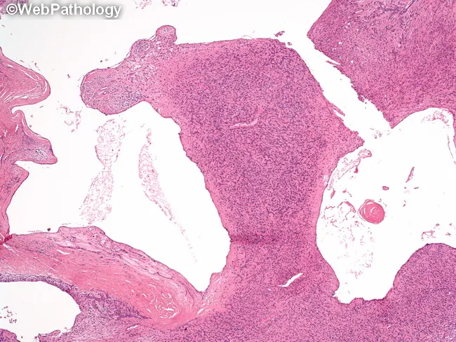
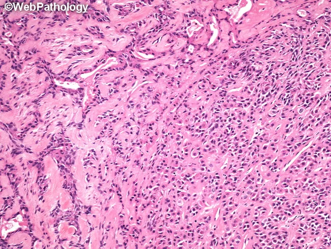
This histopathology image depicts a pleomorphic adenoma of a salivary gland showing prominent cystic change. Prepared as a hematoxylin and eosin stained paraffin section, the sample is examined under light microscopy at low to moderate magnification. The lesion demonstrates the classic biphasic composition of pleomorphic adenoma: ductal epithelial and myoepithelial cells forming primitive acini, embedded in a variable stromal background that ranges from myxoid to chondroid (cartilaginous) stroma. In this instance, extensive cystic degeneration creates large cystic spaces dissecting through glistening, acellular or loosely cellular stromal areas. The cyst lining may be attenuated epithelium or flattened cells with turbulence of mucinous content in the luminal space. The epithelial components show bland cytology without significant pleomorphism or atypia; mitotic activity is low. The myoepithelial cells contribute spindle to plasmacytoid differentiation, contributing to the chondromyxoid stroma. These cystic changes can be primary, or secondary to prior fine-needle aspiration or manipulation, which has diagnostic implications to avoid misinterpretation as a cystic carcinoma or necrotic process. Clinically, pleomorphic adenoma presents as a slow-growing, painless parotid or minor salivary gland mass. Recognition of cystic change is essential to accurate diagnosis, appropriate surgical planning, and margin assessment to minimize recurrence.
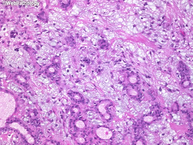
Imaging modality: light microscopy of Hematoxylin and Eosin stained histology section from a salivary gland lesion. Anatomical site: parotid region; histologic plane: peripheral-to-central glandular architecture. The slide reveals classic pleomorphic adenoma features with biphasic tumor components in a variably hyalinized, myxoid stroma. Epithelial nests and anastomosing cords are embedded in a chondromyxoid matrix, while duct-like tubules are evident in places. In the lower-right field, small nests and cords form an Indian file pattern, illustrating cellular diversity within a single tumor. The left third shows compressed and distorted tubular structures amid dense stromal hyalinization, contributing to a mosaic of microarchitectures. The myoepithelial element contributes spindle- to plasmacytoid cells with variable cytoplasmic appearance; the overall cytology lacks significant atypia or invasion. Clinically, these features correlate with a slow-growing, painless parotid mass and favorable prognosis after complete excision. Diagnostic significance rests on recognizing both epithelial and myoepithelial components within a mucoid to cartilaginous stroma, and on identifying variant patterns to avoid misclassification as malignant salivary neoplasms. Differential diagnoses include adenoid cystic carcinoma, mucoepidermoid carcinoma, and basal cell adenoma; accurate histologic interpretation guides management and recurrence risk assessment. Awareness of this histologic diversity improves diagnostic accuracy and informs surgical planning.
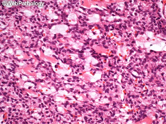
Imaging modality: light microscopy of hematoxylin and eosin stained salivary gland tissue section. The image captures the classic pleomorphic adenoma, a benign mixed tumor, showing a well circumscribed, lobulated neoplasm with a triphasic cellular composition. Epithelial ductal elements form cords and nests of cuboidal to columnar cells, while myoepithelial cells assume plasmacytoid or spindle morphologies surrounding the epithelial structures. The stroma is prominent and variably myxoid to chondromyxoid, creating a cartilaginous or hyaline backdrop. The overall pattern displays heterogeneity yet maintains benign cytology, with uniform nuclei, gentle chromatin, and minimal mitotic activity. No significant pleomorphism or invasion is evident. This combination of epithelial, myoepithelial, and stromal components underlies the tumor’s designation as a pleomorphic (mixed) adenoma. The image aligns with the molecular profile frequently observed: PLAG1 (8q12) and HMGA2 (12q13-15) rearrangements, while TP53 alterations are uncommon. Clinically relevant, pleomorphic adenomas most often arise in the parotid gland and present as slow-growing, painless masses. The diagnosis informs surgical management—complete excision with a margin to prevent recurrence—and highlights a potential, albeit rare, risk of malignant transformation to carcinoma ex pleomorphic adenoma in long-standing cases.
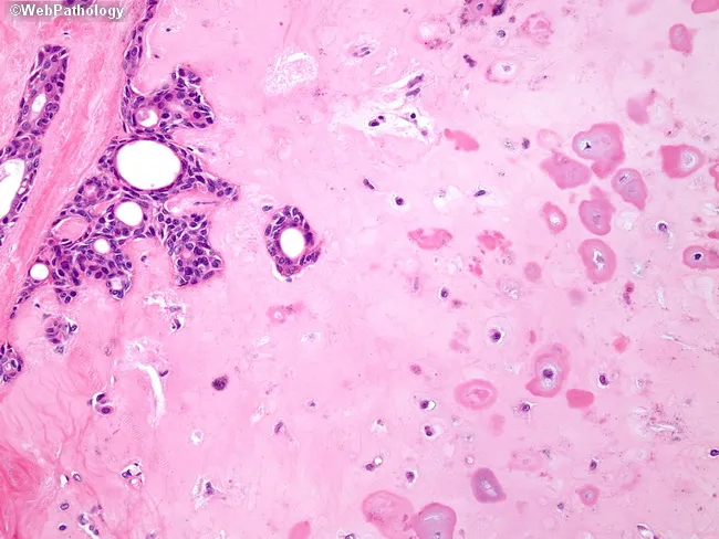
Imaging modality and technique: Bright-field light microscopy of formalin-fixed, paraffin-embedded salivary gland tissue stained with Hematoxylin and Eosin. The specimen represents a pleomorphic adenoma, a benign mixed tumor classically containing epithelial ductal elements and myoepithelial cells within a variably myxoid to chondromyxoid hyalinized stroma. In this high-power field, spindle- to stellate-shaped myoepithelial cells form a loose reticular network around ducts and tubules. The epithelial component yields small duct-like structures and occasional acinar- or tubuloductal formations; the neighboring stroma is abundant, eosinophilic, and matrix-rich, often displaying cartilaginous or myxoid differentiation. Cellular morphology shows spindle-shaped myoepithelial cells with scant cytoplasm and elongated nuclei, embedded in a matrix that may resemble cartilage or fibrous stroma. The lesion is typically well-circumscribed with minimal cytologic atypia and rare mitotic figures. Diagnostic significance resides in recognizing this classic constellation, supporting a benign diagnosis and guiding management toward definitive surgical excision. Clinically, pleomorphic adenoma typically presents as a slow-growing, painless parotid mass; differential diagnoses include adenoid cystic carcinoma and mucoepidermoid carcinoma; radiology and FNA assist preoperative planning. This image also supports AI-based pattern recognition in histopathology, illustrating tumor-stroma interactions and the reticular myoepithelial network associated with chondromyxoid differentiation.
High-power histopathology micrograph of a salivary gland lesion stained with Hematoxylin and Eosin (H&E). The section shows a classic pleomorphic adenoma with biphasic differentiation: ducts lined by a single layer of cuboidal-to-columnar epithelial cells forming small ductal structures, and a dispersed population of plump, bland myoepithelial cells. The stroma is distinctly chondroid and myxoid, producing a cartilaginous, basophilic matrix that mingles with epithelial elements. The epithelial cells display uniform cytomorphology, with minimal mitotic activity and no overt nuclear pleomorphism or atypia. The lesion is well circumscribed within the glandular parenchyma, containing islands of neoplastic epithelium embedded in an abundant chondromyxoid matrix. Some ducts are inconspicuous, others form back-to-back clusters; mucinous secretions are not prominent. The overall impression is of a benign mixed tumor, commonly known as pleomorphic adenoma, characterized by a spectrum of architectural patterns including ductal epithelium and chondroid stroma. Diagnostic significance lies in recognizing the characteristic stromal heterogeneity and bland cytology, which differentiate it from malignant salivary gland neoplasms such as mucoepidermoid or adenoid cystic carcinoma. These findings underpin surgical planning and prognosis, with emphasis on complete excision to minimize recurrence risk. The sample’s architecture underscores the tumor’s benign behavior and guides conservative, organ-preserving resection in this case.
pleomorphic adenoma FNAC fine needle aspiration cytology smear fibrillar matrix epithelial cells
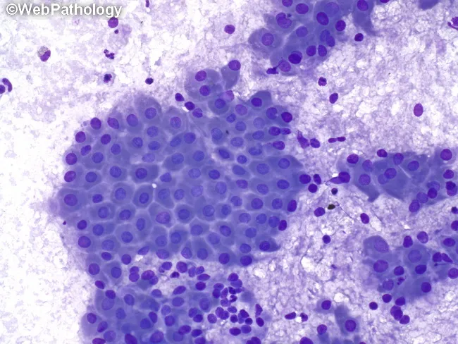
Fine-needle aspiration cytology (FNAC) of a parotid gland lesion demonstrates cohesive sheets and clusters of oncocytic epithelial cells embedded in a lymphoid-rich background. Diff-Quik stained smears reveal cells with abundant granular eosinophilic cytoplasm, round to oval nuclei with smooth nuclear chromatin, and scant cytoplasm at the periphery; occasional mitoses are absent or rare. The inflammatory background consists of mature lymphocytes, plasma cells, and occasional mucus-containing fluid, consistent with Warthin’s tumor (papillary cystadenoma lymphomatosum). The architectural pattern shows double-layered oncocytic epithelium with rosette-like clusters and microcystic spaces; however, cytology often lacks intact glandular ducts, making full histology difficult. Clinically, these findings strongly support a benign parotid tumor; the differential includes pleomorphic adenoma and mucoepidermoid carcinoma, particularly if mucous-producing cells or cytologic atypia are present. The diagnostic significance is high when cytology presents characteristic oncocytic cells and a lymphoid stroma; correlation with imaging and clinical features is essential. Management implications include surgical planning for superficial parotidectomy with facial nerve preservation. In cases with cystic change or hemorrhagic background, FNAC may yield background mucus or thick fluid; the presence of numerous oncocytic cells with dense lymphoid stroma is highly suggestive of Warthin’s tumor. Typical demographic associations include older male patients and parotid localization.
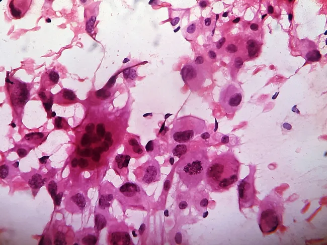
Fine-needle aspiration cytology (FNAC) of a midline anterior neck mass yields a highly cellular smear composed of pleomorphic malignant cells with markedly enlarged, hyperchromatic nuclei, conspicuous nucleoli, and occasional intranuclear inclusions. The tumor cells are variably polygonal to bizarre in shape, often arranged singly or in discohesive clusters, with frequent mitotic figures. Intermixed within the cellular background are numerous osteoclast-like giant cells containing multiple nuclei, creating a characteristic giant cell-rich pattern. Background material shows scant colloid and occasional necrotic debris, consistent with an anaplastic process in the thyroid. The overall cytomorphology is compatible with Anaplastic Thyroid Carcinoma (ATC), a rare, highly aggressive thyroid malignancy; the giant cell-rich variant represents one morphologic spectrum. Immunocytochemical profiling is typically required to confirm thyroid origin and exclude mimics such as lymphoma or metastatic sarcoma. Clinically, ATC presents in elderly patients as a rapidly enlarging neck mass with potential airway compromise and vocal changes; the prognosis remains dismal despite therapy. This image thus documents a high-grade undifferentiated thyroid neoplasm with giant cell differentiation, emphasizing the diagnostic challenge and need for ancillary studies, including thyroid lineage markers (TTF-1, PAX8) and proliferation indices, to support an accurate diagnosis and guide urgent multidisciplinary management for optimal patient care.
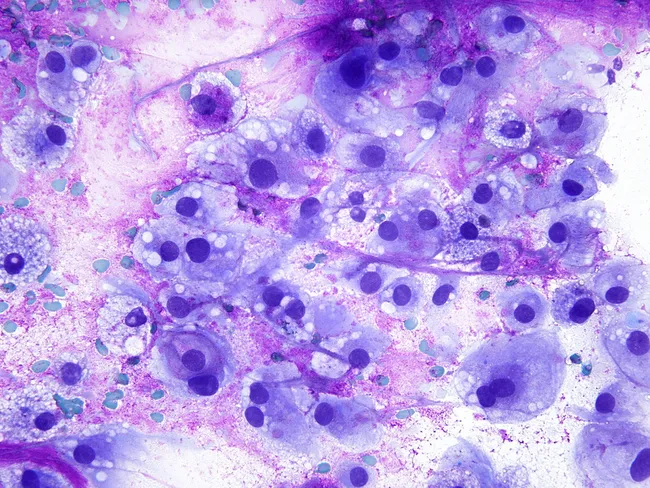
Fine-needle aspiration cytology (FNAC) of a paraspinal soft tissue mass demonstrates cells with voluminous, bubbly cytoplasm and large, bland hyperchromatic nuclei set in a myxoid, occasionally granular background. The cytologic smear is highly cellular, with scattered physaliferous cells featuring vacuolated, sometimes foamy cytoplasm and small-to-moderate nuclei. Many cells appear columnar-oval with a discohesive affinity, and occasional binucleation is seen. The distinctive bubbly cytoplasmic vacuoles and the presence of cartilaginous or chondroid–like matrix support a chordoma diagnosis, though differential includes clear cell renal cell carcinoma and other chondroid neoplasms. The staining used is Giemsa, which highlights the cytoplasmic vacuolization and the delicate, lace-like myxoid stroma around the tumor cells. Overall, the image demonstrates a neoplasm with notochordal differentiation, consistent with chordoma arising in the paraspinal region. Diagnostic significance lies in recognizing physaliferous cells and their abundant cytoplasm, which, in the appropriate clinical and radiologic context, directs confirmation by immunohistochemistry (brachyury positivity) and correlation with imaging to define extent. Differential diagnoses include metastatic RCC, chondrosarcoma, and chordoid variants; immunostaining and radiologic correlation are essential. This cytology image has educational value for surgical pathology, cytology practice, and differential diagnosis training in spine-associated tumors.
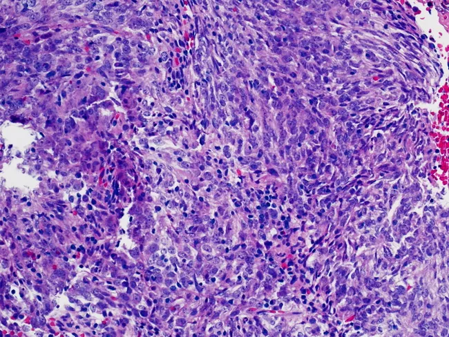
Fine-needle aspiration cytology (FNAC) of an abdominal mass was prepared as a conventional smear and stained with Hematoxylin and Eosin (H&E). The smear is moderately cellular, composed of interlacing sheets and fascicles of spindle-shaped and epithelioid cells with elongated to oval nuclei, finely textured chromatin, and occasional small nucleoli. The cytoplasm is variable, pale to eosinophilic, with occasional binucleation; cells are embedded in a prominent inflammatory background containing lymphocytes and plasma cells, with rare macrophages. Mitoses are infrequent; necrosis is not evident. The overall morphology shows a dendritic cell-like constellation with broad cytoplasmic processes and cohesive clusters that can mimic other spindle cell neoplasms. Immunohistochemistry (performed on ancillary material) reveals strong and diffuse CD21 positivity, supporting a follicular dendritic cell lineage. The cytologic features, together with CD21 expression, favor follicular dendritic cell sarcoma (FDC sarcoma) over other spindle cell tumors such as fibrosarcoma, inflammatory myofibroblastic tumor, sarcomatoid carcinoma, or melanoma variants. Clinically, this diagnosis is important because it informs surgical planning and adjuvant therapy considerations and requires correlative histopathology for definitive confirmation. The image documents cytomorphology with dendritic cell-rich background and immunophenotype concordance; additional markers (CD23, CD35) can increase diagnostic confidence in FDC sarcoma.
pleomorphic adenoma parotid gland FNAC cytology Diff-Quik fibrillar myxoid chondroid matrix plasmacytoid myoepithelial
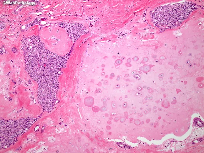
Light microscopy of a hematoxylin and eosin stained section of salivary gland tissue (parotid region) demonstrates a well-defined lobular architecture composed of chondroid (chondromyxoid) material embedded in hyalinized stroma. The epithelial component forms duct-like structures and nests, while the surrounding spindle-to-plasmacytoid myoepithelial cells exhibit diverse morphologies, including epithelial, clear cell, and hyaline (plasmacytoid) differentiation. This biphasic pattern, with intimate admixture of myoepithelial elements and a cartilaginous-like stroma, is characteristic of a benign mixed tumor, pleomorphic adenoma. The chondromyxoid matrix shows variable basophilia and occasional chondral calcifications, though limited cytologic atypia is typical. The lesion is usually well circumscribed within the glandular parenchyma, and mitotic activity is low. Immunohistochemical profiling may reveal p63 and cytokeratin positivity in the myoepithelial cells with S100 protein labeling in the myxoid stroma, supporting diagnosis. Clinically this finding correlates with a slow-growing, painless mass in the salivary gland region, most commonly the parotid. Diagnostic significance lies in distinguishing pleomorphic adenoma from malignant salivary gland tumors such as adenoid cystic carcinoma or mucoepidermoid carcinoma, guiding management toward conservative surgical excision with clear margins. The image is educational for histopathology, surgical pathology, and oral and maxillofacial pathology curricula.
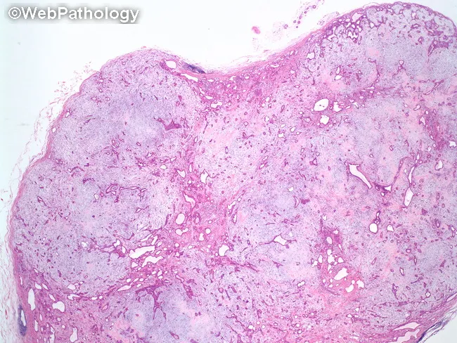
Pleomorphic adenoma (PA) of salivary gland, most commonly the parotid. This low-power histopathology image shows a well-demarcated, lobulated neoplasm with a bland appearance. The tumor is composed of a mixed epithelial and myoepithelial element embedded in a prominent myxoid-to-chondroid stromal background. Epithelial components form variable tubular and acinar structures, sometimes lining duct-like lumina, while myoepithelial cells contribute spindle, plasmacytoid, or clear cytoplasm elements; myxoid stroma gradually transitions to chondroid cartilage-like matrix. The capsule is usually incomplete or thin, allowing some intermingling with adjacent salivary tissue, but the lesion remains well circumscribed in this low magnification field. The histologic pattern is highly pleomorphic, depending on the relative proportion of ductal epithelium, myoepithelial cells, and stroma. Notable features include representation of both epithelial tubules and an abundant myxochondroid stroma, which helps distinguish PA from other salivary gland neoplasms. Clinically, PA is benign, slow-growing, and typically presents as a painless mass; malignant conversion is rare but possible. Immunohistochemistry may reveal cytokeratin-positive epithelial cells and S-100/GFAP-positive myoepithelial components. Treatment is surgical excision with adequate margins. The image exemplifies classic PA morphology: encapsulated lobules, biphasic cell populations, and a chondromyxoid extracellular matrix. The image provides a classic teaching example for salivary gland pathology and differential diagnosis.
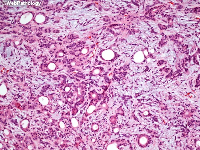
Histopathology of a pleomorphic adenoma from the superficial lobe of the parotid gland. This hematoxylin and eosin stained section demonstrates a benign mixed tumor with triphasic composition: an epithelial component forming cords and duct-like structures, a prominent myoepithelial component with plasmacytoid and spindle cells, and a variably myxoid to chondroid stromal background. The epithelial nests are embedded in a scant to abundant extracellular matrix that ranges from mucoid to cartilaginous, producing the characteristic pleomorphic, or pleomorphic, appearance. The tumor is usually encapsulated or pseudocapsulated with a fibrous capsule; widespread cellular atypia is absent; mitotic activity is rare. The interface with surrounding salivary tissue is well circumscribed, and small satellite nodules may be present at the periphery, accounting for recurrences if the capsule is breached during excision. Clinically, pleomorphic adenoma is the most common benign salivary gland tumor, typically presenting as a slow-growing, painless parotid mass in adults. Surgical management favors superficial parotidectomy with facial nerve preservation; enucleation and simple excision carry high recurrence due to satellite nodules. Adjuvant radiotherapy may be considered if tumor spillage or residual disease occurs. The diagnostic significance rests on recognizing the classic triphasic morphology; differential diagnoses include other benign salivary gland tumors and low-grade carcinomas.
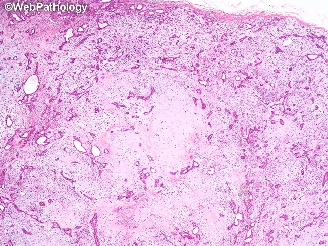
Imaging modality: light microscopy of a hematoxylin and eosin stained salivary gland specimen. This low-power view demonstrates a well-demarcated, lobulated pleomorphic adenoma arising in salivary gland tissue, most consistent with parotid origin. The tumor exhibits a biphasic epithelial/myoepithelial composition with duct-like tubular and acinar structures formed by epithelial cells admixed with variably sized myoepithelial cells. The stroma is abundant and myxoid to chondroid, producing a characteristic, amorphous, basophilic to pink background that supports chondromyxoid differentiation. The lobulated architecture suggests a pseudocapsule and circumscribed growth, while the epithelial elements display ductal differentiation and variable cellularity. Myoepithelial cells may appear spindle- to plasmacytoid-shaped, contributing to a spectrum of morphological patterns within a single lesion. Notable features include prominent stromal components (myxoid matrix, sometimes chondroid-like) alongside epithelial tubules; this histology underlies the classic description of pleomorphic adenoma as a mixed tumor. Clinically, this pattern correlates with a slow-growing, painless parotid mass, favorable prognosis with complete excision, and a need to avoid capsule rupture during surgery to minimize recurrence. Differential considerations include adenoid cystic carcinoma and mucoepidermoid carcinoma, which carry different clinical implications and require distinct management strategies. Overall, the image captures the quintessential PA histomorphology used for diagnostic confirmation and educational reference. Purposes.
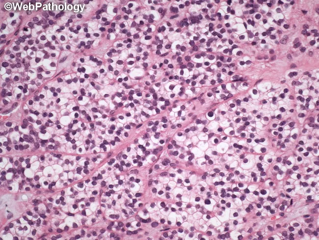
Imaging modality: Light microscopy of a hematoxylin and eosin (H&E) stained salivary gland tissue section, likely parotid, showing microscopic pleomorphic adenoma architecture with epithelial and myoepithelial components embedded in a chondromyxoid stroma. This image highlights a focal clear cell change within the epithelial component: cells with abundant pale, optically clear cytoplasm and centrally placed to slightly eccentric nuclei; the cytoplasmic clearing may reflect glycogen or intracellular mucin. In the background, typical pleomorphic adenoma features include haphazard clusters and nests of duct-like epithelial cells, plasmacytoid myoepithelial cells, and a myxoid to chondroid stroma; stromal regions may appear pink with mucopolysaccharide-rich matrix. The lesion is usually well circumscribed, encapsulated, and shows no invasion into surrounding salivary parenchyma; mitotic activity is low. This histologic variant can mimic other clear cell neoplasms; differential diagnoses include clear cell variant of adenoid cystic carcinoma, mucoepidermoid carcinoma with clear cells, and metastatic clear cell carcinomas; however, the lack of cytologic atypia and invasion supports a benign pleomorphic adenoma with clear cell change. Clinically relevant because recognizing this variant prevents misdiagnosis and guides surgical planning, prognosis, and follow-up in salivary gland tumors. Correlation with imaging and intraoperative frozen section may further aid management.
Pleomorphic Adenoma - Cytopathology (FNAC)
1. Clinical Essentials (Know Before You Aspirate)
| Feature | Detail |
|---|---|
| Most common site | Parotid gland (superficial lobe, tail) |
| Also occurs in | Submandibular gland, minor salivary glands (palate), sublingual |
| Presentation | Slow-growing, painless, mobile, discrete firm lump |
| Age / sex | Adults (30-60 yrs), slight female predominance |
| Firmness | Varies — soft (myxoid-rich) to hard (chondroid/hyaline-rich) |
| Key molecular | PLAG1 rearrangement (8q12); HMGA2 (12q13) |
The preoperative FNAC goal: confirm benign nature, guide surgical planning (superficial parotidectomy, NOT enucleation).
2. Gross / Macroscopic Appearance
- Well-circumscribed, rounded/ovoid, bosselated (lumpy) surface
- Variable but often incomplete capsule (very important surgically)
- Cut surface: grey-white with blue translucent chondroid areas and myxoid zones
- Some examples are nearly semi-fluid (myxoid dominant) - at high risk of capsular rupture during surgery, seeding the operative field
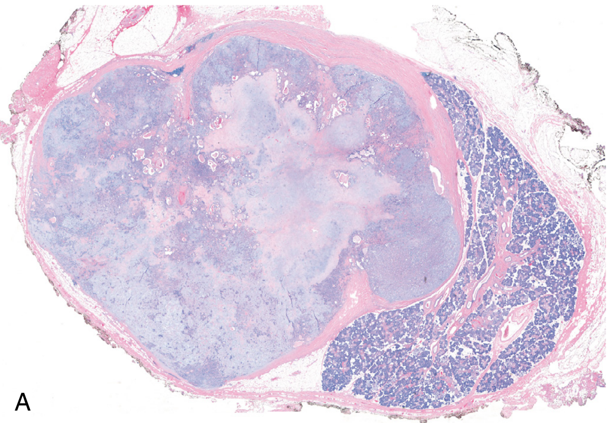
3. Histological Composition - The "Triphasic" Tumour
| Component | Description |
|---|---|
| 1. Ductal epithelial cells | Cuboidal to columnar cells lining tubules/acini; form double-layered ducts |
| 2. Myoepithelial cells | The most important and variable component - see morphologies below |
| 3. Chondromyxoid stroma | Fibrillar/myxoid matrix with islands of cartilage; the hallmark of PA |
Myoepithelial cell morphologies (all in the SAME tumour):
- Spindle cells - elongated, wavy nuclei embedded in fibrillar matrix
- Plasmacytoid (hyaline) cells - eccentric round nuclei, abundant glassy pink cytoplasm - most common
- Clear cells - vacuolated cytoplasm, can mimic clear cell carcinoma
- Stellate cells - within the myxoid matrix, extending into the stroma
- Epithelioid cells - rounded cells in sheets or nests
4. FNAC Cytology - The Diagnostic Triad
1. Epithelial cells + 2. Myoepithelial cells + 3. Fibrillar (chondromyxoid) stromal material
4a. Smear Characteristics
- Cellularity: Moderate to highly cellular
- Background: Magenta-coloured fibrillary/myxoid material (best seen on Diff-Quik / MGG stain); may have chondroid areas
- Stain tip: Romanowsky stains (MGG, Diff-Quik) show the fibrillar stroma brilliantly in magenta-pink. Papanicolaou stain shows epithelial cells better but matrix is pale green/grey.
4b. Epithelial Cell Features
- Cohesive sheets, tubules, or acinar clusters
- Cells are bland, uniform, cuboidal to columnar
- Nuclei: round to oval, smooth membranes, fine chromatin, small nucleolus or inconspicuous
- Cytoplasm: moderate, pale
- No significant nuclear atypia, no mitoses
- May show squamous metaplasia (morules of squamous cells) - normal finding in PA
4c. Myoepithelial Cell Features (KEY component)
| Morphology | Appearance |
|---|---|
| Plasmacytoid (most common) | Eccentric nucleus, abundant glassy/hyaline cytoplasm; resemble plasma cells |
| Spindle | Elongated cells blending into fibrillar matrix; bare spindle nuclei |
| Epithelioid | Round cells in sheets |
| "Naked nuclei" | Stripped myoepithelial nuclei dispersed in the matrix |
Bare oval or spindle nuclei scattered in the fibrillar background = a strong clue to PA on low-power scan.
4d. The Stromal Material - THE Pathognomonic Feature
- Fibrillary / metachromatic matrix - magenta on Diff-Quik, pale on PAP
- Appears as fluffy, irregular, fibrillary pink-grey wisps surrounding epithelial cells
- Transitional zones: cells appear to "melt" into the stroma (cells embedded within the matrix)
- May condense into chondroid globules (translucent blue-grey on Diff-Quik)
- Hyaline globules or acellular homogeneous zones may also be present
5. Cytological Patterns by Stain
| Feature | Diff-Quik / MGG | Papanicolaou (PAP) |
|---|---|---|
| Stroma | Magenta / bright pink fibrillary | Pale grey-green, fibrillar |
| Epithelial cells | Greenish-grey cytoplasm | Well-visualised, translucent |
| Myoepithelial cells | Plasmacytoid cells clearly seen | Less distinct |
| Background | Granular or fibrillary matrix | Cleaner |
| Best for | Matrix identification | Nuclear detail |
6. Diagnostic Summary — What to Report
"Smears show moderate cellularity. Cohesive sheets and clusters of bland ductal epithelial cells are seen along with dispersed plasmacytoid/spindle myoepithelial cells. The background shows abundant fibrillary/chondromyxoid metachromatic material. Cytological features are consistent with pleomorphic adenoma (benign mixed tumour)."
7. Pitfalls and Difficult Cases
7a. Sparse matrix (cellular PA)
- Predominantly epithelial, little stroma
- Can mimic: basal cell adenoma, adenoid cystic carcinoma
- Clue: basaloid cells in PA lack the cribriform cylinders and hyaline globules of AdCC
7b. Predominantly myxoid PA
- Lots of matrix, few cells
- Can mimic: myxoma, mucoepidermoid carcinoma (mucinous type)
- Clue: matrix in PA is fibrillary/chondroid, not mucoid/watery; no mucous cells
7c. Squamous metaplasia within PA
- Islands of squamous cells can appear alarming
- Can mimic: mucoepidermoid carcinoma or squamous cell carcinoma
- Clue: context - surrounded by bland matrix and plasmacytoid cells; no intermediate cells or mucous cells
7d. Atypia in long-standing PA
- Occasional nuclear enlargement, prominent nucleoli
- Do not call malignant unless there is frank carcinoma
- Clue: overall background is classic PA; atypia is focal
7e. Cystic PA
- Cyst fluid ± squamous cells, macrophages, debris
- Mistaken for: Warthin's tumour, mucoepidermoid carcinoma, branchial cyst
- Clue: look for fibrillary material even in small amounts; do cellblock
7f. Carcinoma ex PA
- Suspect when: sudden rapid growth in a long-standing mass, pain, fixity, facial nerve involvement
- FNAC shows: overtly malignant cells - significant nuclear atypia, mitoses, necrosis — superimposed on background of PA
- Report as: "Malignant neoplasm arising in a background of pleomorphic adenoma — carcinoma ex pleomorphic adenoma cannot be excluded"
8. Immunocytochemistry (on cell block)
| Marker | Result |
|---|---|
| CK7, AE1/AE3 | Positive (ductal epithelial cells) |
| S100 protein | Positive (myoepithelial cells + stroma) |
| GFAP | Positive (myoepithelial cells) |
| p63 | Positive (myoepithelial cells) |
| SMA (smooth muscle actin) | Positive (myoepithelial cells) |
| Calponin | Positive (myoepithelial cells) |
| Ki-67 | Low (<5%) — confirms benign behaviour |
S100 + GFAP positivity in the spindle/myoepithelial cells is a strong supporter of PA in difficult cases.
9. Differential Diagnosis on FNAC
| Diagnosis | Overlapping feature | Key distinguishing feature |
|---|---|---|
| Warthin's tumour | Parotid mass | Oncocytic cells + lymphocytes + cystic background; NO fibrillar matrix |
| Adenoid cystic carcinoma | Basaloid cells in matrix | Hyaline globules (NOT fibrillar); cribriform architecture; nuclear angulation/hyperchromasia; malignant atypia |
| Basal cell adenoma | Basaloid epithelial sheets | No fibrillar stroma; nests with peripheral palisading; no plasmacytoid cells |
| Mucoepidermoid carcinoma | Mucoid material | Mucous cells (goblet/columnar mucinous); squamoid + intermediate cells; no fibrillar matrix |
| Myoepithelioma | Plasmacytoid myoepithelial cells | Pure myoepithelial; NO ductal component; NO chondromyxoid stroma |
| Normal salivary gland | Acinar cells | Acinar cells with zymogen granules; no fibrillar matrix; no plasmacytoid cells |
10. The Adenoid Cystic vs PA Matrix: A Critical Distinction
| Feature | PA Matrix | AdCC Matrix |
|---|---|---|
| Appearance | Fibrillary, fluffy, irregular wisps | Rounded globular cylinders (hyaline balls) |
| Stain (DQ) | Magenta fibrillar | Magenta rounded globules |
| Cell-matrix relationship | Cells embedded within/blending into matrix | Cells wrapped around/surrounding globules |
| Cellularity | Moderate | High |
| Nuclear features | Bland, round-oval | Angular, hyperchromatic, "carrot-shaped" |
| Clinical | Slow-growing, benign | Perineural invasion, recurrence, malignant |
11. Recurrence and Malignant Transformation
- Recurrence after enucleation: ~25% (due to satellite nodules in incomplete capsule)
- Recurrence after superficial parotidectomy: ~4%
- Recurrence is typically multifocal (seeding from rupture or satellite nodules)
- Malignant transformation (carcinoma ex PA):
- Overall risk: ~6%
- <5 years duration: 2% risk
-
15 years duration: ~10% risk
- Risk factors: older age, male, long duration, deep lobe location, multiple recurrences
- Resulting carcinoma is highly aggressive: 30-50% mortality at 5 years
- Robbins & Kumar Basic Pathology, p. 543
Quick-Reference Summary Card
PLEOMORPHIC ADENOMA — FNAC TRIAD
━━━━━━━━━━━━━━━━━━━━━━━━━━━━━━━━━━━━━━━━━━━━━
1. DUCTAL EPITHELIAL CELLS
- Bland, cohesive sheets/tubules
- Cuboidal-columnar, small nuclei, no atypia
2. MYOEPITHELIAL CELLS
- Plasmacytoid (glassy hyaline cytoplasm) ← MOST COMMON
- Spindle cells / bare oval nuclei in matrix
3. FIBRILLARY / CHONDROMYXOID STROMAL MATRIX
- THE pathognomonic feature
- Magenta-fibrillar on Diff-Quik
- Cells "embedded" within or blending into it
STAINS: Diff-Quik = matrix identification
PAP = nuclear detail
IHC: S100+, GFAP+, p63+ (myoepithelial)
CK7+ (ductal)
BEWARE: AdCC has globular (not fibrillar) matrix
+ angular hyperchromatic nuclei
━━━━━━━━━━━━━━━━━━━━━━━━━━━━━━━━━━━━━━━━━━━━━
Rosai Dorma n disease
Rosai-Dorfman disease emperipolesis histiocytes lymph node histology
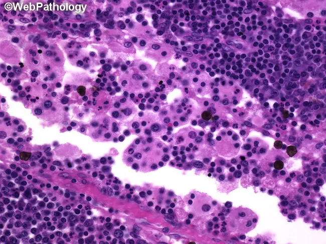
Rosai-Dorfman disease (sinus histiocytosis with massive lymphadenopathy) histology. This light- microscopy image shows a lymph node biopsy with prominent sinus involvement by large histiocytes containing intact lymphocytes and plasma cells within their cytoplasm (emperipolesis). The histiocytes are S-100 positive with abundant pale cytoplasm; non-destructive phagocytosis is evident as clinical hallmark. Erythrophagocytosis may also be present. The surrounding lymphoid paracortex and cortex show a mixed population of small mature lymphocytes and plasma cells, without overt necrosis or significant mitotic activity, indicating benign proliferation rather than malignant lymphoma. The overall architecture of the lymph node is variably preserved apart from the expanded sinuses. Immunohistochemical confirmation (not shown) typically demonstrates histiocytes positive for S-100 and CD68, negative for CD1a, supporting Rosai-Dorfman disease diagnosis. Emperipolesis, although characteristic, is not exclusive to RDD and can be observed in other inflammatory or proliferative conditions. The image is useful for educational differentiation from Langerhans cell histiocytosis, which shows CD1a and langerin positivity and different cellular morphology. This slide exemplifies diagnostic features for pathology residents and hematopathology fellows, complementing clinical correlation of painless lymphadenopathy, fever, and potential extranodal disease. Applications include histopathology teaching, differential diagnosis practice, and research documentation of emperipolesis in nodal tissue for educational use.
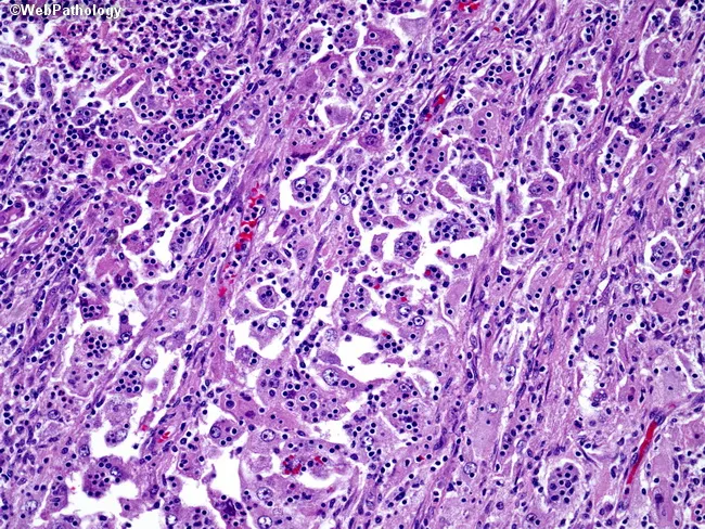
Imaging modality: Light microscopy of an H&E stained lymph node biopsy, captured at high magnification. Anatomical context: Lymphoid tissue within the lymphoreticular system, specifically lymph node parenchyma with sinusoidal expansion. The histology shows Rosai-Dorfman disease features: plump histiocytes with large, vesicular nuclei and prominent central nucleoli; cytoplasm is granular and eosinophilic. These histiocytes are embedded in a dense background of small lymphocytes and scattered plasma cells. A hallmark finding in this image is emperipolesis, where intact lymphocytes and other hematopoietic cells are variably situated within the cytoplasm of the histiocytes without destruction. Immunophenotypic context, although not shown here, typically reveals S-100 positivity in the histiocytes with CD68 positive and CD1a negative staining, supporting Rosai-Dorfman. The architectural pattern is that of sinus histiocytosis with histiocytes occupying sinuses and occasionally effacing normal nodal architecture. Clinically, Rosai-Dorfman presents with painless, often massive cervical lymphadenopathy and possible fever; the lesion is benign but can be progressive and extranodal involvement may occur. Diagnostic significance lies in recognizing emperipolesis and the characteristic histiocytic profile, which differentiates Rosai-Dorfman disease from Langerhans cell histiocytosis and infectious/inflammatory lymphadenitis. Potential educational and research use includes pathology training, differential diagnosis practice, and clinical-pathological correlation.
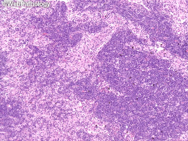
Rosai-Dorfman disease is illustrated by a cervical lymph node specimen in which nodal architecture is markedly effaced by expanded sinusoids filled with abundant pale-staining histiocytes. The histiocytes commonly display large, round to polygonal morphology with ample cytoplasm, and may exhibit emperipolesis, wherein intact lymphocytes or plasma cells are seen within the histiocyte cytoplasm. Hematoxylin and eosin staining produces a pink-purple parenchyma with scattered multinucleated histiocytes and a mixed inflammatory background of lymphocytes and plasma cells. The architectural distortion contrasts with benign reactive nodes and contrasts with Langerhans cell histiocytosis, which lacks prominent emperipolesis. Immunohistochemistry typically demonstrates S-100 protein positivity and CD68 positivity in the histiocytes, supporting the diagnosis together with clinical features. The lesion is usually part of a systemic process; extranodal involvement may occur but massive cervical adenopathy remains a common presentation in pediatric and young adult patients. The image highlights sinusoids expanded by histiocytes rather than neoplastic lymphoid proliferation, suggesting a nonmalignant sinus histiocytosis pattern. Clinically, RDD should be considered in the differential diagnosis of persistent cervical lymphadenopathy with systemic symptoms; differentiate from lymphoma and infectious etiologies. The demonstrated histology is essential for confirming Rosai-Dorfman disease and guiding management and prognosis.
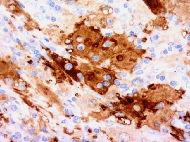
Immunohistochemical micrograph of Rosai-Dorfman disease demonstrating S-100 positive histiocytes highlighting emperipolesis within lymph node–derived tissue. The specimen is prepared as a formalin-fixed paraffin section, stained with S-100 antibody (DAB chromogen) and counterstained with hematoxylin, yielding brown cytoplasmic labeling of histiocytes and blue nuclei in adjacent lymphocytes. Histiocytes are large, with abundant eosinophilic to pale cytoplasm and vesicular nuclei arranged in sinusoids or clusters, and they actively contain intact lymphocytes and plasma cells within their cytoplasm, a feature termed emperipolesis. The immunophenotype is supportive, with S-100 positivity and strong CD68 positivity; CD1a is typically negative or rarely positive, helping distinguish Rosai-Dorfman from Langerhans cell histiocytosis. Additional non-specific markers such as CD4, CD11c, CD14, CD33, CD64, CD163, lysozyme, alpha-1-antitrypsin, and alpha-1-antichymotrypsin may be expressed in histiocytes, whereas CD3 and CD20 are absent in the lesional histiocytes. This image emphasizes the diagnostic hallmark of Rosai-Dorfman disease: emperipolesis by S-100 positive histiocytes. Clinically, this finding supports a nodal lymphohistiocytic disorder with potential extranodal involvement; integration with clinical presentation and imaging is essential for differential diagnosis and appropriate management. Keywords for search include Rosai-Dorfman, sinus histiocytosis, emperipolesis, S-100 positivity, histiocytes, immunophenotype, CD68, CD1a negative, Langerhans cell histiocytosis differential, nodal disease, pediatric/adolescent presentation, extranodal Rosai-Dorfman, DAB immunostain, bright-field microscopy.
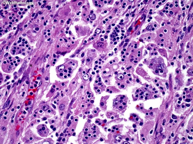
Imaging Modality: Light microscopy of a hematoxylin and eosin-stained lymph node biopsy section examined at high magnification. Primary subject: Rosai-Dorfman disease (sinus histiocytosis with massive lymphadenopathy), demonstrated by large pale histiocytes within nodal sinuses containing internalized, non-degrading lymphocytes. Morphology: prominent emperipolesis with intact lymphocytes traversing histiocyte cytoplasm; histiocytes show ample cytoplasm and irregular, vesicular nuclei amid a lymphoplasmacytic background. Immunophenotype (inferred from diagnosis, not visible in this image): S-100 positive and CD68 positive; CD1a negative, distinguishing from Langerhans cell histiocytosis. Architectural pattern: nodal sinus expansion with scattered histiocytes, occasional plasma cells, and preserved lymphoid architecture. Notable features: absence of necrosis or significant mitotic activity; absence of Birbeck granules on EM is not shown here. Diagnostic significance: emperipolesis within histiocytes is pathognomonic for Rosai-Dorfman when combined with supportive clinical context; differential diagnoses include LCH, reactive lymphadenitis, and other histiocytic disorders. Clinical correlation: commonly presents in children or young adults with generalized painless lymphadenopathy; extranodal involvement may occur. Potential clinical use: educational reference for histiocyte biology, emperipolesis, and differential diagnosis of histiocytic proliferations; guides immunohistochemical workup and clinical management planning. This image serves as a concise reference for learners to recognize emperipolesis and differentiate Rosai-Dorfman from other histiocytic disorders in education and diagnostic practice.
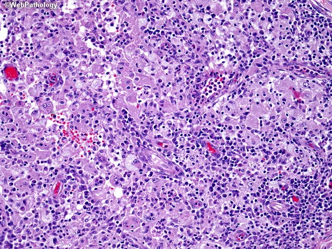
This histopathology slide depicts classic Rosai-Dorfman disease (sinus histiocytosis with massive lymphadenopathy) seen on light microscopy of a lymph node biopsy stained with Hematoxylin and Eosin. The tissue shows a dense, cellular infiltrate composed of large, pale histiocytes with vesicular or central nuclei and prominent nucleoli, intermingled with a background of lymphocytes and plasma cells. A key diagnostic feature is emperipolesis, wherein intact lymphocytes or plasma cells are observed within the cytoplasm of histiocytes, creating a striking, non-destructive intracytoplasmic engulfment. The architecture may be preserved or disrupted by proliferating histiocytes, and mitotic activity, if present, is typically low. Immunophenotype is not shown here but in standard practice Rosai-Dorfman histiocytes are S-100 positive and CD68 positive, helping distinguish from Langerhans cell histiocytosis and other histiocytoses. Clinically, the disease often presents with lymphadenopathy but may involve extranodal sites, including the gastrointestinal tract, where involvement is frequently incidental. Etiology remains uncertain; proposed triggers include infectious agents or immune dysregulation, yet no conclusive cause exists. Management varies from observation in self-resolving cases to chemotherapy in selected, progressive disease; many cases can follow a protracted course spanning years to decades. This image is well-suited for education on histiocytic disorders, differential diagnosis, and learning objectives related to emperipolesis.
Rosai-Dorfman Disease (RDD)
Also known as:
- Sinus Histiocytosis with Massive Lymphadenopathy (SHML)
- Destombes-Rosai-Dorfman Syndrome
1. Background and Definition
2. Epidemiology
| Feature | Detail |
|---|---|
| Prevalence | ~5 per 100,000 (rare) |
| Peak age | 2nd and 3rd decades (children and young adults) |
| Sex | More common in males (nodal form); females predominate in skin-limited form |
| Race | Higher incidence in West Indies; reported more in Black individuals in older registries |
| Geography | Worldwide distribution |
- H syndrome (SLC29A3 mutation) - nodal RDD in 20%
- Autoimmune lymphoproliferative syndrome (ALPS) type Ia (TNFRSF6 / Fas mutation) - nodal RDD in 41%
- Immune disorders occur in ~15% of RDD patients (associated with worse prognosis)
- Association with Hodgkin and non-Hodgkin lymphomas reported
- Goldman-Cecil Medicine, p. 1785
3. Pathogenesis
Molecular/Genetic:
- Somatic mutations activating the MAPK pathway — found in a subset:
- NRAS, KRAS, ARAF, MAP2K1/MEK1, MET — most common
- BRAF — rarely involved (unlike Langerhans cell histiocytosis)
- This MAPK activation links RDD to the broader family of histiocytic neoplasms
- Germline mutations: SLC29A3 (familial form); FAS/TNFRSF6 (ALPS-associated)
Viral hypothesis:
- Postulated association with HHV-6, CMV, EBV, parvovirus B19, polyomavirus
- No definitive causal link proven to date
Immunological:
- Polyclonal hypergammaglobulinemia and abundant plasma cells suggest immune dysregulation
- The histiocytes are polyclonal (not a true neoplasm in the traditional sense)
- IgG4-positive plasma cells are frequent within the infiltrate (can mimic IgG4-related disease)
Current understanding places RDD within the "R group" of the revised histiocytosis classification (2016), alongside Erdheim-Chester disease. - Dermatology 2-Volume Set, p. 1954
4. Histopathology — The Diagnostic Core
Architecture (low power):
- Lymph node sinuses are massively dilated and packed with large pale histiocytes
- Residual lymphoid follicles remain at the periphery (architecture partially preserved)
- Background: abundant plasma cells and mature lymphocytes between sinuses
- Alternating light and dark zones = pale histiocyte-filled sinuses alternating with dark lymphoid areas (classic low-power pattern)
The RDD Histiocyte — Morphology (high power):
| Feature | Description |
|---|---|
| Size | Very large (30-100 µm diameter) |
| Nucleus | Large, vesicular, hypochromatic (pale), with prominent central nucleolus |
| Cytoplasm | Abundant, pale/eosinophilic or vacuolated |
| Nuclear membranes | Smooth, regular |
| Mitoses | Rare or absent |
5. Emperipolesis — THE Pathognomonic Feature
What gets engulfed:
- Lymphocytes (most common)
- Plasma cells
- Erythrocytes (erythrophagocytosis)
- Neutrophils
- Other histiocytes (occasionally)
Key distinction — Emperipolesis vs. Phagocytosis:
| Feature | Emperipolesis | Phagocytosis |
|---|---|---|
| Engulfed cell state | Intact, viable (nucleus well preserved) | Degenerate, fragmented |
| Phagosome | Absent (cell sits free in cytoplasm) | Present (cell enclosed in vacuole) |
| Process | Active, non-destructive | Destructive digestion |
| Significance | Pathognomonic of RDD | Non-specific (macrophage function) |
6. Immunohistochemistry
| Marker | Result | Significance |
|---|---|---|
| S100 | Strongly positive (cytoplasmic + nuclear) | Most important marker; highlights both histiocytes AND emperipolesis |
| CD68 | Positive | Macrophage lineage confirmation |
| CD163 | Positive | Macrophage/histiocyte marker |
| CD14 | Positive | Monocyte/macrophage marker |
| HLA-DR | Positive | Antigen-presenting cell activation |
| Fascin | Positive | Actin-bundling protein in histiocytes |
| Stabilin-1 | Positive | Scavenger receptor; relatively specific for RDD histiocytes |
| α1-antitrypsin | Positive | Histiocyte marker |
| HAM-56 | Positive | Human alveolar macrophage marker |
| CD1a | Negative | Excludes Langerhans cell histiocytosis |
| CD207 (Langerin) | Negative | Excludes LCH |
| OCT2 | Positive (nuclear) | Recently reported in all RDD cases (nonspecific) |
| Cyclin D1, pERK | Positive | Reflects MAPK pathway activation |
The combination of S100+ / CD68+ / CD1a- / CD207- is the defining immunophenotype. The IgG4+ plasma cells within the infiltrate can raise suspicion of IgG4-related disease, but clinical features are distinct. - Fitzpatrick's Dermatology, p. 117
7. Clinical Manifestations
Nodal RDD (classic form):
| Feature | Detail |
|---|---|
| Lymphadenopathy | Massive, painless, bilateral cervical — hallmark presentation |
| Other nodal sites | Axilla, mediastinum, retroperitoneum, inguinal (any node group) |
| Constitutional | Fever, elevated ESR/CRP |
| Blood | Mild anemia (occasionally severe), neutrophilia |
| Serology | Polyclonal hypergammaglobulinemia |
| Complications | Nasal or tracheal obstruction |
Extranodal RDD (43% of sporadic cases):
- Skin and soft tissue (~10% of systemic form)
- Eyelids and orbit (proptosis)
- Upper respiratory tract / nasal cavity / sinuses
- Major salivary glands
- Central nervous system — dura (mimics meningioma on imaging)
- Bone
- Others: breast, kidney, thyroid, testis, GI tract
Intracranial RDD mimics meningioma radiographically but characteristic cells may be seen in CSF. - Goldman-Cecil Medicine, p. 1786
Skin-limited RDD (distinct form):
- Occurs primarily in adult women
- Single or multiple lesions: red, red-brown, or xanthomatous macules, papules, nodules, or plaques
- Favours eyelids and malar regions
- Classified in "histiocytosis group C" (not group R like systemic RDD)
- No associated lymphadenopathy
8. Clinical Course
- Immunologic abnormalities (~15% of cases) — associated with most fatalities
- Disseminated nodal disease
- Liver, kidney, or lower respiratory tract involvement
- ~10 of 14 fatal cases in one analysis had immune dysfunction
9. Diagnosis
- Expanded sinuses filled with large pale histiocytes
- Emperipolesis (engulfed intact lymphocytes/plasma cells in histiocyte cytoplasm)
- Immunophenotype: S100+ / CD68+ / CD1a-
- Plasma cell-rich background
10. Differential Diagnosis
| Condition | Overlap | Key Distinguishing Feature |
|---|---|---|
| Langerhans Cell Histiocytosis | S100+ histiocytes, lymphadenopathy | CD1a+, CD207(Langerin)+; Birbeck granules on EM; grooved "coffee-bean" nuclei; NO true emperipolesis |
| Lymphoma (Hodgkin/NHL) | Massive lymphadenopathy | Atypical lymphoid cells, Reed-Sternberg cells (HL); CD30+, CD15+; no emperipolesis |
| Juvenile Xanthogranuloma (JXG) | Can have large S100+ macrophages | Touton giant cells; CD1a-; emperipolesis rare; no massive lymphadenopathy |
| IgG4-Related Disease | IgG4+ plasma cells in infiltrate | Fibrosing/sclerosing pattern; no emperipolesis; different organ distribution |
| Hemophagocytic Syndrome (HLH) | Erythrophagocytosis, systemic inflammation | Erythrophagocytosis inside phagosomes (destructive); fever, cytopenias, hyperferritinemia; no emperipolesis |
| Erdheim-Chester Disease | Histiocytic infiltration, extranodal | CD68+, CD1a-, S100- (usually negative); foam cells; BRAF V600E common; bone involvement |
| Kikuchi-Fujimoto Disease | Cervical lymphadenopathy, fever | Necrotic areas with karyorrhectic debris; histiocytes DO NOT show emperipolesis |
| Reactive Sinus Histiocytosis | Sinus expansion, histiocytes | Histiocytes small, lack vesicular nuclei/prominent nucleoli; no true emperipolesis; S100 negative |
| Infectious Lymphadenitis | Enlarged tender nodes, fever | Suppurative foci, organisms on culture/PCR; no emperipolesis |
11. Treatment
| Modality | Use |
|---|---|
| Observation | Asymptomatic/mild disease; spontaneous resolution common |
| Corticosteroids | First-line for symptomatic cases |
| Clofarabine | Refractory cases |
| Radiation therapy | Localised symptomatic disease |
| Surgical resection | Localised extranodal lesions (especially CNS, orbital, ENT) |
| MEK inhibitors (cobimetinib) | KRAS- or MAP2K1-variant disease — targeted therapy |
| Cryotherapy | Refractory cutaneous RDD (case reports) |
12. FNAC / Cytology Findings
- Highly cellular smear
- Large histiocytes with abundant pale cytoplasm and large vesicular nuclei with prominent nucleoli
- Emperipolesis visible on smear — intact lymphocytes or plasma cells within histiocyte cytoplasm (the most diagnostic cytological feature)
- Background: abundant mature lymphocytes, plasma cells
- No necrosis, no mitoses, no atypical lymphoid cells
Cytological diagnosis is possible but confirmatory IHC on cell block (S100+, CD1a-) is strongly recommended.
13. Summary Mnemonics
- Emperipolesis (pathognomonic)
- Elevated ESR + hypergammaglobulinemia
- Extranodal involvement (43%)
S100 + CD68 + CD163 + Stabilin-1 | CD1a − CD207− = Rosai-Dorfman ✓ (not LCH)
Quick Reference Card
ROSAI-DORFMAN DISEASE — AT A GLANCE
━━━━━━━━━━━━━━━━━━━━━━━━━━━━━━━━━━━━━━━━━
Synonym: Sinus Histiocytosis with Massive Lymphadenopathy
Classic: Young adults, painful → NO; Massive bilateral cervical LN
Mutation: MAPK pathway (NRAS, KRAS, MAP2K1); BRAF rare
HISTOLOGY TRIAD:
1. Dilated sinuses packed with large pale histiocytes
2. EMPERIPOLESIS (intact lymphocytes/plasma cells inside histiocytes)
3. Plasma cell-rich background
HISTIOCYTE: Large, vesicular hypochromatic nucleus,
prominent nucleolus, abundant pale cytoplasm
IHC: S100 ✓ CD68 ✓ CD163 ✓ Stabilin-1 ✓
CD1a ✗ CD207 ✗ (excludes LCH)
KEY DDIFF vs LCH:
LCH = CD1a+, CD207+, grooved coffee-bean nuclei, Birbeck granules
RDD = S100+, CD1a−, emperipolesis, plasma cell background
TREATMENT: Mostly self-limited → observe
Symptomatic: steroids / clofarabine / RT
MAPK-mutant: cobimetinib
━━━━━━━━━━━━━━━━━━━━━━━━━━━━━━━━━━━━━━━━━