
explain me in detail about tetratology of fallots
tetralogy of fallot heart anatomy diagram
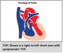
Summary : This illustration demonstrates the cardiac anatomy and blood flow in Tetralogy of Fallot (TOF), highlighting the right-to-left shunt characteristic of symptomatic TOF. illustration: # Scene Overview : • Main subject: Diagram of the human heart showing abnormal blood flow in Tetralogy of Fallot. • Perspective: Frontal cross-sectional view of the heart. • Colour palette: Blue and red regions indicate deoxygenated and oxygenated blood, respectively. • Key anatomical features: Right ventricle, left ventricle, aorta, pulmonary artery, and ventricular septal defect. # Technical Details : • On-image text: "Tetralogy of Fallot" at the top. • Caption below: "TOF: Shown is a right-to-left shunt seen with symptomatic TOF." • No scale bar or magnification indicated. # Spatial Relationships : • Blue (deoxygenated) blood flows from the right ventricle through a ventricular septal defect into the aorta, mixing with red (oxygenated) blood. • The shunt direction is right-to-left, bypassing the pulmonary circulation. # Analysis : • The figure visually explains the pathophysiology of Tetralogy of Fallot, specifically the right-to-left shunt that leads to mixing of deoxygenated and oxygenated blood, a hallmark of symptomatic TOF. • The abnormal flow pattern is clearly depicted, supporting the educational message in the caption.
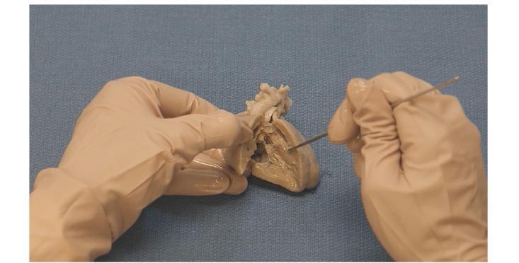
Anatomical Diagram/Clinical Photograph: This image depicts a gross anatomical dissection of a human heart specimen, specifically prepared to demonstrate the pathophysiology of Tetralogy of Fallot (TOF). The cadaveric specimen exhibits a preserved, tan, and fibrous texture, having been bisected to reveal the internal cardiac chambers. Two gloved hands are shown; one stabilizes the specimen while the other utilizes a metal surgical probe to indicate specific structural defects. The educational focus is on visualizing the four key components of TOF: ventricular septal defect (VSD), pulmonary stenosis, right ventricular hypertrophy, and the overriding aorta. The clinical significance of this specimen lies in its use for pediatric cardiology education, providing a three-dimensional perspective on congenital heart disease (CHD) morphology. The image is intended for intermediate to advanced medical learners to understand the spatial relationships between the malaligned interventricular septum and the great vessels.
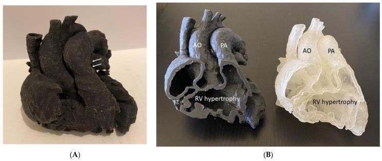
This educational image displays 3D-printed heart models used to demonstrate the complex anatomy of Tetralogy of Fallot (ToF). Panel A shows a monolithic, dark-colored model from an external perspective; white arrows point to the pulmonary artery, highlighting pulmonary artery stenosis. Panel B illustrates the same heart model printed in two sagittal halves using different material colors (opaque dark grey and translucent white). This cross-sectional view provides a detailed internal anatomical study of the four hallmark features of ToF: the overriding aorta (labeled AO), pulmonary artery stenosis (labeled PA), and significant right ventricular hypertrophy (labeled RV hypertrophy). The sectioned view allows for clear visualization of the thickened right ventricular myocardial wall and the spatial relationship between the great vessels and the ventricular chambers. These models serve as high-fidelity educational tools for surgical planning and understanding congenital heart disease (CHD) manifestations.
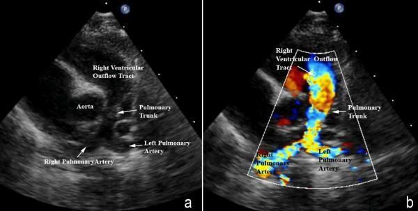
Two-panel diagnostic ultrasound image demonstrating Tetralogy of Fallot anatomy and hemodynamics. Panel (a) is a 2D grayscale echocardiogram in a modified parasternal short-axis view showing the right ventricular outflow tract (RVOT), the aorta, and the pulmonary arteries. There is significant hypoplasia of the pulmonary trunk and the right and left pulmonary arteries, resulting from the antero-cephalad deviation of the outlet septum. The pulmonary valve leaflets are not clearly visualized. Panel (b) utilizes color Doppler imaging to map blood flow through the same structures. It reveals marked turbulence and flow acceleration, represented by a mosaic of red, yellow, and blue colors, beginning at the level of the deviated outlet septum in the RVOT. This high-velocity turbulent flow continues through the stenotic, hypoplastic pulmonary trunk into the pulmonary artery branches. This clinical imaging is essential for assessing the severity of subpulmonary obstruction and hypoplasia in congenital heart disease management.
"tetralogy of fallot"[MeSH Terms] AND management
Tetralogy of Fallot (TOF) - A Comprehensive Overview
What Is It?
The Four Defects (The "Tetrad")
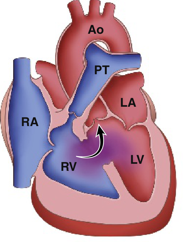
| # | Defect | Details |
|---|---|---|
| 1 | Ventricular Septal Defect (VSD) | Large, unrestrictive, misaligned defect near the membranous portion of the interventricular septum |
| 2 | Right Ventricular Outflow Tract (RVOT) Obstruction | Subpulmonary (infundibular) stenosis is most common; pulmonary valve stenosis can also contribute; in extreme cases, pulmonary atresia |
| 3 | Overriding Aorta | The aortic root straddles the VSD and receives blood from both ventricles |
| 4 | Right Ventricular Hypertrophy (RVH) | Secondary to the pressure load imposed by RVOT obstruction |
Embryological Basis
-
Abnormal septation between the pulmonary trunk and the aortic root
-
Unequal division of the truncus arteriosus - the pulmonary side becomes stenotic/small while the aorta enlarges
-
The VSD is a direct consequence of the malaligned septum failing to meet the rest of the interventricular septum
-
The Developing Human: Clinically Oriented Embryology, p. 847
-
Robbins & Kumar Basic Pathology, p. 283
Gross Morphology (Pathological Anatomy)
-
The heart is enlarged and boot-shaped ("coeur en sabot") - a classic X-ray finding - due to right ventricular hypertrophy
-
The proximal aorta is dilated; the pulmonary trunk is hypoplastic
-
Left-sided chambers are normal in size
-
The right ventricular wall is markedly hypertrophied - sometimes exceeding the thickness of the left ventricle
-
The VSD is large, near the membranous septum, with the aortic valve immediately overriding it
-
RVOT obstruction is most commonly infundibular (subpulmonic), but can also be valvular or combined
-
Robbins & Kumar Basic Pathology, p. 288-290
Associated Anomalies
-
Right aortic arch: present in ~25% of patients
-
Anomalous coronary arteries: most commonly the left anterior descending (LAD) arises from the right coronary artery (RCA) and crosses the RVOT - has important surgical implications
-
ASD (creating a pentalogy of Fallot)
-
Pulmonary artery hypoplasia: reported in up to 50%
-
Down syndrome (Trisomy 21): can coexist with AVSD variant
-
MAPCAs (Major Aortopulmonary Collateral Arteries): present in ~15% - an extreme TOF variant (TOF with pulmonary atresia) where no direct heart-to-pulmonary artery connection exists
-
Braunwald's Heart Disease, p. 2776-2778
Pathophysiology
Right-to-Left Shunt
-
Mild RVOT obstruction: high left-sided pressure causes a left-to-right shunt - patient is acyanotic ("Pink Tet" or "acyanotic TOF"). Clinically resembles an isolated VSD.
-
Severe RVOT obstruction: deoxygenated blood from the RV is forced right-to-left across the VSD into the overriding aorta - producing cyanosis
-
This causes: decreased pulmonary blood flow + increased aortic volumes of mixed (deoxygenated) blood
-
Robbins & Kumar Basic Pathology, p. 292; Rosen's Emergency Medicine, p. 1384
Clinical Features
In Infants and Children
| Feature | Details |
|---|---|
| Cyanosis | Varies - may be absent at birth; worsens with crying and feeding; progressive with age |
| Clubbing | Fingers and toes - from chronic hypoxemia |
| Polycythemia | Compensatory response to chronic hypoxia |
| Systolic ejection murmur | Along the left sternal border (from RVOT obstruction - not from VSD itself) |
| Single S2 | Soft or absent pulmonary component due to low PA pressure |
| Squatting posture | Older children squat after exertion - increases SVR, reduces right-to-left shunt |
In Adults with Repaired TOF
-
Shortness of breath on exertion
-
Palpitations and syncope
-
Diastolic to-and-fro murmur in the pulmonary area = pulmonary regurgitation
-
RV heave + single S2 when PR is severe
-
Overt right heart failure (hepatomegaly, elevated JVP, edema) is uncommon
-
Braunwald's Heart Disease, p. 2787-2789; Rosen's Emergency Medicine, p. 1384-1388
The "Tet Spell" (Hypercyanotic / Hypoxic Spell)
Mechanism (Vicious Cycle):
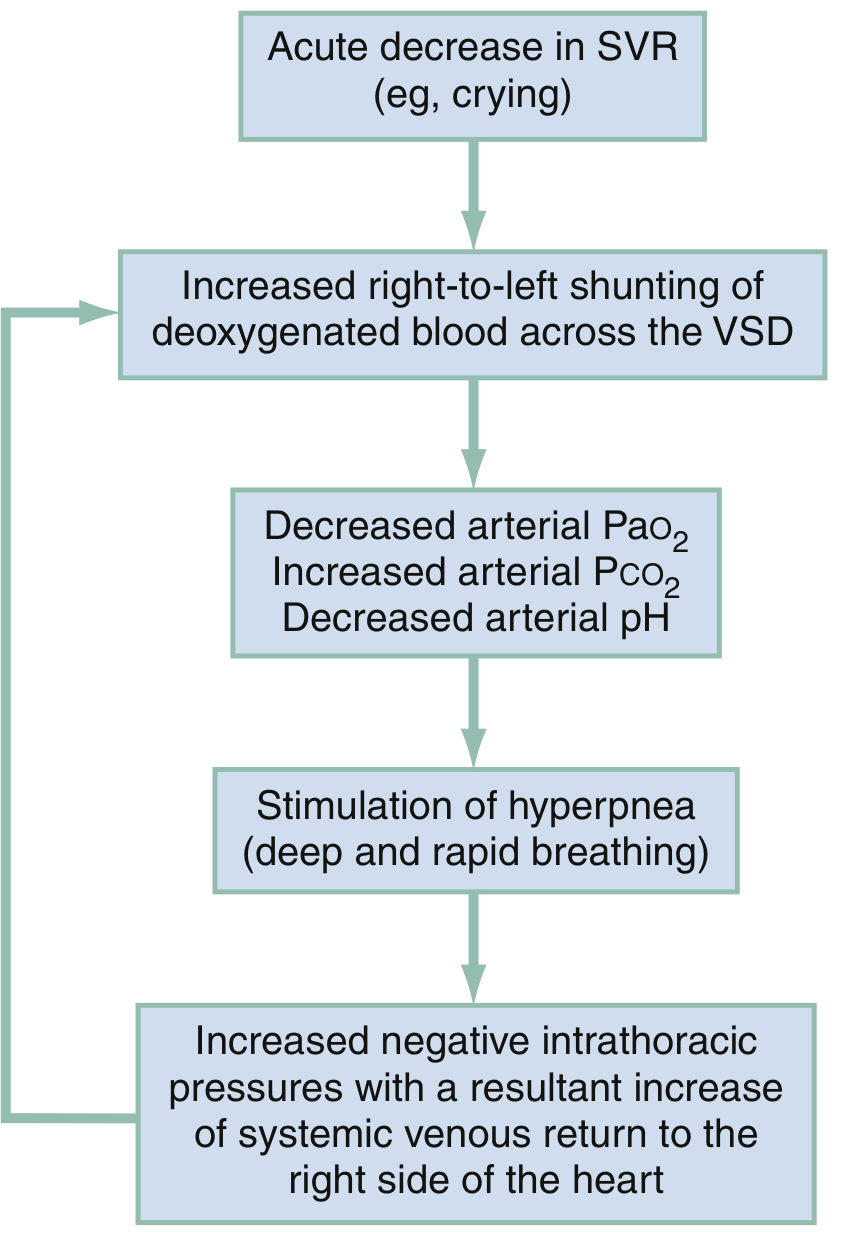
Emergency Management of Tet Spell:
| Step | Intervention | Rationale |
|---|---|---|
| 1 | Knee-to-chest position | Increases SVR → reduces R→L shunt |
| 2 | Supplemental O₂ | Limited value alone, but helpful |
| 3 | Morphine 0.1-0.2 mg/kg IV/IM | Reduces hyperpnea and infundibular spasm |
| 4 | Fentanyl 1 μg/kg IV/IM (or 1.5-2 μg/kg intranasal) | Alternative to morphine |
| 5 | Sodium bicarbonate 1 mEq/kg IV | If acidosis documented or suspected |
| 6 | Ketamine 1-2 mg/kg IV or 3-5 mg/kg IM | Increases SVR, provides sedation |
| 7 | Propranolol 0.1-0.2 mg/kg | Reduces infundibular spasm |
| 8 | Phenylephrine 0.01-0.02 mg/kg IV | Pure alpha agonist - rapidly raises SVR |
- Rosen's Emergency Medicine, Box 165.8, p. 1414-1436
Investigations
Chest X-Ray
- Boot-shaped heart (coeur en sabot) - from RVH with uplifted apex
- Decreased pulmonary vascular markings
- Right aortic arch in 25%
- Normal heart size
ECG
- Right ventricular hypertrophy (tall R in V1, deep S in V5-V6)
- Right axis deviation
- Complete right bundle branch block (RBBB) - common in surgically repaired patients
Echocardiography
- Gold standard for diagnosis
- Identifies all four components of the tetrad
- Assesses severity of RVOT obstruction, VSD size, aortic override degree
- Color Doppler shows direction and magnitude of shunt
- Monitors pulmonary regurgitation and RV function post-repair
Cardiac MRI (CMR)
- Best for quantifying RV volume and function post-repair
- Measures pulmonary regurgitant fraction (significant PR = regurgitant fraction ≥40%)
- Assesses branch pulmonary arteries, RVOT aneurysms, and coronary anatomy
- Late gadolinium enhancement correlates with adverse outcomes
Cardiac Catheterization
-
Rarely needed for diagnosis
-
Used pre-operatively in complex cases (TOF with pulmonary atresia, MAPCAs assessment)
-
Braunwald's Heart Disease, p. 2793-2807
Surgical Management
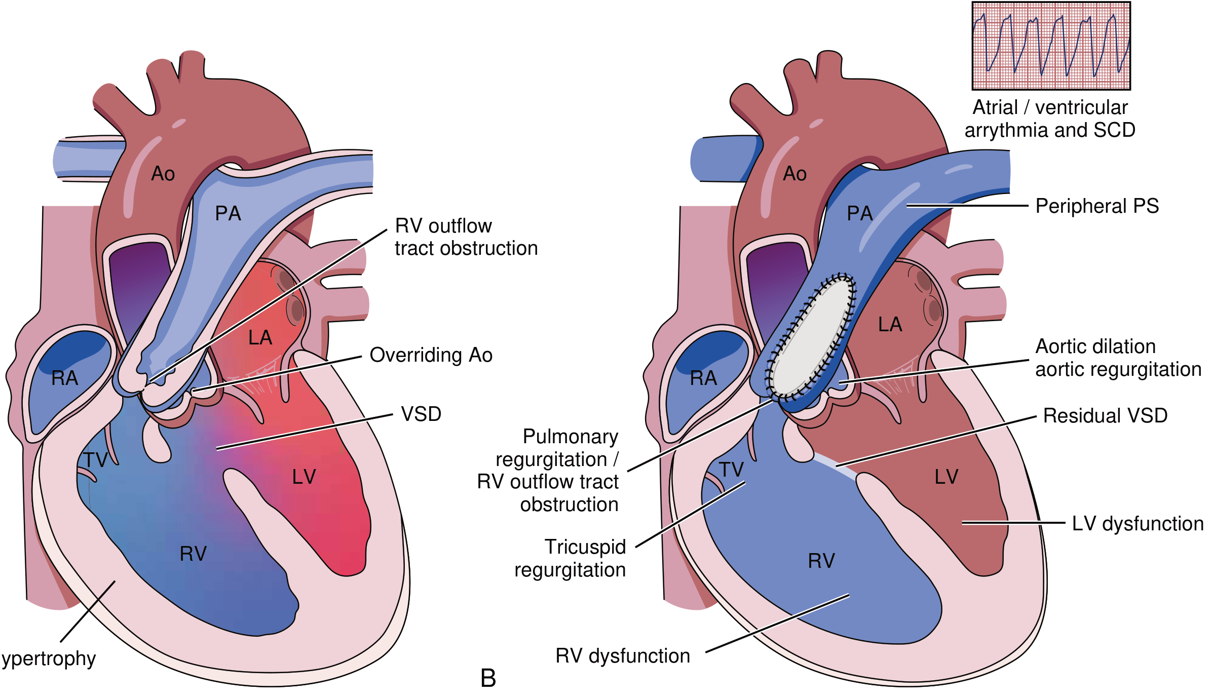
Primary Repair (Definitive)
- VSD closure with a patch
- RVOT reconstruction - resection of RV muscle bundles; may include pulmonary valvotomy or transannular patch
- Goal: avoid transannular patch where possible to preserve pulmonary valve integrity (reduces long-term pulmonary regurgitation)
Staged Repair (Historical / Selected Cases)
- Blalock-Taussig-Thomas (BTT) shunt: subclavian artery to pulmonary artery - palliative, increases pulmonary blood flow
- Still used in very small or complex anatomy cases before definitive repair
- TOF with pulmonary atresia + MAPCAs: requires unifocalization of MAPCAs + VSD closure + RV-to-PA conduit
Transcatheter Pulmonary Valve Replacement
Long-Term Complications After Repair
| Complication | Details |
|---|---|
| Pulmonary regurgitation (PR) | Most common sequela, especially after transannular patch repair |
| RV dilatation and dysfunction | Consequence of chronic PR - leads to arrhythmia, heart failure |
| RVOT aneurysm | Akinetic areas at repair site |
| Residual VSD | At patch edges |
| Aortic root dilatation + aortic regurgitation | Progressive in adults |
| Arrhythmias | Atrial and ventricular tachyarrhythmias; sustained VT risk increases with RV dilatation |
| Sudden cardiac death (SCD) | ~2% over 20 years post-repair; associated with VT, large RV, QRS duration >180 ms |
| Endocarditis | Lifelong risk |
| Pulmonary valve replacement | Indicated when RV ESV index >80 mL/m² or symptomatic severe PR |
- Braunwald's Heart Disease, p. 2823-2835
Prognosis
- Without surgery: ~25% die in the first year, ~40% by age 3, ~70% by age 10
- With timely surgical repair: life expectancy is near-normal for uncomplicated anatomy
- All patients require lifelong follow-up at ACHD (Adult Congenital Heart Disease) centers
- BNP level is predictive of mortality in adults with repaired TOF
Quick Summary Table
| Feature | Key Point |
|---|---|
| Most common cyanotic CHD | Yes (beyond infancy) |
| Prevalence | ~5% of all CHD |
| Root cause | Anterocephalad deviation of outlet septum |
| Degree of cyanosis depends on | Severity of RVOT obstruction |
| "Pink Tet" | Mild RVOT obstruction - acyanotic |
| Classic CXR | Boot-shaped heart, decreased pulmonary markings |
| Tet spell trigger | Drop in SVR (crying, defecation) |
| First-line tet spell maneuver | Knee-to-chest position |
| Definitive treatment | Complete surgical repair (VSD patch + RVOT reconstruction) |
| Most common post-repair complication | Pulmonary regurgitation |
- Braunwald's Heart Disease: A Textbook of Cardiovascular Medicine (10e), Chapter 82
- Robbins & Kumar Basic Pathology, Chapter 9
- Rosen's Emergency Medicine, Chapter 165
- The Developing Human: Clinically Oriented Embryology
- Recent meta-analyses: PMID 39444206, PMID 40659877, PMID 40122137