
Teach me pediatrics topics ASD , VSD, PDA - comparison table with etiology, pathogenesis,& hemodynamics, signs and symptoms, investigations, management, complications, Differentials
Reading File
Now let me search the medical library for all three conditions simultaneously.
Finding Sources
Finding Sources
Finding Sources
Excellent results. Now let me read the most comprehensive sources - Sabiston for ASD/VSD and Schwartz's for VSD/PDA, plus Robbins for pathology, and Goldman-Cecil for clinical aspects.
Reading File
Reading File
Reading File
Good content coming in. Let me now read more detailed content on all three conditions from the most comprehensive sources.
Reading File
Reading File
Reading File
Let me now get more VSD and PDA clinical details, and fetch the key diagram image from Robbins showing all three defects together.
Reading File
Reading File
Loading Image
Excellent! I have a beautiful diagram showing all three defects. Now let me get the Goldman-Cecil PDA section and Robbins VSD/PDA pathology details to complete the picture.
Reading File
Reading File
I now have comprehensive data from multiple authoritative textbooks. Let me compile the full detailed comparison table response.
ASD vs VSD vs PDA - Complete Pediatric Comparison
Sources: Robbins & Kumar Pathologic Basis of Disease, Goldman-Cecil Medicine, Sabiston Textbook of Surgery, Schwartz's Principles of Surgery, Creasy & Resnik's Maternal-Fetal Medicine
Hemodynamic Overview Diagram
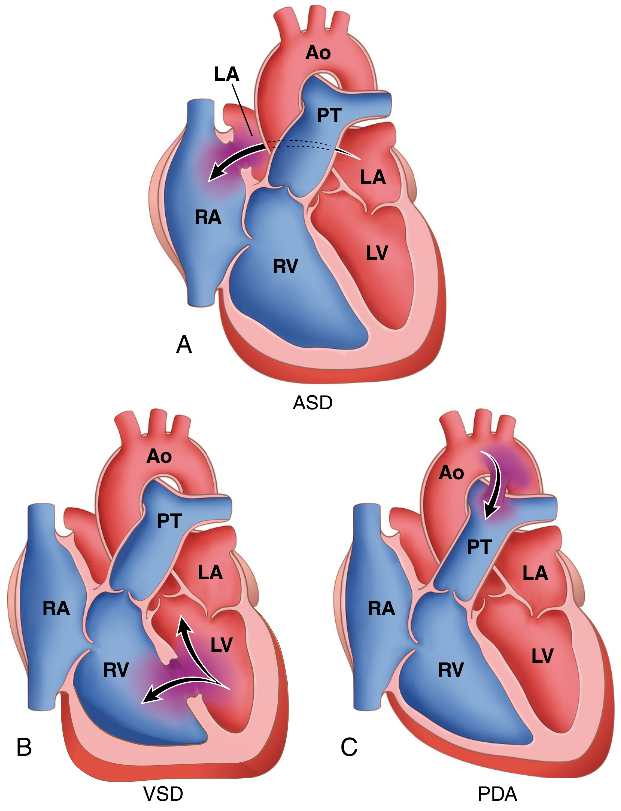
Fig. 12.3 - Robbins, Cotran & Kumar Pathologic Basis of Disease: Common congenital causes of left-to-right shunts
1. ETIOLOGY & CLASSIFICATION
| Feature | ASD | VSD | PDA |
|---|---|---|---|
| Incidence | 13 per 10,000 live births; ~10% of all CHD | Most common CHD - 20-30% of all CHD | ~1 per 2000 births; incidence dramatically increases with prematurity (up to 75% at 28-30 weeks gestation) |
| Sex predilection | Female > Male (2:1) | Equal | Female > Male (2:1) |
| Embryologic basis | Failure of closure of the interatrial septum during development; septum primum/secundum anomaly | Incomplete closure of the interventricular septum | Persistence of the 6th aortic arch derivative connecting pulmonary artery to descending aorta |
| Types / Classification | Secundum (90%) - deficient septum secundum near fossa ovalis; Primum (5%) - near AV valves, often + AV valve defects; Sinus venosus (5%) - near SVC entry, may have anomalous pulmonary venous drainage | Perimembranous / paramembranous (80%) - membranous septum below aortic valve; Muscular (most common by number, multiple possible - "Swiss-cheese"); Inlet/AV canal - below tricuspid valve; Outlet/Supracristal - conal septum below pulmonary valve | Isolated (90%); also seen with complex CHD where duct-dependent circulation may be critical |
| Associated conditions | Down syndrome (primum type); Holt-Oram syndrome; Ellis-van Creveld syndrome | Trisomy 21 (Down), Trisomy 18; Tetralogy of Fallot (component); DiGeorge syndrome; Turner syndrome | Rubella syndrome; prematurity; Trisomy 21; high-altitude birth |
| Genetic/risk factors | Maternal rubella, alcohol, TBX5 mutation (Holt-Oram) | GATA4 mutations; maternal diabetes, alcohol | Prematurity is the biggest risk factor; maternal rubella |
2. PATHOGENESIS & HEMODYNAMICS
| Feature | ASD | VSD | PDA |
|---|---|---|---|
| Shunt direction (initial) | Left atrium → Right atrium (L-to-R) | Left ventricle → Right ventricle (L-to-R) | Aorta → Pulmonary artery (L-to-R) - continuous, both systole and diastole |
| Driving pressure | Left atrial pressure slightly > Right atrial pressure (low pressure difference ~5 mmHg) | LV pressure >> RV pressure during systole (~120 vs 25 mmHg - large gradient) | Aortic systolic AND diastolic pressure > Pulmonary artery pressure (continuous gradient) |
| Primary hemodynamic consequence | Right heart volume overload → RV/RA enlargement; increased pulmonary blood flow | Left heart volume overload → LA and LV dilation; ± pulmonary hypertension depending on size | Left heart volume overload (LA, LV); reduced systemic diastolic pressure due to diastolic runoff |
| Qp:Qs | Can be >3:1 with large defects | >1.5:1 in moderate-severe defects | Depends on duct size; continuous flow in both phases |
| Pulmonary vascular effects | Pulmonary hypertension is uncommon/late in secundum ASD; PVR generally normal | Large defects → pulmonary HTN early in life; irreversible changes possible within first year | Large ducts → pulmonary HTN; Eisenmenger in 5% |
| Eisenmenger development | Rare in secundum ASD; possible with large or primum defects | ~10% of patients with large/unrestrictive VSDs | ~5% of isolated PDAs |
| Special hemodynamic feature | Transient R-to-L shunting with increased intrathoracic pressure (Valsalva, coughing) - paradoxical embolism risk | Small VSDs: negligible; Large VSDs: equal LV-RV pressures; diastolic filling abnormalities in LA | Diastolic shunt lowers aortic diastolic pressure → bounding pulses; "diastolic steal" from coronary/systemic circulation |
| Spontaneous closure | Rare (small secundum may reduce in size) | ~50% of small muscular VSDs; 80% close by 1 month; only 25% still closing by 12 months | Functional closure in 10-15 hours post-term birth; common in premature infants with indomethacin treatment |
3. SIGNS & SYMPTOMS
| Feature | ASD | VSD | PDA |
|---|---|---|---|
| Presentation timing | Often asymptomatic until adulthood (3rd-5th decade); >70% symptomatic by 5th decade | Large defects: symptoms from birth/infancy; small defects: asymptomatic for years | Narrow PDA: asymptomatic; large PDA: failure to thrive, recurrent respiratory infections; premature infants: respiratory distress |
| Key symptoms | Exercise intolerance, dyspnea on exertion, fatigue, palpitations, recurrent respiratory infections in children | Heart failure symptoms: tachypnea, poor feeding, failure to thrive, recurrent respiratory infections, excessive sweating during feeds | Tachypnea, poor feeding, failure to thrive; dyspnea/palpitations in older patients |
| Cyanosis | Absent initially; appears only with Eisenmenger (differential cyanosis NOT typical for ASD) | Absent initially; cyanosis with Eisenmenger reversal | Absent initially; with Eisenmenger: differential cyanosis - lower extremity cyanosis/clubbing more than upper extremity (PA blood flows to descending aorta below left subclavian) |
| Heart sounds | Wide, FIXED splitting of S2 (hallmark - does not vary with respiration); P2 may be loud | Loud P2 if pulmonary hypertension; variable S2 splitting | Normal S2 obscured by murmur; P2 loud with pulmonary HTN |
| Murmur | Soft ejection systolic murmur at 2nd left intercostal space (increased flow across pulmonary valve, NOT across the defect itself); mid-diastolic rumble at lower left sternal border (increased tricuspid flow) if large shunt | Loud, harsh pan-systolic (holosystolic) murmur at left lower sternal border (3rd-4th ICS); thrill often palpable; small "restrictive" VSDs may have louder murmur (maladie de Roger) | Continuous "machinery" murmur at left infraclavicular area / 1st-2nd left ICS (Gibson murmur) - begins after S1, peaks at S2, continues through diastole; louder in left infraclavicular region |
| Pulse character | Normal | Normal or prominent if large shunt | Bounding/waterhammer pulses with wide pulse pressure (diastolic runoff into pulmonary circulation) |
| Precordial findings | Right ventricular heave (parasternal); dilated pulmonary artery palpable at 2nd left ICS | Palpable thrill at left lower sternal border; right ventricular heave if large | Left ventricular heave; bounding peripheral pulses |
| Other signs | Signs of right heart failure in adults | Hepatomegaly, failure to thrive in large VSDs | Tachycardia; hyperactive precordium |
4. INVESTIGATIONS
| Investigation | ASD | VSD | PDA |
|---|---|---|---|
| ECG | Incomplete RBBB (rSr' pattern in V1, V2) - hallmark; Right axis deviation (secundum); Left axis deviation (primum); PR prolongation; atrial fibrillation/flutter in adults | Right ventricular hypertrophy (large defects); left atrial enlargement; left ventricular hypertrophy | Left ventricular hypertrophy; left atrial enlargement in large PDA; normal in small PDA |
| Chest X-ray | Pulmonary plethora (increased vascular markings both lung fields); dilated main PA and branches; RA and RV enlargement; aortic knuckle normal/small | Cardiomegaly; pulmonary plethora; LA and LV enlargement; prominent pulmonary artery | Cardiomegaly; pulmonary plethora; prominent ascending aorta and pulmonary artery; LA/LV enlargement; calcification at duct in older patients |
| Echocardiography | Diagnostic - identifies defect size/location; Doppler quantifies shunt (Qp:Qs) and PA pressures; may miss sinus venosus ASD on TTE → use TEE; bubble study (agitated saline) for PFO | Diagnostic - visualizes defect, quantifies shunt; identifies associated lesions; Doppler for PA pressure estimation | May not directly visualize duct but Doppler signal identifies it; assesses LA/LV size, PA pressure |
| Cardiac catheterization | "Step-up" in O2 saturation at right atrial level; Qp:Qs calculation; pulmonary vascular resistance | "Step-up" in O2 saturation at right ventricular level; Qp:Qs; PA pressure measurement; needed pre-operatively for older patients | "Step-up" at PA level; Qp:Qs ratio; catheterization for pulmonary vascular resistance assessment before closure in borderline cases |
| Special tests | TEE for sinus venosus or ambiguous TTE; cardiac MRI for complex anatomy | Cardiac MRI for complex VSDs; TEE intraoperatively | Cardiac catheterization if Eisenmenger suspected; CT angiography |
5. MANAGEMENT
| Aspect | ASD | VSD | PDA |
|---|---|---|---|
| Medical (conservative) | Observation for small defects; manage arrhythmias with beta-blockers or calcium channel blockers; infective endocarditis (IE) prophylaxis NOT routinely needed | Observation for small VSDs (high spontaneous closure rate); diuretics + ACE inhibitors for heart failure; anti-failure treatment to allow spontaneous closure | Premature infants: Indomethacin (COX inhibitor - reduces prostaglandin E2) or Ibuprofen IV - accelerates closure; Acetaminophen (alternative); surgical ligation if pharmacotherapy fails |
| Interventional / Catheter-based | Transcatheter closure (now >60% of ASD interventions) - Amplatzer Septal Occluder for secundum defects up to 3.5 cm with adequate rims; requires adequate septal rims | Amplatzer device closure - good results; risk of heart block with paramembranous device closure (proximity to conduction system) | Transcatheter coil or device occlusion - treatment of choice in most centers; Amplatzer duct occluder |
| Surgical | Surgical patch closure (direct suture, autologous pericardium, or prosthetic patch) on cardiopulmonary bypass - indicated for primum, sinus venosus, and defects not suitable for device closure | Patch repair on CPB with moderate hypothermia and cardioplegic arrest; right atrial approach for most; right ventriculotomy for apical muscular VSDs; pulmonary artery band as temporizing for "Swiss-cheese" VSDs | Surgical ligation/division via left posterolateral thoracotomy - gold standard in premature infants not responding to indomethacin; video-assisted thoracoscopic surgery (VATS) available |
| Indications for closure | Right-sided heart enlargement with or without symptoms; Qp:Qs >1.5:1; before school age recommended | Large defects: repair in infancy to prevent irreversible pulmonary vascular changes; moderate defects: close if Qp:Qs >1.5:1; small restrictive: observe | Any hemodynamically significant PDA; small PDA: close due to endarteritis risk (0.45%/year after 2nd decade) |
| Contraindication to closure | Eisenmenger syndrome (irreversible pulmonary HTN) | Eisenmenger syndrome | Eisenmenger with R-to-L shunt; severe irreversible pulmonary HTN |
6. COMPLICATIONS
| Complication | ASD | VSD | PDA |
|---|---|---|---|
| Pulmonary hypertension / Eisenmenger | Late, less common in secundum; possible with large/primum defects | ~10% of large unrepaired VSDs; develops early (first year of life with large defects) | ~5% of isolated PDAs |
| Arrhythmias | Atrial fibrillation, atrial flutter, SVT - common in adults; intraatrial reentrant tachycardia post-surgical repair | Less common; conduction defects post-surgery (LBBB, heart block) | Atrial arrhythmias with large shunts |
| Heart failure | Right heart failure (RV volume overload); leading long-term morbidity | Biventricular failure with large VSDs; leading cause of death if large defect untreated | Congestive heart failure - leading cause of death in untreated isolated PDA (~30% mortality untreated) |
| Infective endocarditis / endarteritis | Low risk for unclosed ASD (IE prophylaxis not routinely recommended) | Risk present, especially with small VSDs (turbulent jet); prophylaxis considered | Endarteritis - ~0.45%/year especially with small PDA; rarely fatal with early antibiotic treatment |
| Paradoxical embolism / Stroke | R-to-L shunting (even transient with Valsalva) enables venous thrombus crossing to systemic circulation → CVA | Less significant | Differential embolism to lower extremities with Eisenmenger |
| Aortic regurgitation | Uncommon | Supracristal/outlet VSDs → aortic cusp prolapse → aortic regurgitation | Uncommon |
| Recurrent respiratory infections | Common in children with large shunts | Common with large left-to-right shunts | Common |
| Post-repair complications | Arrhythmias (scar-related AFL), residual shunt, device embolization | Residual VSD, heart block (esp. paramembranous device closure), arrhythmias | Recurrent PDA, chylothorax (surgical), vocal cord palsy (left recurrent laryngeal nerve injury) |
7. DIFFERENTIALS
| Differential | Why it mimics | How to distinguish |
|---|---|---|
| Patent Foramen Ovale (PFO) | Like ASD but no fixed shunt at rest | PFO: no fixed shunt on TTE; only R-to-L with Valsalva on TEE + bubble study; no RA/RV enlargement |
| Pulmonary stenosis | Ejection systolic murmur at 2nd left ICS like ASD | PS: S2 is normally split but NOT fixed; harsh murmur with ejection click; RV hypertrophy on ECG; echo confirms |
| Mitral stenosis | Mid-diastolic murmur (large ASD may have tricuspid flow rumble) | MS: murmur at apex, opening snap, no fixed S2 split; LA not RA enlargement |
| VSD vs ASD | Both are left-to-right shunts | VSD: harsh holosystolic murmur LLSB with thrill; LV enlargement; O2 step-up at RV level; no fixed S2 split |
| VSD vs AVSD | Both septal defects | AVSD: left axis deviation on ECG; associated AV valve abnormality; Down syndrome common |
| PDA vs Aortopulmonary window | Both continuous murmurs | AP window: murmur at upper sternal border; diagnosed on echo/angio; requires surgical repair (no catheter closure) |
| PDA vs Peripheral pulmonic stenosis | Continuous murmur | PPS: radiates to axillae and back; no bounding pulses; seen in neonates, Williams syndrome, Alagille syndrome |
| PDA vs Aortic stenosis + AR (to-and-fro) | Continuous-sounding murmur | AS+AR: murmur changes with posture; maximal at aortic area; echocardiography diagnostic |
| Eisenmenger syndrome (any) | Cyanosis + pulmonary HTN may obscure original defect | History; differential cyanosis in PDA (toes > fingers); cardiac catheterization for O2 step-up level |
Quick Memory Aid: The "3 Left-to-Right Shunts"
| ASD | VSD | PDA | |
|---|---|---|---|
| Where | Atrial septum | Ventricular septum | Aorta to PA |
| Murmur | Soft ESM (2nd LICS) + Fixed split S2 | Harsh PSM (LLSB) + thrill | Continuous "machinery" (left infraclavicular) |
| O2 step-up at | Right atrium | Right ventricle | Pulmonary artery |
| Heart enlarged | RV + RA | LV + LA (± RV with PHT) | LV + LA |
| ECG | IRBBB + RAD | RVH / LVH | LVH |
| Spontaneous closure | Rare | Common (small muscular) | Yes (term infants); indomethacin (premature) |
| Special feature | Fixed S2 split | Restrictive small VSD = louder murmur | Bounding pulses + wide pulse pressure |
| Eisenmenger | Rare | 10% (large) | 5% |
Key Exam Points to Remember:
- ASD hallmark: Wide, fixed S2 splitting that does not change with respiration - this is pathognomonic
- VSD: Most common CHD overall; harsh holosystolic murmur with thrill at LLSB; large VSDs can have equal LV/RV pressures (so murmur may be softer than expected)
- PDA: Indomethacin works in premature infants (prostaglandin-mediated closure); doesn't work well in term infants where functional closure has already failed; differential cyanosis (feet > hands) is diagnostic of PDA + Eisenmenger
- Eisenmenger: Irreversible pulmonary HTN with shunt reversal - closure is contraindicated once established
- All three start as L-to-R shunts → pulmonary overcirculation → pulmonary HTN → R-to-L reversal → cyanosis (Eisenmenger)
Next is cyanotic congenital heart diseases -etiolgy, pathogenesis & hemodynamics, cigns and symptoms, investigations management and complications comparison table
Finding Sources
Finding Sources
Finding Sources
Finding Sources
Finding Sources
Finding Sources
Now let me read from the best sources in parallel - TOF from Rosen's + Robbins, TGA + TOF from Sabiston, and TAPVC + Tricuspid Atresia from Schwartz's:
Reading File
Reading File
Reading File
Reading File
Reading File
Loading Image
Reading File
Reading File
Loading Image
I now have excellent content from multiple authoritative sources, plus two key diagrams. Let me compile the comprehensive comparison table.
Cyanotic Congenital Heart Diseases - Complete Comparison
Sources: Robbins & Kumar Basic Pathology, Rosen's Emergency Medicine, Sabiston Textbook of Surgery, Schwartz's Principles of Surgery, Robbins, Cotran & Kumar Pathologic Basis of Disease
Classification by Pulmonary Blood Flow
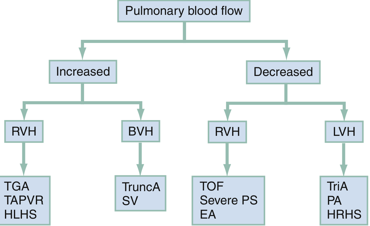
Clinical diagnostic framework: Increased vs Decreased pulmonary blood flow, and ventricular hypertrophy pattern
The "5 T's" of Cyanotic CHD
| TOF | TGA | Tricuspid Atresia | Truncus Arteriosus | TAPVR | |
|---|---|---|---|---|---|
| Full name | Tetralogy of Fallot | Transposition of Great Arteries (D-TGA) | Tricuspid Atresia | Persistent Truncus Arteriosus | Total Anomalous Pulmonary Venous Return |
| Incidence / Frequency | Most common cyanotic CHD beyond infancy; ~5% of all CHD | 2-3 per 10,000 live births; most common cyanotic CHD in neonates | 2-3% of all CHD; most common form of functionally single-ventricle physiology | 1-2% of all CHD; rare | 1-2% of all cardiac malformations |
| Pulmonary blood flow | Decreased | Increased | Decreased (usually) | Increased (massively) | Increased (but obstructed = emergency) |
| ECG pattern | RVH | RVH | LVH (unique - left ventricle does all the work) | BVH (biventricular hypertrophy) | RVH |
TOF Anatomy Diagram
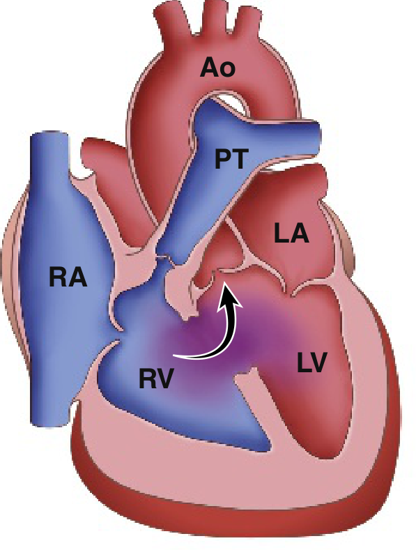
Fig. 9.4A - Robbins & Kumar Basic Pathology: Classic Tetralogy of Fallot. Arrow = R-to-L shunt from RV through VSD into overriding aorta
1. ETIOLOGY & EMBRYOLOGY
| Feature | TOF | TGA (D-TGA) | Tricuspid Atresia | Truncus Arteriosus | TAPVR |
|---|---|---|---|---|---|
| Embryologic defect | Anterosuperior displacement of the infundibular septum → abnormal septation between pulmonary trunk and aortic root; single embryologic error causing all 4 features | Failure of the aortopulmonary septum to spiral during division; results in aorta arising from RV and PA from LV (ventriculoarterial discordance with concordant AV connections) | Unequal division of the AV canal → absent tricuspid valve; mitral valve becomes larger than normal; RV is underdeveloped (hypoplastic) | Arrest of separation of the embryonic truncus arteriosus into aorta and PA; single great vessel overrides ventricular septum (VSD always present); neural crest cell migration failure | Failure of the pulmonary vein evagination from the posterior LA surface to fuse with pulmonary venous plexus around lung buds; persistent connection to splanchnic plexus |
| Associated conditions | Right-sided aortic arch (25%); ASD; anomalous coronary origin; 22q11 deletion (DiGeorge); Down syndrome | Usually isolated; associated with VSD (TGA-VSD) or intact ventricular septum (TGA-IVS); aortic arch hypoplasia/coarctation with VSD variant | ASD (obligatory - PFO/ASD needed to survive); VSD (variable); PDA (pulmonary blood flow often duct-dependent) | DiGeorge syndrome / 22q11 deletion (neural crest association); truncal valve abnormalities (bi- or quadricuspid 30-40%); anomalous coronary arteries (50%) | ASD/PFO (obligatory - only route for oxygenated blood to reach LA); associated with asplenia/polysplenia syndromes |
| Risk factors | Maternal diabetes; alcohol; phenylketonuria; rubella; genetic (22q11) | Maternal diabetes (strongest association); male sex predominance | Sporadic; occasional association with TGA | 22q11 microdeletion; sporadic | Sporadic; associated with asplenia syndrome |
| Sex ratio | M = F | Male predominance (2:1) | M = F | M = F | M = F |
2. PATHOGENESIS & HEMODYNAMICS
| Feature | TOF | TGA (D-TGA) | Tricuspid Atresia | Truncus Arteriosus | TAPVR |
|---|---|---|---|---|---|
| Core anatomical defect | 4 components: (1) large unrestrictive VSD; (2) RVOTO (subpulmonic stenosis most common, also valvular or pulmonary atresia); (3) overriding aorta receiving blood from both ventricles; (4) RVH | Aorta arises from RV (anterior, rightward); PA arises from LV (posterior, leftward) - parallel circulations instead of normal series connection | No connection between RA and RV (tricuspid valve atretic); hypoplastic RV; obligatory ASD for survival; pulmonary flow via PDA or small VSD | Single great artery (truncus) overrides the VSD and gives rise to both aorta and PA; coronary arteries also from truncus | All 4 pulmonary veins drain into systemic venous system (RA or its tributaries) instead of LA; oxygenated blood reaches LA only via ASD/PFO |
| Shunt direction | R-to-L across VSD (due to RVOTO raising RV pressure above LV); deoxygenated blood enters aorta | Parallel circuits - no true shunting; pulmonary and systemic circulations run in parallel, not series; mixing only at level of ASD, VSD, or PDA | R-to-L at atrial level (RA → LA through ASD) | Obligatory mixing at truncal level (VSD + truncal valve); then non-restrictive L-to-R shunt into pulmonary circulation | Obligatory mixing at RA level; both venous streams mix in RA → L-to-R at atrial level (ASD/PFO) for LA filling |
| Mechanism of cyanosis | RVOTO diverts deoxygenated RV blood through VSD into overriding aorta → systemic desaturation | Aorta receives only deoxygenated systemic venous return (from RV); PA receives only oxygenated pulmonary venous return (from LV) - circulations are disconnected unless mixing occurs | Deoxygenated blood from RA crosses ASD into LA, mixes with pulmonary venous return, enters systemic circulation | Mixing of systemic and pulmonary venous blood at truncal level; SpO2 ~85% (depends on Qp:Qs ratio) | All oxygenated pulmonary venous blood diverted to RA; mixed (partially oxygenated) blood crosses ASD to LA |
| Pulmonary blood flow | Decreased (RVOTO restricts flow) | Increased (entire LV output goes to PA) - leads to pulmonary overcirculation | Decreased (usually duct-dependent) | Massively increased - entire cardiac output passes through both lungs; rapid development of pulmonary vascular disease | Depends on obstruction: Unobstructed TAPVR = increased (pulmonary overcirculation + CHF); Obstructed TAPVR = decreased (pulmonary edema, severe cyanosis - surgical emergency) |
| Hemodynamic consequence | RV pressure = LV pressure (unrestrictive VSD equalizes pressures); degree of cyanosis proportional to RVOTO severity; LV normal size | LV faces low pulmonary vascular resistance → LV may become thin and incapable of supporting systemic circulation over time; RA-LA mixing determines survival saturation | LV volume-overloaded (receives both systemic and pulmonary return); RA enlarged; RV hypoplastic | Volume + pressure overload of both ventricles; rapid pulmonary HTN development (may develop by 6 months); truncal valve stenosis/regurgitation adds to load | Right heart dilated; pulmonary hypertension common; with obstruction: pulmonary venous HTN, pulmonary edema |
| Tet spells (TOF only) | Paroxysmal hypercyanotic spells: acute RVOTO worsening (infundibular spasm) → more R-to-L shunting → severe cyanosis → hypoxia → acidosis → worsens spasm (vicious cycle) | N/A | N/A | N/A | N/A |
| Special physiology | "Pink TOF" - if RVOTO is mild, initial L-to-R shunt with no cyanosis (resembles VSD); RVOTO worsens as child grows → increasing cyanosis over time | Survival depends entirely on mixing at ASD, VSD, or PDA; TGA-IVS = most critical (no mixing without intervention) | Pulmonary blood flow duct-dependent in most; some (with large VSD + no RVOTO) may have excess pulmonary flow + CHF | Pulmonary HTN earliest of all 5 Ts - irreversible changes possible by 6 months | Infracardiac type TAPVR with obstruction = TRUE surgical emergency (only cardiac surgery emergency with no palliation option) |
3. SIGNS & SYMPTOMS
| Feature | TOF | TGA (D-TGA) | Tricuspid Atresia | Truncus Arteriosus | TAPVR |
|---|---|---|---|---|---|
| Age of presentation | Not always at birth; worsening cyanosis over weeks-months; "pink TOF" may be asymptomatic initially | First days of life (especially TGA-IVS) - profound neonatal cyanosis | Within days of birth (duct-dependent pulmonary flow); some not until PDA closes | Neonatal period - CHF + cyanosis from birth | Unobstructed: CHF in first weeks; Obstructed: Severe cyanosis + respiratory distress at birth |
| Cyanosis | Progressive; worse with crying, feeding, exertion; relieved by squatting | Profound from birth (especially TGA-IVS); paradoxically "blue" despite normal-sounding heart initially | From birth; profound | Moderate; SpO2 ~85% (mixing at truncal level prevents extreme cyanosis) | Variable; obstructed type = profound cyanosis from birth |
| Squatting (TOF only) | Classic - child squats to relieve cyanosis: increases systemic vascular resistance → reduces R-to-L shunting → more blood goes to lungs | Not applicable | Not applicable | Not applicable | Not applicable |
| Tet spells | Hypercyanotic (Tet) spells: sudden onset intense cyanosis, irritability, hyperpnea, limpness, LOC; worst in morning/after feeding; may → syncope, CVA, death | No | No | No | No |
| Heart failure symptoms | Uncommon early (decreased pulmonary flow); failure to thrive | Tachypnea, poor feeding, tachycardia with large VSD variant (TGA-VSD); otherwise cardiac failure not prominent early | Uncommon (decreased flow); failure to thrive | Prominent CHF: tachypnea, poor feeding, diaphoresis, failure to thrive; CHF + cyanosis is classic combination | Unobstructed: CHF symptoms; Obstructed: severe respiratory distress, pulmonary edema |
| Murmur | Ejection systolic murmur at left sternal border (RVOTO); VSD itself is usually silent (unrestrictive = equal pressures); paradox: louder murmur = less severe TOF (more obstruction = softer murmur as less flow crosses RVOT) | Absent or soft murmur in TGA-IVS; murmurs relate to associated defects (VSD, PS) | Soft; related to associated VSD or ASD | Systolic murmur + sometimes diastolic component from truncal regurgitation; single S2 (only one semilunar valve) | Non-specific murmur or absent; features of right heart enlargement |
| S2 | Single S2 (pulmonary component absent/soft due to hypoplastic PA) | Single loud S2 (aorta is anterior - A2 is loud and P2 is posterior/obscured) | Variable | Single S2 (single semilunar valve) | May be widely split |
| Clubbing | Present - chronic hypoxemia | Present with uncorrected TGA | Present | Less prominent (SpO2 not as low) | Present with chronic uncorrected disease |
| Polycythemia | Present - compensatory; raises risk of cerebral venous thrombosis | Present in chronic cases | Present | Less marked | Present in unobstructed (long-standing) |
| Special signs | Infants: hyperpnea during spells; older children: growth retardation, exercise intolerance | "Egg on a string" appearance on CXR; profound cyanosis out of proportion to respiratory distress | Hepatomegaly; signs of RA enlargement | Bounding pulses (wide pulse pressure from pulmonary overcirculation); signs of CHF | Obstructed type: severe respiratory distress mimicking RDS |
4. INVESTIGATIONS
| Investigation | TOF | TGA (D-TGA) | Tricuspid Atresia | Truncus Arteriosus | TAPVR |
|---|---|---|---|---|---|
| ECG | RVH (right axis deviation, tall R in V1); right ventricular strain pattern | RVH (right axis deviation - paradoxically RV is "systemic" ventricle); may look normal at birth | LVH (hallmark - LV does all work); Left axis deviation (superior QRS axis); RA enlargement | BVH (biventricular hypertrophy); combined ventricular enlargement | RVH; right axis deviation; P pulmonale; RBBB pattern |
| Chest X-ray | "Boot-shaped" heart (coeur en sabot): RVH elevates cardiac apex; concavity at pulmonary artery segment; decreased pulmonary vascular markings (oligemic lung fields); right-sided aortic arch in 25% | "Egg on a string" / "egg on its side": narrow superior mediastinum (great vessels overlapping = no thymic shadow), oval-shaped cardiac silhouette; increased pulmonary vascular markings (plethoric lung fields) | Cardiomegaly (RA + LV enlarged); normal or decreased pulmonary markings depending on pulmonary flow; left-sided cardiac apex (due to LV dominance) | Cardiomegaly (biventricular); increased pulmonary vascular markings; right-sided aortic arch in 30%; absent main PA segment (replaced by truncal artery) | "Snowman"/"Figure-of-8" appearance (supracardiac type - left SVC + left innominate vein form top of snowman + cardiac shadow = bottom); increased markings in unobstructed; white-out/pulmonary edema in obstructed |
| Echocardiography | Diagnostic: VSD, overriding aorta (>50% override), RVOTO, RVH; PA size assessment; coronary artery anatomy | Diagnostic: demonstrates AV concordance + ventriculoarterial discordance; identifies associated VSD, PS, ASD; LV morphology assessment for arterial switch timing | Shows absent tricuspid valve, hypoplastic RV, ASD, VSD; LV dilation; great vessel relationship; PDA assessment | Single truncal valve (trileaflet, bicuspid, or quadricuspid); VSD; PA origin from truncus; truncal valve regurgitation/stenosis | Identifies site of anomalous pulmonary venous drainage; presence/absence of obstruction; ASD/PFO; right heart dilation |
| Cardiac catheterization | O2 saturation step-DOWN at RV level; PA pressure low; coronary anatomy if echo inconclusive; not routine for diagnosis | O2 saturation "step-up" pattern complex; useful to assess pulmonary vascular resistance; less needed if echo diagnostic | Angiography shows anatomy; PA pressure and resistance measurement | PA pressures and resistance measurement (critical for surgical planning); truncal valve assessment | PA pressure and resistance; differentiate types; obstructed vs unobstructed |
| Cardiac MRI/CT | Coronary artery anatomy, PA branch sizes pre-operatively | LV mass and function assessment (pre-arterial switch); post-operative baffle assessment | Pre-Fontan anatomical mapping; pulmonary artery sizes | Pulmonary artery anatomy; coronary origins (50% anomalous) | Pulmonary vein anatomy; drainage pathway assessment |
| Hyperoxia test | PaO2 fails to rise significantly above 150 mmHg on 100% O2 (distinguishes cardiac from pulmonary cyanosis) | PaO2 fails to rise | PaO2 fails to rise | PaO2 minimal rise | PaO2 minimal rise |
5. MANAGEMENT
| Aspect | TOF | TGA (D-TGA) | Tricuspid Atresia | Truncus Arteriosus | TAPVR |
|---|---|---|---|---|---|
| Emergency stabilization | PGE1 if duct-dependent (severe RVOTO/pulmonary atresia variant); Tet spell management: knee-chest position, IV morphine, IV beta-blocker (propranolol), IV phenylephrine (↑SVR), O2, fluids, correct acidosis | PGE1 immediately to maintain/open PDA for mixing; balloon atrial septostomy (Rashkind procedure) to create/enlarge ASD for adequate atrial mixing; achieves SpO2 70-80% | PGE1 if duct-dependent; stabilize with ASD if too small | PGE1 usually NOT used (would worsen pulmonary overcirculation); anti-failure therapy (diuretics, digoxin) | Obstructed TAPVR = true surgical emergency - no palliation possible; immediate surgery; PGE1 contraindicated (would worsen pulmonary overcirculation) |
| Palliative surgery | Blalock-Taussig (BT) shunt (modified - subclavian artery to PA with Gore-Tex conduit) if complete repair not feasible (small infant, hypoplastic PA); increases pulmonary blood flow | Balloon atrial septostomy (Rashkind) is itself palliative to bridge to definitive repair | Staged palliation (mandatory for single ventricle physiology): Stage 1: Blalock-Taussig shunt (if too little PBF) OR PA banding (if too much PBF); Stage 2: Glenn shunt (SVC to PA, bidirectional cavopulmonary connection) at ~6 months; Stage 3: Fontan completion (IVC to PA) at 2-4 years | PA banding if early repair not feasible | N/A for obstructed type (emergency repair only) |
| Definitive surgery | Complete intracardiac repair on CPB: VSD patch closure + RVOTO relief (resection of infundibular muscle, pulmonary valvotomy/valvectomy ± transannular patch); ideally in first 6 months | Arterial Switch Operation (ASO) / Jatene procedure - definitive repair; aorta re-anastomosed to LV, PA re-anastomosed to RV; coronary buttons transferred; must be done within first 2-3 weeks of life before LV "deconditions" to low pulmonary pressure | Fontan procedure (total cavopulmonary connection): IVC connected to PA; RA excluded from systemic circulation; single ventricle (LV) pumps to systemic and pulmonary circuits in series | Complete repair on CPB in neonatal period: (1) Pulmonary arteries detached from truncus + connected to RV via conduit; (2) VSD closed to direct LV output to truncus (aorta); early repair essential (pulmonary HTN by 6 months) | Surgical re-anastomosis of pulmonary veins to LA; ASD closure; resection of obstructing membrane; emergent in obstructed type |
| Historical operations (TGA) | N/A | Mustard/Senning procedure (atrial switch - now historical): intraatrial baffles redirect venous blood; physiologically corrected but morphologic RV remains systemic ventricle → long-term RV failure | N/A | N/A | N/A |
| Medical management | Propranolol for Tet spell prophylaxis; iron supplementation (iron deficiency worsens polycythemia complications) | PGE1 infusion preoperatively; anti-failure therapy for TGA-VSD with CHF | Diuretics + ACE inhibitors for volume overload; anti-coagulation post-Fontan (warfarin/aspirin) | Diuretics, digoxin, ACE inhibitors preoperatively; rapid deterioration means early surgical planning | Furosemide for pulmonary edema in obstructed type (bridge to surgery only) |
| Timing of definitive repair | Complete repair: ideally 3-6 months (avoid polycythemia complications); symptomatic neonates with duct-dependent flow: emergency BT shunt or primary repair | ASO: within first 2-3 weeks of life (before LV regression); TGA-VSD: up to 6-8 weeks | Glenn: ~6 months; Fontan: 2-4 years | Neonatal period (within first few weeks); do NOT delay - PVR irreversible by 6 months | Obstructed: immediate surgical emergency (hours); unobstructed: within first few weeks |
6. COMPLICATIONS
| Complication | TOF | TGA (D-TGA) | Tricuspid Atresia | Truncus Arteriosus | TAPVR |
|---|---|---|---|---|---|
| If untreated | Death; cerebral abscess (paradoxical emboli), cerebral venous thrombosis (polycythemia), progressive hypoxia, infective endocarditis | Death within weeks (TGA-IVS); progressive cyanosis and hypoxia | Death from hypoxia; high early mortality | Rapidly progressive CHF + pulmonary HTN; death usually in infancy | Obstructed: death in hours/days; unobstructed: CHF → death within months-years |
| Post-repair | Pulmonary regurgitation (most common long-term issue after transannular patch repair) → progressive RV dilation → RV failure → arrhythmias; residual RVOTO; residual VSD; ventricular arrhythmias / sudden death (RBBB + left anterior fascicular block = bifascicular block is common post-repair marker); pulmonary valve replacement often needed in adulthood | Atrial switch (historical): RV failure (morphologic RV as systemic ventricle), atrial arrhythmias, baffle obstruction/leak, sudden death; ASO: coronary artery problems (kinking/stenosis), neoaortic root dilation, pulmonary stenosis at anastomosis | Fontan circulation complications: protein-losing enteropathy (PLE - 10%), plastic bronchitis, Fontan failure, arrhythmias (atrial flutter), thromboembolism, hepatic fibrosis/cirrhosis (long-term), lymphatic dysfunction | Truncal valve regurgitation (progressive - most important); conduit failure/obstruction (RV-to-PA conduit requires replacement as child grows); pulmonary HTN if late repair | Pulmonary vein stenosis (most feared post-repair complication - difficult to treat); recurrent obstruction; arrhythmias; residual ASD |
| Infective endocarditis | Risk present (repaired/unrepaired) - IE prophylaxis recommended | Risk post-operatively | Risk present | Risk present | Risk present |
| Polycythemia/hyperviscosity | Cerebral venous thrombosis; cerebral abscess (paradoxical embolism through VSD) | Present | Present | Mild | Present |
| Arrhythmias | Ventricular arrhythmias post-repair (scarring from ventriculotomy); RBBB common; sudden cardiac death (lifetime risk) | Atrial arrhythmias (especially post-atrial switch) | Atrial arrhythmias (especially post-Fontan) | Conduction abnormalities | Arrhythmias post-repair |
Quick Comparison Summary Table
| Feature | TOF | TGA | Tricuspid Atresia | Truncus Arteriosus | TAPVR |
|---|---|---|---|---|---|
| Cyanosis onset | Weeks-months | Day 1-2 of life | Within days | Birth | Birth (obstructed) or weeks (unobstructed) |
| Pulmonary flow | Decreased | Increased | Decreased | Massively increased | Increased (or obstructed = emergency) |
| CXR finding | Boot-shaped heart | Egg on string | Cardiomegaly + LAH | Cardiomegaly + plethora + no main PA shadow | Snowman sign (supracardiac type) |
| ECG hallmark | RVH | RVH | LVH + LAD | BVH | RVH |
| Emergency Rx | PGE1 (severe); Knee-chest for Tet spell | PGE1 + Rashkind septostomy | PGE1 | Anti-failure (NOT PGE1) | Immediate surgery (obstructed) |
| Definitive Rx | Complete repair + RVOTO relief | Arterial Switch Op (within 2-3 weeks) | Staged Fontan (Glenn → Fontan) | Neonatal complete repair | Pulmonary vein re-anastomosis to LA |
| Key complication | PR → RV failure; VT/SCD | RV failure (Mustard/Senning era); coronary stenosis (ASO) | Fontan failure, PLE | Truncal valve regurgitation; conduit failure | Pulmonary vein stenosis (post-repair) |
| Murmur character | Ejection systolic (RVOTO); single S2 | Absent/soft; single loud S2 | Variable | Systolic ± diastolic; single S2 | Non-specific or absent |
| Unique sign | Squatting; Tet spells | Cyanosis out of proportion to respiratory distress | LVH on ECG (paradox) | CHF + cyanosis together | Snowman CXR; "white-out" if obstructed |
Key Memory Mnemonics
The 5 T's: Truncus, Transposition, Tricuspid atresia, Tetralogy of Fallot, TAPVR
TOF "PROVE": Pulmonary stenosis (RVOTO), Right ventricular hypertrophy, Overriding aorta, VSD, (E) = all from one Embryologic defect (infundibular septal displacement)
TGA rule: "Arteries are transposed" = Aorta from RV (wrong!), PA from LV (wrong!) = parallel circuits = death unless mixing
Tet spell management "MOPKF": Morphine, O2, Propranolol/Phenylephrine, Knee-chest position, Fluids + bicarbonate
TAPVR CXR snowman = only in supracardiac type (left vertical vein + left innominate vein + SVC forms "head"; cardiac shadow = "body")
Tricuspid Atresia LVH on ECG = because morphologic LV pumps EVERYTHING (both systemic and pulmonary circuits)
Sources: Rosen's Emergency Medicine p. 1372-1411; Robbins & Kumar Basic Pathology p. 271-325; Sabiston Textbook of Surgery p. 2696-2750; Schwartz's Principles of Surgery p. 398-700; Robbins, Cotran & Kumar Pathologic Basis of Disease p. 990-1003
Now teach me MS, MR, AS, AR, - etiology, pathogenesis hemodynamics, signs and symptoms, investigations management and complications, differential in comparison table
Finding Sources
Finding Sources
Finding Sources
Finding Sources
Excellent. Braunwald's Heart Disease and Grainger & Allison's Radiology have the most comprehensive sections. Let me read all four simultaneously.
Reading File
Reading File
Reading File
Reading File
Good content from Grainger & Allison's. Now let me read the Braunwald's clinical sections and Grainger's AR/MR sections in parallel:
Reading File
Reading File
Reading File
Now let me get the detailed clinical sections from Braunwald's for MR and AS/AR, plus fetch key images:
Reading File
Reading File
Loading Image
I now have rich content from multiple sources. Let me compile the comprehensive comparison table:
Valvular Heart Disease: MS, MR, AS, AR - Complete Comparison
Sources: Braunwald's Heart Disease, Grainger & Allison's Diagnostic Radiology, Textbook of Clinical Echocardiography, Creasy & Resnik's Maternal-Fetal Medicine
Mitral Stenosis - Echo (Parasternal Long Axis)
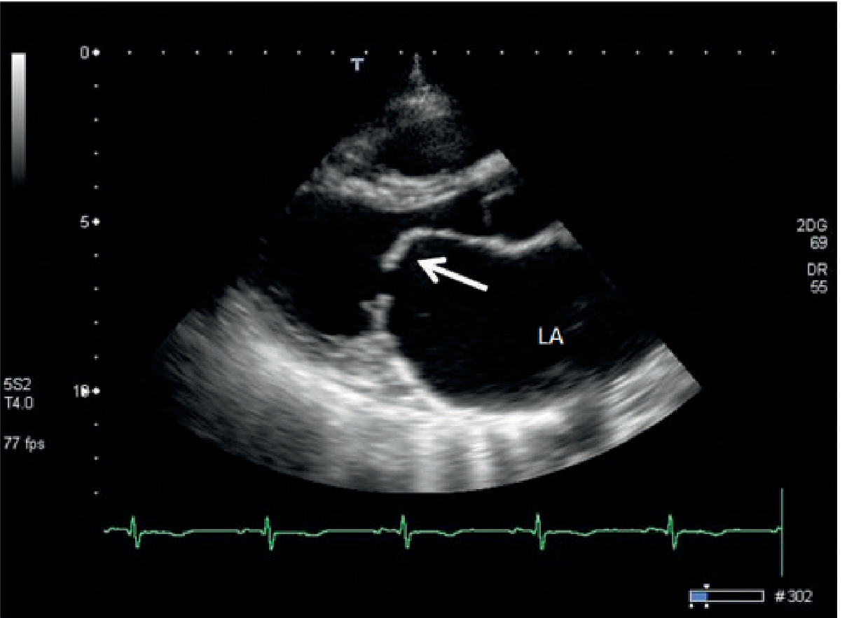
Fig. 14.38 - Grainger & Allison's Diagnostic Radiology: MS with LA enlargement and doming anterior leaflet (arrow)
1. ETIOLOGY
| Feature | MS (Mitral Stenosis) | MR (Mitral Regurgitation) | AS (Aortic Stenosis) | AR (Aortic Regurgitation) |
|---|---|---|---|---|
| Most common cause | Rheumatic fever (by far - 60% give history of RF; 40% of all rheumatic HD is MS) | Myxomatous degeneration (mitral valve prolapse) in developed countries; Rheumatic in developing countries | Degenerative calcific disease (trileaflet) in elderly (>65 yrs); Bicuspid aortic valve in younger adults (presents 1-2 decades earlier) | Bicuspid aortic valve (congenital); Rheumatic fever; Aortic root dilatation (Marfan, HTN, annuloaortic ectasia) |
| Rheumatic mechanism | Commissural fusion, leaflet thickening, chordal shortening/fusion, "fish-mouth" valve | Leaflet destruction + scarring; acute MR reversible; chronic MR → mixed MS/MR usually | Commissural fusion (less common now); fibrosis | Slow destruction of free cusp edges; commissural fusion |
| Degenerative / Calcific | Severe mitral annular calcification (MAC) in elderly | MAC stretching leaflets; papillary muscle dysfunction | Calcification of normal trileaflet cusps (most common in West); ~15% of those ≥65 yrs have AS | Dystrophic calcification of bicuspid valve |
| Congenital | Rare; congenital mitral stenosis | Mitral valve prolapse (MVP); cleft mitral valve (AVSD) | Bicuspid AV (commonest congenital AS) | Bicuspid AV; isolated congenital AR |
| Acute causes | N/A | Acute MR: infective endocarditis (IE), papillary muscle rupture post-MI, chordal rupture (MVP), trauma | N/A | Acute AR: infective endocarditis, aortic dissection, trauma; Takayasu arteritis |
| Other / Rare | Cor triatriatum (mimics MS), LA myxoma (ball-valve), radiation-induced, mucopolysaccharidosis | Functional MR: LV dilation → annular dilation; ischemic MR (papillary muscle dysfunction); carcinoid syndrome; radiation | Sub-aortic membrane (HOCM), supra-aortic stenosis (congenital) | Marfan syndrome, Ehlers-Danlos, syphilitic aortitis, Ankylosing spondylitis, Reactive arthritis (Reiter's), Rheumatoid arthritis, Takayasu |
| Sex predilection | Female > Male (2:1) for isolated MS | Female > Male for MVP; equal for other causes | Male > Female for calcific AS | Male > Female |
2. PATHOGENESIS & HEMODYNAMICS
| Feature | MS | MR | AS | AR |
|---|---|---|---|---|
| Core mechanism | Mechanical obstruction to LV inflow → pressure overload of LA | Systolic backflow from LV → LA → volume overload of LA and LV | Mechanical obstruction to LV outflow → pressure overload of LV | Diastolic backflow from aorta → LV → volume overload of LV |
| Valve area (normal) | Normal MV area = 4-6 cm² | N/A (competence, not area) | Normal AV area = 2-4 cm² | N/A |
| Severity grading | Mild: >1.5 cm², gradient <5 mmHg; Moderate: 1.0-1.5 cm², 5-10 mmHg; Severe: <1.0 cm², >10 mmHg | Mild/Moderate/Severe based on regurgitant fraction; Severe: RF >50%, regurgitant vol >60 mL | Mild: AVA >1.5 cm²; Moderate: 1.0-1.5 cm²; Severe: <1.0 cm² or <0.6 cm²/m² BSA, mean gradient ≥40 mmHg, Vmax ≥4 m/s | Mild/Moderate/Severe; Severe: PHT <200 ms, regurgitant fraction >50%, vena contracta >6 mm |
| Primary chamber affected | LA (pressure rises; LA dilates) | LA + LV (both volume overloaded) | LV (hypertrophies concentrically) | LV (dilates eccentrically - largest hearts in medicine) |
| LV function | LV underfilled (starved); LV size normal or small; LV function preserved until late | LV volume overloaded; LV dilates; EF appears falsely high early (due to low afterload ejecting into LA) | LV hypertrophies (concentric); normal or small LV cavity; EF preserved until late | LV massively dilates (eccentric hypertrophy); EF eventually falls |
| Pulmonary circulation | Elevated LA pressure → pulmonary venous HTN → pulmonary arterial HTN → RV pressure overload → RV failure → TR | LA pressure elevated → pulmonary HTN in severe/chronic MR | LV hypertrophy → diastolic dysfunction → raised LVEDP → back-pressure to pulmonary circulation in late/severe disease | Increased LV volume → elevated LVEDP in decompensated disease → pulmonary HTN |
| Cardiac output | Reduced (LV underfilling); falls more with exercise or tachycardia (less diastolic filling time) | Maintained early (compensated); falls with decompensation | Maintained until late; critical reduction with decompensation; fixed low cardiac output = angina + syncope + failure | Maintained until decompensation |
| Pulse character | Low volume; tapping apex | Brisk, sustained displaced apex | Slow-rising, small volume (pulsus parvus et tardus) - in severe AS | Collapsing / waterhammer pulse (Corrigan's pulse) - wide pulse pressure due to diastolic runoff back into LV |
| Acute vs chronic hemodynamics | Always chronic | Acute MR: sudden massive LA pressure rise (normal-sized, non-compliant LA cannot accommodate) → acute pulmonary edema; Chronic MR: LA compliance increases, less pressure rise, tolerates large regurgitant volume | Usually chronic; LV adapts with hypertrophy over years | Acute AR: LV cannot dilate rapidly → LVEDP rises acutely → acute pulmonary edema + cardiogenic shock (emergency); Chronic AR: LV slowly dilates over years, well-tolerated until decompensation |
| Key physiologic difference | Inflow obstruction; RV affected | Volume overload; LV eccentric hypertrophy | Outflow obstruction; LV concentric hypertrophy | Volume overload + reduced coronary perfusion (low diastolic pressure) |
3. SIGNS & SYMPTOMS
| Feature | MS | MR | AS | AR |
|---|---|---|---|---|
| Onset / Course | Latent period after RF (10-20 yrs); slowly progressive; symptoms often triggered by AF or pregnancy | Chronic: slow and insidious; Acute: sudden, catastrophic (APO) | Very long asymptomatic phase; sudden onset of symptoms marks critical transition | Chronic: long asymptomatic phase; Acute: sudden cardiovascular collapse |
| Symptoms triad / Cardinal symptoms | Dyspnea (most common); hemoptysis (pulmonary venous HTN); palpitations (AF) | Dyspnea on exertion; fatigue; palpitations; orthopnea | SAD triad: Syncope (exertional), Angina (exertional), Dyspnea (exertional/orthopnea); symptoms mark a critical prognostic threshold | Dyspnea on exertion (most common chronic symptom); palpitations (awareness of hyperdynamic circulation); angina (coronary ischemia from low diastolic pressure); acute AR: pulmonary edema, shock |
| Prognosis after symptoms | Progressive CHF; AF complications | Depends on cause; chronic MR: decline with LV dysfunction | Angina: mean survival 5 yrs; Syncope: 3 yrs; Heart failure: 1-2 yrs (classic mnemonic: "A5-S3-F1") | Chronic AR: 10+ yrs if managed; Acute AR: rapidly fatal if not treated |
| Apex beat | Tapping apex (accentuated S1 palpable); undisplaced; no heave | Displaced, hyperdynamic, thrusting apex (LV enlargement) | Heaving (sustained), non-displaced apex initially; displaced only with LV dilation (decompensation) | Displaced, hyperdynamic, thrusting apex (massive LV dilation) - "water-hammer" apex; visible neck pulsations (Corrigan's sign) |
| Heart sounds | S1 loud (snapping/tapping) - if pliable leaflet; S1 soft if calcified immobile valve; S2 normal or P2 loud (pulmonary HTN) | S1 soft (leaflets don't coapt fully); S3 gallop (volume overload, rapid early filling); P2 loud with pulmonary HTN | S1 normal; S2 soft/absent A2 (calcified, immobile leaflet); S4 gallop (reduced compliance of hypertrophied LV); paradoxical S2 split in severe AS (delayed LV emptying) | S1 soft; S2 soft A2 (poor leaflet coaptation); S3 in decompensated AR; Austin Flint murmur (see below) |
| Opening snap (OS) | Present - high-pitched, early diastolic snap immediately after S2; heard at apex/left sternal border; pliable non-calcified valve; A2-OS interval inversely proportional to severity (shorter A2-OS = more severe MS = higher LA pressure) | Absent | Absent | Absent |
| Murmur | Low-pitched, rumbling mid-diastolic murmur at apex; best heard in left lateral decubitus position with bell of stethoscope; presystolic accentuation (in sinus rhythm - atrial contraction); murmur intensity does NOT correlate with severity | Pansystolic (holosystolic) murmur at apex radiating to axilla (rheumatic/organic MR); or posterior (MVP with posterior leaflet prolapse); in MVP: mid-systolic click + late systolic murmur; murmur severity correlates with LA vs LV pressure gradient | Harsh, crescendo-decrescendo ejection systolic murmur at right upper sternal border (aortic area) radiating to carotids and apex; late-peaking = more severe AS; may be harsh at base but musical ("cooing dove") at apex (Gallavardin phenomenon) | High-pitched, blowing, decrescendo diastolic murmur at left sternal border (3rd-4th ICS); best heard with patient leaning forward in expiration; early diastolic = acute/severe AR (rapid pressure equalization); Austin Flint murmur: low-pitched mid-diastolic rumble at apex (regurgitant jet vibrates anterior mitral leaflet - mimics MS but no OS, no loud S1) |
| Peripheral signs | Mitral facies (malar flush - peripheral cyanosis + pink cheeks); AF irregular pulse | Brisk carotid upstroke (hyperdynamic); signs of heart failure | Pulsus parvus et tardus (slow-rising, small volume carotid pulse, especially in young); anacrotic pulse; systolic thrill in aortic area; carotid shudder | Corrigan's pulse (collapsing/waterhammer); de Musset's sign (head bobbing); Traube's sign (pistol-shot femorals); Müller's sign (uvular pulsations); Duroziez's sign (to-and-fro bruit over femoral artery); Quincke's sign (capillary pulsation in nailbed); Hill's sign: popliteal SBP exceeds brachial SBP by >20 mmHg |
| JVP | Elevated with pulmonary HTN + RV failure; large 'a' wave (if in SR) | Elevated with pulmonary HTN | Usually normal until decompensation | Usually normal until decompensation; may be elevated in acute AR |
4. INVESTIGATIONS
| Investigation | MS | MR | AS | AR |
|---|---|---|---|---|
| ECG | P mitrale (bifid P wave, >0.12 sec, best in lead II) = LA enlargement; AF (most common arrhythmia - LA dilation); RVH if pulmonary HTN | P mitrale (LA enlargement); LVH (volume overload); AF in severe/chronic MR | LVH (voltage criteria + strain pattern: deep ST depression + T inversion in lateral leads V4-V6, I, aVL) = "LV strain pattern"; LBBB sometimes; first-degree AV block | LVH (tall QRS + strain); left axis deviation; LBBB in chronic severe AR |
| Chest X-ray | LA enlargement (double shadow at right heart border; splaying of carina - "carina angle >70°"; left atrial appendage bulge on left heart border = "straightening" of left heart border); Pulmonary venous congestion (upper lobe blood diversion, Kerley B lines, pulmonary edema); Pulmonary arterial HTN (main PA prominent); RV enlargement; NO LV enlargement (LV underfilled); valve calcification on lateral view | LA enlargement; LV enlargement (displaced apex); pulmonary vascular congestion in decompensated MR; Acute MR: pulmonary edema with near-normal cardiac size | LV hypertrophy (rounded LV contour); Post-stenotic dilatation of ascending aorta (prominent first right cardiac arch); Aortic valve calcification on lateral CXR (best seen fluoroscopy); cardiac size normal until decompensation | LV enlargement (massive - "cor bovinum" = ox heart in severe chronic AR); aortic root/ascending aorta dilation; Normal or enlarged LA; signs of pulmonary edema only in decompensation |
| Echocardiography - key findings | 2D: leaflet thickening, "hockey stick" doming of anterior leaflet, "fish-mouth" orifice on short axis, commissural fusion, subvalvular fusion; MVA by planimetry (most accurate); Doppler: mean gradient, pressure half-time (MVA = 220/PHT); Wilkins score (leaflet mobility, thickening, calcification, subvalvular involvement - guides PBMV eligibility) | Color Doppler: regurgitant jet size and direction; vena contracta width; proximal isovelocity surface area (PISA/ERO); LV size and function (EDD, ESD, EF); grading severity: mild, moderate, severe; MVP: posterior displacement of leaflet >2mm below mitral annular plane in parasternal long axis | 2D: calcified, restricted, thickened leaflets (bicuspid or trileaflet); reduced leaflet excursion; Doppler: peak velocity (Vmax), mean pressure gradient, AVA by continuity equation (AVA = (LVOT area × LVOT VTI)/AV VTI); Severe AS: AVA <1.0 cm², mean gradient ≥40 mmHg, Vmax ≥4 m/s | Color Doppler: regurgitant jet (vena contracta ≥6 mm = severe); jet width/LVOT width ratio; PHT (pressure half-time <200 ms = severe AR); pandiastolic flow reversal in descending aorta; LV dimensions (key for surgery timing: EDD >70 mm or ESD >50 mm) |
| Cardiac catheterization | Mitral valve gradient (LA-LV pressure difference); Gorlin formula for MVA; pulmonary artery pressure; still gold standard for hemodynamics; indicated when echo inconclusive or pre-operatively for coronary assessment | Left ventriculogram (LV injection shows contrast opacification of LA - grades 1-4); pulmonary artery wedge pressure; cardiac output | AVA by Gorlin formula: AVA = CO/44.3 × HR × SEP × √mean AV gradient; peak-to-peak gradient (aortic pullback); pressure equalization time; coronary angiography pre-AVR (most pts >40 yrs) | Aortography (regurgitant contrast fills LV - grades 1-4); pressure measurements; coronary angiography pre-AVR |
| Severity grading summary | Mild >1.5 cm²; Mod 1.0-1.5 cm²; Severe <1.0 cm² | Mild RF <30%; Mod 30-49%; Severe RF ≥50%, ERO ≥0.4 cm² | Mild AVA >1.5 cm²; Mod 1.0-1.5 cm²; Severe <1.0 cm², gradient ≥40 mmHg | Mild PHT >500 ms; Mod 200-500 ms; Severe PHT <200 ms, vena contracta ≥6 mm |
5. MANAGEMENT
| Aspect | MS | MR | AS | AR |
|---|---|---|---|---|
| Medical - AF management | Rate control (beta-blockers, digoxin, CCBs) to prolong diastolic filling time; Anticoagulation (warfarin - AF + MS = very high stroke risk; target INR 2-3); rhythm control if symptomatic; no role for anticoagulation in pure sinus rhythm MS (but consider with spontaneous echo contrast or LA thrombus) | Rate control in AF; anticoagulation in AF | No specific role | Same as MS |
| Medical - symptoms | Diuretics for pulmonary congestion; beta-blockers for symptom control (heart rate reduction prolongs filling time); avoid vasodilators (preload-dependent state) | Diuretics for pulmonary congestion; ACE inhibitors/ARBs reduce afterload (beneficial in chronic severe MR with LV dysfunction); Beta-blockers for LV dysfunction | Diuretics cautiously (preload-dependent); avoid vasodilators/nitrates (may cause syncope - preload dependent); no proven medical therapy delays surgical need in AS | Vasodilators (ACE inhibitors, ARBs, nifedipine, hydralazine): reduce afterload and regurgitant fraction; indicated in chronic AR with LV dysfunction/symptoms not ready for surgery; diuretics for volume overload |
| Interventional / Percutaneous | PBMV (Percutaneous Balloon Mitral Valvuloplasty / Inoue technique) - treatment of choice if Wilkins score ≤8, no LA thrombus, no significant MR: balloon catheter inflated across MV through transseptal puncture; 1-year outcomes excellent in good candidates | MitraClip (TEER - Transcatheter Edge-to-Edge Repair): clip approximates anterior and posterior leaflet (Alfieri technique); indicated for high surgical risk patients with primary or secondary MR; EVEREST trial + COAPT trial evidence | TAVR (Transcatheter Aortic Valve Replacement): 1st line for high/intermediate/prohibitive surgical risk; PARTNER trials showed mortality benefit; now expanding to low-risk (younger) patients; risks include paravalvular leak, need for pacemaker, stroke, vascular complications | No percutaneous option for isolated AR currently (TAVR for AR with specific devices under evaluation) |
| Surgery - type | Open mitral commissurotomy (pliable non-calcified valve, no MR, no LA thrombus - ideal); Mitral Valve Replacement (MVR) - mechanical (young, needs anticoagulation) or bioprosthetic (elderly, RF) | Mitral Valve Repair (preferred over replacement - better survival, preserves LV function, no anticoagulation for bioprosthesis); Repair not possible: MVR | Surgical AVR (SAVR): gold standard for appropriate surgical risk; tissue vs mechanical valve; concurrent CABG if CAD; TAVR now preferred in many centers for intermediate-low risk elderly | Aortic Valve Replacement (AVR) - surgical (SAVR); TAVR emerging; aortic root replacement if significant aortic dilation (>5.0-5.5 cm in Marfan) |
| Indications for intervention | Symptomatic severe MS (MVA ≤1.5 cm²); Asymptomatic severe MS with systolic PAP >50 mmHg or new AF or recurrent emboli; PBMV if anatomy favorable (Wilkins ≤8) | Symptomatic severe primary MR (class I); Asymptomatic severe MR with: EF ≤60% OR ESD ≥40 mm; surgery preferred when repair feasible | Symptomatic severe AS (class I): any of SAD symptoms; Asymptomatic severe AS with: EF <50%, mean gradient >60 mmHg, exercise test showing symptoms, very severe AS (Vmax >5 m/s) | Symptomatic severe AR (class I); Asymptomatic severe AR with: EF <55% (class I) OR EDD >70 mm or ESD >50 mm (class IIa); concurrent cardiac surgery |
| Special considerations | Pregnancy with MS: PBMV can be done in 2nd trimester with lead shielding (radiation); rate control critical; high-risk delivery | Acute MR: emergency surgery; vasodilators (nitroprusside), IABP as bridge | Low-flow low-gradient AS (EF <50% + AVA <1 cm² + gradient <40 mmHg): dobutamine stress echo to distinguish truly severe AS from pseudosevere AS | Acute AR: emergency AVR; vasodilators + inotropes as bridge; IABP contraindicated (worsens AR by augmenting diastolic pressure → more regurgitation) |
6. COMPLICATIONS
| Complication | MS | MR | AS | AR |
|---|---|---|---|---|
| Atrial fibrillation | Very common (LA dilation); once AF develops → embolic risk rises dramatically; may cause acute decompensation (loss of atrial kick) | Common in chronic severe MR | Less common; may precipitate decompensation | Less common in chronic; can develop with decompensation |
| Systemic embolism / Stroke | Most feared - LA thrombus (especially appendage) → stroke; risk highest when AF develops; anticoagulation mandatory in MS + AF | Thrombus risk in AF; paradoxical embolism | Emboli less common than MS | Emboli possible (endocarditis) |
| Pulmonary hypertension | Very common - chronic LA → pulmonary venous HTN → reactive pulmonary arterial HTN → RV failure → TR | In severe chronic MR; secondary PHT | Late complication of decompensated AS | Late complication |
| Infective endocarditis (IE) | Relatively low risk for pure MS (no turbulent regurgitant jet); risk if associated MR | Higher risk - regurgitant turbulent jet damages leaflets; IE can cause chordal rupture → acute severe MR | Risk present - prophylaxis historically recommended; current guidelines selective | Higher risk - turbulent regurgitant jet; IE can perforate cusps → acute severe AR |
| Heart failure | Biventricular failure in advanced MS (RV fails secondary to PHT); also "cardiac cachexia" | LV failure; acute pulmonary edema in decompensation or acute MR | LV failure (late) - onset of HF = median survival 1-2 years untreated | LV failure - massive dilated LV eventually fails; acute AR = cardiovascular collapse |
| Hemoptysis | Classic - from pulmonary venous hypertension; epistaxis-like hemoptysis to frank pulmonary hemorrhage | Less prominent | Uncommon | Uncommon |
| Sudden cardiac death | Uncommon (more from PHT/RV failure) | Uncommon | Risk present - especially with severe symptomatic AS and exertion; AVR reduces SCD risk | Risk in decompensated LV failure |
| Post-surgical complications | PBMV: cardiac tamponade (5%), embolism (3%), death (3%), acute MR (2-5%); MVR: thromboembolism, paravalvular leak, endocarditis, structural deterioration (bioprosthetic) | MVR/repair: LV dysfunction, residual MR, prosthetic valve complications | SAVR/TAVR: stroke, pacemaker requirement (TAVR >SAVR), paravalvular leak (TAVR), prosthesis-patient mismatch | AVR: LV reverse remodeling (good sign); LV may not recover if too dilated too long before surgery |
7. DIFFERENTIAL DIAGNOSES
| Differentials | MS | MR | AS | AR |
|---|---|---|---|---|
| Mimics the murmur | Austin Flint murmur (severe AR → mid-diastolic rumble at apex mimics MS); LA myxoma (tumor plop + diastolic murmur); Tricuspid stenosis (diastolic rumble at left sternal border, increases with inspiration); Cor triatriatum (membrane obstructs LA); Ball-valve LA thrombus | Tricuspid regurgitation (pansystolic at LLSB, increases with inspiration); VSD (pansystolic, LLSB, thrill); Aortic stenosis (Gallavardin effect - AS heard at apex); HOCM (ejection murmur at LSB) | HOCM (ejection systolic murmur at LSB; increases with Valsalva, standing; decreases with squatting - opposite to AS); Pulmonary stenosis (ejection systolic at 2nd LICS, with ejection click); Supravalvular AS (Williams syndrome); Innocent flow murmur in children | Pulmonary regurgitation (Graham Steell murmur - early diastolic at left sternal border, increases with inspiration, due to PHT); Austin Flint murmur of severe AR (mimics MS at apex, but AR itself has early diastolic murmur at LSB) |
| Key differentiator from each other | MS vs MR: MS = opening snap + loud S1 + mid-diastolic rumble; MR = soft S1 + pansystolic murmur | MR vs AS (apex murmur): MR = pansystolic, radiates to axilla; AS (Gallavardin) = crescendo-decrescendo, no radiation to axilla | AS vs HOCM: AS murmur decreases with Valsalva/standing; HOCM murmur increases; HOCM has systolic anterior motion (SAM) on echo | AR vs Graham Steell: Graham Steell has signs of severe PHT (RV heave, loud P2, TR), no signs of AR peripheral pulses |
Quick Summary Table
| Feature | MS | MR | AS | AR |
|---|---|---|---|---|
| Valve problem | Doesn't open fully (diastole) | Doesn't close fully (systole) | Doesn't open fully (systole) | Doesn't close fully (diastole) |
| Timing of lesion | Diastolic obstruction | Systolic regurgitation | Systolic obstruction | Diastolic regurgitation |
| Primary overload | LA pressure | LA + LV volume | LV pressure | LV volume |
| LV | Small, underfilled | Dilated (eccentric) | Hypertrophied (concentric) | Massively dilated |
| Murmur | Mid-diastolic rumble (apex) + OS | Pansystolic (apex → axilla) | Ejection systolic (aortic area → carotids) | Early diastolic decrescendo (LSB) |
| Special sound | Opening snap; loud S1 | S3; soft S1 | Soft A2; S4; ejection click (bicuspid) | Austin Flint murmur; soft A2 |
| Pulse | Low volume | Brisk, hyperdynamic | Pulsus parvus et tardus | Collapsing/Corrigan's |
| CXR clue | LA enlargement, no LV enlargement | LA + LV enlargement | Post-stenotic aortic dilation, AV calcification | Massive LV enlargement |
| ECG | P mitrale, AF, RVH | P mitrale, LVH | LVH + strain | LVH, LBBB |
| Most common cause | Rheumatic fever | MVP (West) / Rheumatic (developing) | Calcific/degenerative (elderly), bicuspid (young) | Bicuspid AV / Rheumatic / Aortic root disease |
| Key intervention | PBMV (if favorable anatomy) | Repair > Replacement | SAVR / TAVR | AVR (no PBMV option) |
| Surgery trigger | Symptomatic MVA ≤1.5 cm² | EF ≤60% or ESD ≥40 mm | Any SAD symptom | EF <55% or EDD >70 mm |
| Do NOT use | Vasodilators (preload dependent) | - | Nitrates/vasodilators (syncope risk) | IABP (worsens AR) |
Memory Aids
AS - "SAD" triad with survival:
- Syncope → 3 years mean survival
- Angina → 5 years mean survival
- Dyspnea (heart failure) → 1-2 years mean survival Once symptoms develop in AS = urgent AVR!
AR peripheral signs = alphabet:
- Corrigan's pulse (waterhammer)
- De Musset's (head bobbing)
- Duroziez's (femoral to-and-fro bruit)
- Hill's sign (popliteal > brachial >20 mmHg)
- Müller's (uvula pulsations)
- Quincke's (nailbed capillary pulsation)
- Traube's (pistol-shot femorals)
MS Wilkins Score (for PBMV eligibility) - score 1-4 each:
- Leaflet mobility
- Thickening
- Calcification
- Subvalvular involvement
- Total score ≤8 = suitable for PBMV; >8 = consider surgery
Austin Flint vs True MS:
- Both: mid-diastolic rumble at apex
- Austin Flint (AR): NO opening snap, NO loud S1, peripheral signs of AR (collapsing pulse, etc.)
- True MS: Opening snap present, Loud S1, Tapping apex, signs of RF
Sources: Braunwald's Heart Disease p. 2762-2780; Grainger & Allison's Diagnostic Radiology p. 1576-1833; Textbook of Clinical Echocardiography p. 1258-3055; Creasy & Resnik's Maternal-Fetal Medicine p. 1300-1410
This is a shared conversation. Sign in to Orris to start your own chat.