

6afbc0b2-0e25-4d4a-b161-ee8bfa2e647a.jpeg
Provide a detailed explanation of the topics in image for mbbs 1st tear student
analyse_media
Reading File
Now let me search the medical library for these four topics in parallel.
Finding Sources
Finding Sources
Finding Sources
Reading File
Reading File
Finding Sources
Finding Sources
Reading File
Finding Sources
Finding Sources
Reading File
Finding Sources
Finding Sources
I now have excellent material from the textbooks. Let me compile the full detailed explanation for the MBBS 1st year student.
Loading Image
I have all the material I need. Here is the full, detailed explanation of all four chapters from Unit XII - The Integrated Physiology for MBBS 1st year students.
Unit XII: The Integrated Physiology
MBBS 1st Year - Complete Chapter Notes
Chapter 102: Regulation of Body Temperature in Humans
Normal Body Temperature
The body maintains two distinct temperature zones:
- Core temperature (deep tissues): highly regulated, normally 97°F - 99.5°F (36°C - 37.5°C). This remains stable within ±0.6°C in health.
- Skin (shell) temperature: fluctuates freely with the environment, and is important for heat exchange.
The average normal oral temperature is 98.0-98.6°F (37°C); rectal temperature is ~1°F higher.
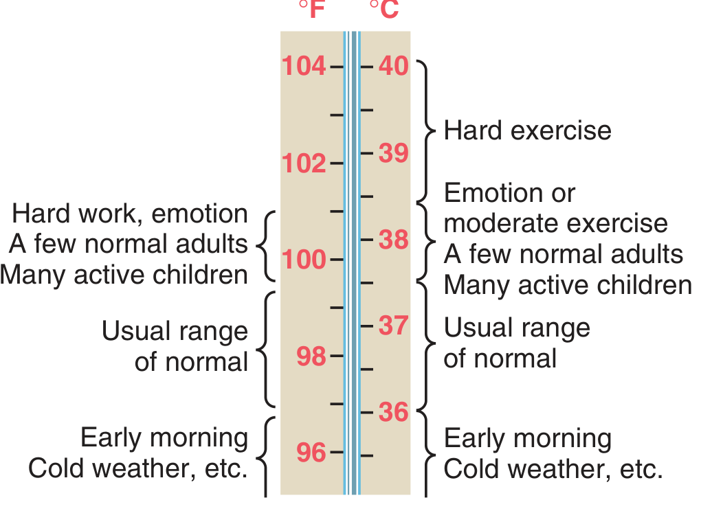
Figure: Estimated normal range of body core temperatures under various conditions - Guyton & Hall, p. 897
Core temperature can transiently rise to 101-104°F during strenuous exercise or fall below 96°F in extreme cold.
Factors Affecting Body Temperature
Heat production in the body depends on:
- Basal metabolic rate (BMR) of all cells
- Muscle activity - exercise and shivering are major sources
- Thyroid hormones (thyroxine) - increase cellular metabolism
- Epinephrine/norepinephrine and sympathetic stimulation
- Cell temperature - higher temperature accelerates chemical reactions
- Thermogenic (specific dynamic) effect of food - digestion generates heat
Other factors affecting body temperature include: time of day (lowest at 6 AM, highest at 6 PM - diurnal variation), age (higher in children, lower in elderly), exercise, emotions, and environmental temperature.
Temperature Regulating Mechanisms
The body maintains thermal balance by balancing heat production vs. heat loss:
"When the rate of heat production in the body is greater than the rate at which heat is being lost, heat builds up in the body, and the body temperature rises. Conversely, when heat loss is greater than heat production, body temperature decreases." - Guyton & Hall
The Hypothalamic Thermostat:
- The anterior hypothalamus is the body's thermoregulatory center (the "thermostat")
- It receives input from:
- Peripheral thermoreceptors in the skin (warm & cold receptors)
- Central thermoreceptors in the anterior hypothalamus itself monitoring blood/core temperature
- The hypothalamus compares sensed temperature against a set-point (~37°C) and activates corrective responses
Heat-Generating Mechanisms (when core temperature falls below set-point):
- Shivering - rapid involuntary muscle contractions generate heat
- Vasoconstriction of skin blood vessels (sympathetic tone increase) - reduces heat loss from skin
- Increased metabolic rate via thyroid hormones
- Piloerection (goosebumps) - traps insulating air layer
Heat-Dissipating Mechanisms (when core temperature rises above set-point):
- Sweating - evaporation is the most powerful heat loss mechanism
- Vasodilation of skin vessels (reduced sympathetic tone) - brings warm blood to skin surface for radiation, conduction, convection
- Increased respiration (minor effect in humans; major in panting animals)
Routes of Heat Loss:
| Method | Mechanism |
|---|---|
| Radiation | Infrared waves from skin (~60% at rest) |
| Conduction | Direct contact with cooler objects |
| Convection | Air currents carry heat away |
| Evaporation | Sweat evaporation (~22% at rest, dominant during exercise) |
Applied Aspects
Fever:
- Defined as core temperature above normal due to resetting of the hypothalamic set-point upward by pyrogens
- Pyrogens (e.g., bacterial lipopolysaccharide endotoxins) stimulate macrophages to release cytokines, especially Interleukin-1 (IL-1), which is also called endogenous pyrogen
- IL-1 acts on the anterior hypothalamus to increase local prostaglandin synthesis, which raises the set-point
- The body then activates heat-generating mechanisms (shivering, vasoconstriction) until the new, higher set-point is reached - this is the "chills" phase
- Aspirin reduces fever by inhibiting cyclooxygenase (COX), blocking prostaglandin synthesis - this lowers the set-point back to normal, triggering heat-loss mechanisms (sweating, vasodilation) - Costanzo Physiology 7th Ed., p. (block2)
Hypothermia:
- Core temperature below 35°C
- At <32°C: loss of shivering (heat production fails), cardiac arrhythmias risk
- At <20°C: potentially fatal
- Causes: exposure to extreme cold, immersion in cold water, anesthesia
Heat Stroke:
- Occurs when core temperature rises to levels causing tissue damage, due to failure of heat-dissipating mechanisms
- If sweating fails, heat cannot be released and temperature rises uncontrolled
- Medical emergency requiring rapid cooling
Malignant Hyperthermia:
- A rare but life-threatening condition triggered by certain inhalation anesthetics in susceptible individuals
- Causes massive increase in skeletal muscle metabolic rate, extreme heat production that overwhelms dissipating mechanisms
Cold Injuries:
- Frostnip - mild, reversible freezing of superficial skin
- Frostbite - ice crystal formation in tissues causing cell damage and vascular injury
Chapter 103: Physiology of Exercise
Grading of Exercise
Exercise intensity is classified using MET (Metabolic Equivalent of Task) values or VO₂max percentage:
- Light exercise: <3 METs (e.g., slow walking)
- Moderate exercise: 3-6 METs (e.g., brisk walking, cycling)
- Vigorous/Hard exercise: >6 METs (e.g., running, sports)
The motor unit is the fundamental functional unit - it consists of one motor neuron and all muscle fibers it innervates. Motor units are recruited progressively:
- Type I (slow-twitch) fibers - recruited first, high oxidative capacity, fatigue-resistant - used for sustained activities
- Type IIa (fast-twitch oxidative) - intermediate
- Type IIx (fast-twitch glycolytic) - recruited last, powerful but fatigue quickly
"Physical exercise is often the greatest stress that the body encounters in the course of daily life... the body must make rapid, integrated adjustments at the level of cells and organ systems." - Medical Physiology (Boron & Boulpaep)
Skeletal muscle converts only ~25% of stored chemical energy into mechanical work; the remaining ~75% appears as heat - which is why exercise raises body temperature and demands active thermoregulation.
Cardio-respiratory Adaptation to Exercise
Immediate (Acute) Cardiovascular Responses:
- Heart rate (HR) increases - due to withdrawal of vagal tone and increased sympathetic activity
- Stroke volume increases - due to increased venous return (Frank-Starling mechanism) and sympathetic stimulation of the heart
- Cardiac output (CO = HR × SV) can increase from 5 L/min at rest to 20-25 L/min during maximal exercise
- Blood pressure rises - systolic increases more than diastolic
- Vasodilation in active muscles (due to local metabolites: CO₂, H⁺, adenosine, K⁺) and vasoconstriction in visceral organs - blood is redirected to muscles
Immediate Respiratory Responses:
- Respiratory rate and tidal volume both increase
- Minute ventilation can rise from 5-6 L/min at rest to >100 L/min during maximal exercise
- Increased ventilation matches increased CO₂ production and O₂ demand
- Ventilatory threshold (anaerobic threshold): point at which lactic acid accumulates faster than it can be cleared; ventilation rises disproportionately to clear excess CO₂ from lactate buffering
Long-term (Chronic) Adaptations with Training:
| Parameter | Trained Effect |
|---|---|
| Resting HR | Decreases (athlete's bradycardia - HR may be 40-50 bpm) |
| Heart size | Cardiac hypertrophy (especially left ventricle) |
| Stroke volume | Increases at rest and during exercise |
| VO₂max | Increases (best measure of cardiovascular fitness) |
| Muscle capillary density | Increases |
| Mitochondria in muscle | Increase in number and size |
| Oxidative enzyme activity | Increases in skeletal muscle |
Physiological Effects of Physical Training
Systematic physical training produces beneficial adaptations across multiple systems:
- Musculoskeletal: increased muscle strength, bone density, joint flexibility
- Cardiovascular: lower resting HR, larger stroke volume, reduced cardiovascular disease risk
- Metabolic: improved insulin sensitivity, better glucose and lipid metabolism, increased fat oxidation
- Respiratory: improved efficiency, larger vital capacity
- Psychological: reduced anxiety and depression, improved mood (endorphin release)
Exercise under Heat and Cold
Exercise in Heat:
- Competing demands between muscles (needing blood flow) and skin (needing blood flow for cooling) create cardiovascular strain
- Core temperature rises faster; risk of heat exhaustion and heat stroke
- Fluid replacement (hydration) and acclimatization are essential
- Acclimatization over 7-14 days increases plasma volume, improves sweating efficiency and earlier onset of sweating
Exercise in Cold:
- Shivering increases heat production
- Vasoconstriction protects core temperature but peripheral extremities are at risk of cold injury
- Wet cold and wind greatly increase heat loss
Consequences of Sedentary Lifestyle
Physical inactivity is now recognized as a major independent risk factor for chronic disease. Key consequences include:
- Cardiovascular disease: sedentary lifestyle is associated with increased inflammation, insulin resistance, decreased wall shear stress, and heightened vascular disease risk
- Obesity and metabolic syndrome: reduced caloric expenditure, impaired glucose metabolism
- Osteoporosis: reduced mechanical loading leads to bone loss
- Muscle atrophy (disuse atrophy)
- Depression and anxiety
- Increased all-cause mortality
Regular physical activity of moderate intensity (at least 150 minutes/week) is recommended by WHO for adults.
Chapter 104: Physiology of Yoga
General Introduction
Yoga is a holistic discipline originating in ancient India that integrates physical postures (asanas), breathing techniques (pranayama), and meditative practices (dhyana). From a physiological standpoint, yoga produces measurable effects on the autonomic nervous system, cardiovascular system, musculoskeletal system, endocrine system, and mental health.
Types of Yogic Exercises
| Type | Description |
|---|---|
| Asanas (postures) | Static or dynamic body positions improving flexibility, balance, and strength |
| Pranayama (breathing) | Controlled breathing patterns affecting autonomic and respiratory function |
| Dhyana (meditation) | Mental focus and mindfulness, reduces stress hormone levels |
| Shatkarmas | Cleansing techniques (e.g., Kapalbhati for respiratory tract) |
Major categories of asanas:
- Relaxative (e.g., Shavasana)
- Meditative (e.g., Padmasana)
- Cultural/corrective - for posture and organ function
Health Benefits of Yoga Practice
Cardiovascular:
- Reduces resting heart rate and blood pressure
- Improves heart rate variability (reflects improved parasympathetic tone)
- Lowers cholesterol and triglycerides
Respiratory:
- Pranayama improves lung volumes - vital capacity (VC) and tidal volume increase
- Improves respiratory muscle strength and efficiency
Musculoskeletal:
- Increases flexibility and range of joint motion
- Strengthens postural muscles and core stability
- Helps prevent and manage low back pain
Endocrine/Metabolic:
- Reduces cortisol (stress hormone) levels
- Improves insulin sensitivity; beneficial in Type 2 diabetes
- Balances thyroid function
Neurological/Psychological:
- Stimulates parasympathetic nervous system (rest-and-digest), countering chronic sympathetic over-activation
- Reduces anxiety, depression, and perceived stress
- Improves sleep quality
- Increases GABA levels in the brain (anti-anxiety)
Yoga in Health and Disease
- Hypertension: yoga's parasympathomimetic effect reduces sympathetic drive and lowers BP
- Diabetes mellitus: improves glycemic control and insulin sensitivity
- Asthma and COPD: pranayama improves breathing efficiency
- Depression/anxiety: meditative practices reduce cortisol, increase serotonin
- Obesity: combined effect of physical activity + stress reduction + improved eating behaviors
- Osteoporosis: weight-bearing asanas stimulate bone formation
- Arthritis: gentle movement improves joint lubrication and reduces stiffness
Yoga vs. Conventional Exercises
| Parameter | Yoga | Conventional Exercise |
|---|---|---|
| Primary focus | Mind-body integration | Physical performance |
| Intensity | Low-moderate | Variable (low to very high) |
| Caloric expenditure | Moderate | Higher |
| Cardiovascular training | Moderate | High (aerobics/HIIT) |
| Flexibility | Excellent | Moderate (unless stretched) |
| Stress/autonomic effects | Strong parasympathetic shift | Sympathetic activation during; parasympathetic after |
| Injury risk | Low | Higher (especially high-intensity) |
| Suitability | All ages, chronic illness | Primarily healthy/active populations |
The two are complementary rather than mutually exclusive. Yoga excels in stress management and flexibility; conventional aerobic exercise excels in cardiovascular fitness and caloric expenditure.
Chapter 105: Physiology of Infancy and Aging
Physiology of Infancy
Infancy spans birth to 1 year of age and is a period of rapid physiological adaptation and development.
At birth (transition from fetal to neonatal life):
- Circulatory transition: The foramen ovale and ductus arteriosus close (functional closure within hours, anatomical within weeks), converting from parallel fetal circulation to adult serial circulation
- Respiratory onset: First breath is triggered by hypoxia, hypercarbia, cold stimulation, and tactile stimulation at birth
- Thermoregulation: Neonates are poikilothermic - they lose heat rapidly due to high surface area:body mass ratio and thin subcutaneous fat. Brown adipose tissue (BAT) - unique to neonates - provides non-shivering thermogenesis via uncoupling of oxidative phosphorylation
Key physiological features of infants vs. adults:
| Parameter | Infant | Adult |
|---|---|---|
| Heart rate | 120-160 bpm | 60-100 bpm |
| Respiratory rate | 40-60 breaths/min | 12-18 breaths/min |
| Blood pressure | 60-90/30-60 mmHg | 120/80 mmHg |
| Hemoglobin type | Fetal HbF (higher O₂ affinity) shifts to HbA | Adult HbA |
| Renal function | Immature - poor concentrating ability | Fully mature |
| Immune system | Passive immunity from maternal IgG; active immunity developing | Fully active |
Neonatal brain: Rapidly growing; myelination of nerve fibers continues until early adulthood. The blood-brain barrier is less complete in neonates, making them more susceptible to certain drugs and toxins.
Growth: Infants double their birth weight by 5-6 months and triple it by 1 year. Growth hormone and thyroid hormone are critical regulators.
Aging and Free Radicals and Antioxidants
The Free Radical Theory of Aging:
The most widely accepted cellular theory of aging proposes that reactive oxygen species (ROS) - also called free radicals - accumulate over a lifetime and progressively damage cellular components:
Sources of Free Radicals (ROS):
- Normal byproduct of mitochondrial oxidative phosphorylation (electron leakage)
- Inflammatory reactions
- UV radiation, ionizing radiation
- Environmental toxins, cigarette smoke
- Ischemia-reperfusion injury
Damage Caused by ROS:
- DNA damage: strand breaks, base modifications - leads to mutations and cancer
- Lipid peroxidation: damage to cell membranes
- Protein oxidation: inactivates enzymes
- Mitochondrial damage: creates a vicious cycle of more ROS production
Antioxidant Defense Systems:
The body combats oxidative damage through:
| Antioxidant | Type | Mechanism |
|---|---|---|
| Superoxide dismutase (SOD) | Enzyme | Converts O₂•⁻ to H₂O₂ |
| Catalase | Enzyme | Converts H₂O₂ to H₂O + O₂ |
| Glutathione peroxidase | Enzyme | Neutralizes H₂O₂ and lipid peroxides |
| Vitamin E (tocopherol) | Dietary | Scavenges lipid radicals in membranes |
| Vitamin C (ascorbate) | Dietary | Water-soluble radical scavenger |
| Beta-carotene | Dietary | Quenches singlet oxygen |
| Alpha-lipoic acid | Dietary | Potent antioxidant in dermis; may reduce both intrinsic and extrinsic aging from free radical damage |
Aging Changes Across Systems:
- Cardiovascular: Reduced cardiac output, arterial stiffening (atherosclerosis), hypertension
- Respiratory: Reduced vital capacity, decreased elastic recoil of lungs
- Renal: Reduced GFR (decreases ~1% per year after age 40), reduced tubular function
- Nervous system: Neuronal loss, reduced neurotransmitter levels, slowed conduction velocities, memory decline
- Musculoskeletal: Sarcopenia (muscle wasting), osteoporosis, joint degeneration
- Immune system: Immunosenescence - reduced T-cell function, increased autoimmunity and infection susceptibility
- Hormonal: Menopause (women), andropause (men), reduced growth hormone and IGF-1
Brain Death
Definition: Brain death is the irreversible cessation of all brain functions, including the brainstem, while the heart may continue to beat with ventilatory support. It is legally and medically accepted as the definition of death in most countries.
Criteria for Brain Death (must fulfill all):
- Coma - completely unresponsive to all external stimuli
- Absence of all brainstem reflexes:
- No pupillary light reflex (pupils fixed and dilated)
- No corneal reflex
- No oculovestibular (caloric) reflex
- No gag or cough reflex
- No oculocephalic reflex (doll's eye absent)
- Apnea test positive - no spontaneous breathing even when PaCO₂ rises to ≥60 mmHg, confirming complete loss of respiratory drive
- Irreversibility confirmed - known cause, reversible causes ruled out (hypothermia, drug intoxication, metabolic disturbances excluded)
Confirmatory tests (when clinical tests are inconclusive):
- EEG - shows electrocerebral silence (flat trace)
- Cerebral angiography - no intracranial blood flow
- Transcranial Doppler - absent cerebral blood flow
- Radionuclide scan (SPECT) - no cerebral perfusion
Persistent Vegetative State (PVS) vs. Brain Death:
- In PVS, the brainstem functions are preserved (breathing, reflexes intact), but higher cortical functions are lost
- Brain death is complete and irreversible loss including brainstem - PVS is not brain death
Clinical Significance:
- Brain death declaration allows discontinuation of mechanical ventilation
- It is the medical and legal prerequisite for organ donation
- Two independent physicians must certify brain death
Quick Revision Summary for Exams
| Chapter | Key Concept | Remember |
|---|---|---|
| 102 - Thermoregulation | Anterior hypothalamus is the thermostat | Pyrogen → IL-1 → PGs → raise set-point → fever |
| 103 - Exercise | CO can rise 4-5x during exercise | VO₂max = best index of cardiorespiratory fitness |
| 104 - Yoga | Parasympathetic activator | Yoga + conventional exercise = complementary |
| 105 - Aging | Free radical theory | Antioxidant enzymes: SOD, Catalase, Glutathione peroxidase |
| 105 - Brain death | Irreversible loss of all brain + brainstem function | Apnea test is key clinical test |
Sources: Guyton and Hall Textbook of Medical Physiology, Costanzo Physiology 7th Ed., Medical Physiology (Boron & Boulpaep)
Are following topics covered in the above chat Thermoregulation Role of sleep Consequence of sedentary life styles Oxidative stress Mechanism of heat gain and heat loss aging Role of antioxidant
Here's a clear checklist of which topics were covered in the previous explanation:
| # | Topic | Covered? | Where |
|---|---|---|---|
| 1 | Thermoregulation | ✅ Yes | Chapter 102 - in full detail (hypothalamic thermostat, set-point, heat gain/loss mechanisms, fever, heat stroke, hypothermia) |
| 2 | Role of Sleep | ❌ No | Not mentioned anywhere in the explanation |
| 3 | Consequences of Sedentary Lifestyle | ✅ Yes | Chapter 103 - briefly covered (CVD, obesity, osteoporosis, muscle atrophy, depression) |
| 4 | Oxidative Stress | ⚠️ Partially | Chapter 105 - covered under "Free Radical Theory of Aging" (ROS sources, DNA/lipid/protein damage) but not labeled as "oxidative stress" explicitly |
| 5 | Mechanism of Heat Gain and Heat Loss | ✅ Yes | Chapter 102 - heat production factors listed, heat loss routes (radiation, conduction, convection, evaporation) with a table |
| 6 | Aging | ✅ Yes | Chapter 105 - covered across all systems (cardiovascular, renal, nervous, musculoskeletal, immune, hormonal) |
| 7 | Role of Antioxidants | ✅ Yes | Chapter 105 - full table of antioxidant enzymes (SOD, Catalase, Glutathione peroxidase) and dietary antioxidants (Vit C, E, beta-carotene, alpha-lipoic acid) |
Summary:
- 5 out of 7 topics are fully or substantially covered.
- Role of Sleep is not covered at all - it is not part of the Unit XII chapters shown in your image.
- Oxidative stress is covered by another name (free radical damage/ROS) - the concepts are identical, just the terminology differs.
Would you like me to add a detailed explanation of the Role of Sleep as a supplementary topic for your notes?
Teach me these 7 topics based on Physiology mbbs 1st year student for upcoming University exams
Finding Sources
Finding Sources
Finding Sources
Finding Sources
Finding Sources
Finding Sources
Reading File
Reading File
Finding Sources
Reading File
Reading File
Reading File
Reading File
Reading File
Reading File
Reading File
Here are all 7 topics taught in full detail, exam-ready for MBBS 1st year:
UNIT XII - INTEGRATED PHYSIOLOGY
7 Topic Master Notes | MBBS 1st Year University Exam
TOPIC 1: THERMOREGULATION
Definition: Thermoregulation is the ability of the body to maintain its core temperature within a narrow normal range (36-37.5°C) despite wide variations in environmental temperature.
Normal Body Temperature
- Core temperature (rectal/esophageal): 37°C (98.6°F) - most accurate
- Oral temperature: ~0.5°C lower than rectal
- Axillary temperature: ~1°C lower than rectal
- Diurnal variation: Lowest at 4-6 AM, highest at 6-8 PM (up to 0.5°C difference)
- Temperature rises with exercise, emotions, menstrual cycle (rises at ovulation)
The Hypothalamic Thermostat (Master Controller)
The anterior hypothalamus / preoptic area acts as the body's thermostat:
THERMORECEPTORS ──────────────────────────────────────────────────────────►
                 ↗ Peripheral receptors (warm/cold receptors in skin)        ANTERIOR
                 ↘ Central receptors (in anterior hypothalamus itself)   ──► HYPOTHALAMUS
                                                                              (Set-point ~37°C)
                                                                                  ↓
                                          ┌─────────────────────────────────────┤
                                          ↓                                     ↓
                          TEMP BELOW SET-POINT                     TEMP ABOVE SET-POINT
                          (Heat Conservation)                       (Heat Dissipation)
When temp falls below set-point:
- Shivering (rapid involuntary muscle contractions)
- Vasoconstriction of skin vessels (less blood to skin)
- Piloerection (goosebumps - trap insulating air)
- Increased thyroid hormone secretion (raises BMR)
- Behavioral: seeking warmth, curling up
When temp rises above set-point:
- Sweating (main mechanism in humans)
- Vasodilation of skin vessels (more blood to skin)
- Inhibition of shivering
- Behavioral: seeking shade, removing clothes
"The temperature-regulating center in the anterior hypothalamus receives information about environmental temperature from thermoreceptors in the skin and about core temperature from thermoreceptors in the anterior hypothalamus itself. The anterior hypothalamus then orchestrates the appropriate responses." - Costanzo Physiology
TOPIC 2: MECHANISMS OF HEAT GAIN AND HEAT LOSS
A. MECHANISMS OF HEAT GAIN (Heat Production)
Heat is produced as a by-product of metabolism. The major sources are:
| Source | Details |
|---|---|
| 1. Basal Metabolic Rate (BMR) | Minimum heat produced by all cells at rest |
| 2. Muscle activity | Exercise and shivering - most powerful generator |
| 3. Thyroxine (T4/T3) | Increases cellular metabolic rate across all tissues |
| 4. Epinephrine / Sympathetic stimulation | Rapid but short-lived heat increase |
| 5. Specific Dynamic Action (SDA) of food | Heat produced during digestion and absorption |
| 6. Temperature effect | Higher cell temperature speeds up biochemical reactions (Q10 effect) |
| 7. Growth hormone, testosterone | Minor but contributing roles |
Brown Adipose Tissue (BAT) - Non-shivering Thermogenesis:
- Especially important in neonates
- Contains uncoupling protein-1 (thermogenin) in mitochondria
- Instead of making ATP, energy is released directly as heat
- Stimulated by noradrenaline
B. MECHANISMS OF HEAT LOSS
Most heat is produced in deep organs (liver, brain, heart, active muscles) and must be transferred to skin for dissipation. Two stages:
- Core → Skin (via blood flow)
- Skin → Environment (via physical processes)
Stage 1: Core to Skin - Blood as Heat Carrier
- Skin has a rich venous plexus supplied by capillaries and arteriovenous anastomoses (especially in hands, feet, ears)
- Skin blood flow can range from near-zero to 30% of total cardiac output
- Vasodilation = high skin flow = efficient heat transfer to skin surface
- Vasoconstriction = low skin flow = body retains heat
Stage 2: Skin to Environment - The 4 Physical Methods
| Method | Mechanism | % at Rest | Key Notes |
|---|---|---|---|
| Radiation | Infrared electromagnetic waves from skin | ~60% | Most important at rest; does not need physical contact |
| Conduction | Direct contact with cooler objects/air | ~3% | Enhanced by water (water conducts 25× better than air) |
| Convection | Air/water currents carry heat away | ~12% | Wind chill increases convective loss |
| Evaporation | Sweating - latent heat of vaporization | ~22% at rest; ~80% during exercise | Only method that works when environmental temp > body temp |
Insulator system of the body:
- Skin + subcutaneous tissue + fat act as natural insulation
- Fat conducts heat only 1/3 as readily as other tissues
- This is why insulation is better in women (more subcutaneous fat)
"The rate at which heat is lost is determined almost entirely by two factors: (1) how rapidly heat can be conducted from where it is produced in the body core to the skin, and (2) how rapidly heat can then be transferred from the skin to the surroundings." - Guyton & Hall
Sweating - Key Details (Exam Favourite)
- Sweat glands innervated by cholinergic sympathetic fibers (unusual - sympathetic but ACh-secreting)
- Hypothalamus → spinal cord → sympathetic outflow → sweat glands
- Precursor secretion is like plasma (Na⁺ 142 mEq/L) but protein-free
- As sweat passes through the duct, Na⁺ and Cl⁻ are reabsorbed
- Acclimatization to heat improves aldosterone-mediated Na⁺ reabsorption → sweat becomes more dilute
TOPIC 3: ROLE OF SLEEP
Definition: Sleep is a state of unconsciousness from which a person can be aroused by sensory or other stimuli - this distinguishes it from coma (from which one cannot be aroused). - Guyton & Hall
Types of Sleep - Two Major Types
1. NREM Sleep (Non-Rapid Eye Movement = Slow-Wave Sleep)
- Makes up ~75% of total sleep time
- Deep, restful sleep
- Associated with decreased heart rate, BP, respiratory rate, metabolic rate
- EEG shows slow, high-voltage waves (delta waves) in deep stages
- Occurs predominantly in the first half of the night
- 4 stages (Stage 1 → lightest; Stage 4 → deepest)
| NREM Stage | EEG Pattern | Features |
|---|---|---|
| Stage 1 (N1) | Low voltage, mixed | Light sleep, easily awakened |
| Stage 2 (N2) | Sleep spindles, K-complexes | True sleep begins |
| Stage 3 (N3) | Delta waves (20-50%) | Deep slow-wave sleep |
| Stage 4 (N4) | Delta waves (>50%) | Deepest; growth hormone released |
2. REM Sleep (Rapid Eye Movement = Paradoxical Sleep)
- Makes up ~25% of total sleep time
- Occurs in episodes every 90 minutes, each lasting 5-30 minutes
- REM periods get longer as the night progresses
Key features of REM sleep (exam MCQs):
- Rapid, conjugate eye movements (EOM active)
- Vivid dreaming occurs
- EEG resembles wakefulness (beta waves - desynchronized) - "paradoxical"
- Peripheral muscle atonia - spinal muscle inhibition (body is paralyzed)
- Heart rate and respiratory rate are irregular
- Brain metabolism may increase by 20%
- Person is more difficult to arouse than in deep NREM, yet wakes spontaneously in morning during REM
"REM sleep is also called paradoxical sleep because it is a paradox that a person can still be asleep despite the presence of marked activity in the brain." - Guyton & Hall
Neuroscience of Sleep: Brain Waves (EEG Patterns)
| Wave | Frequency | Associated State |
|---|---|---|
| Beta | >14 Hz | Alert wakefulness, mental activity |
| Alpha | 8-13 Hz | Quiet, relaxed wakefulness (eyes closed) |
| Theta | 4-7 Hz | Drowsiness, early sleep, emotional stress |
| Delta | <3.5 Hz | Deep NREM sleep, infancy, brain disease |
Functions of Sleep (Physiological Role)
Sleep serves multiple physiological purposes:
- Neural restoration - restores balance among neuronal centers
- Memory consolidation - facilitates learning; synaptic strengthening
- Synaptic pruning - erases unimportant information ("synaptic homeostasis theory")
- Metabolic waste clearance - the glymphatic system in the brain is most active during sleep, clearing metabolic waste products (including beta-amyloid)
- Hormonal regulation:
- Growth hormone (GH) - peak release during deep NREM (Stage 4) sleep
- Cortisol - rises in early morning hours (pre-waking)
- Prolactin - rises during sleep
- Melatonin - signals sleep onset (from pineal gland, in darkness)
- Immune function - cytokine release during sleep enhances immune response; sickness promotes sleep to divert energy to healing
- Cardiovascular rest - HR and BP drop during NREM, reducing cardiac workload
- Energy conservation
Effects of Sleep Deprivation
- Impaired cognition, concentration, and memory
- Irritability, mood disturbances; psychosis with prolonged deprivation
- Increased cortisol (chronic stress response)
- Impaired glucose tolerance (increased diabetes risk)
- Increased risk of hypertension and cardiovascular disease
- Immunosuppression
- Increased leptin resistance (obesity risk)
"Sleep deprivation affects the functions of the central nervous system. Prolonged wakefulness is often associated with progressive malfunction of the thought processes and sometimes even causes abnormal behavioral activities." - Guyton & Hall
TOPIC 4: CONSEQUENCES OF SEDENTARY LIFESTYLE
Definition: A sedentary lifestyle is one with little or no physical activity, typically <150 minutes of moderate-intensity activity per week.
Cardiovascular Consequences
- Decreased wall shear stress on arterial walls → endothelial dysfunction
- Increased inflammatory mediators → accelerates atherosclerosis
- Insulin resistance → metabolic syndrome
- Hypertension - reduced cardiovascular fitness
- Increased risk of coronary artery disease, heart failure, stroke
- Physical inactivity is now the 4th leading risk factor for global mortality (WHO)
Metabolic Consequences
- Obesity - reduced caloric expenditure with unchanged/increased intake
- Type 2 Diabetes Mellitus - impaired glucose uptake by muscles, insulin resistance
- Dyslipidemia - elevated LDL, triglycerides; reduced HDL
- Non-alcoholic fatty liver disease (NAFLD)
Musculoskeletal Consequences
- Sarcopenia - progressive loss of skeletal muscle mass and strength
- Osteoporosis - reduced mechanical loading = reduced osteoblast stimulation = bone loss
- Joint stiffness - reduced synovial fluid production and cartilage nutrition
- Low back pain - weak postural and core muscles
Respiratory Consequences
- Reduced vital capacity and lung compliance
- Decreased respiratory muscle endurance
Neurological/Psychological Consequences
- Depression and anxiety - reduced endorphin and serotonin release
- Cognitive decline - reduced neuroplasticity and BDNF (Brain-Derived Neurotrophic Factor)
- Poor sleep quality
Cancer Risk
- Sedentary behavior is associated with increased risk of colon, breast, and endometrial cancers (chronic inflammation, insulin, and estrogen as mediators)
Summary Table - Key Numbers to Remember
| Risk | Increase with Sedentary Lifestyle |
|---|---|
| Cardiovascular disease | +35% risk |
| Type 2 Diabetes | +91% risk |
| All-cause mortality | +17-30% |
| Depression | Significant association |
Prevention: WHO recommends 150-300 minutes/week of moderate-intensity or 75-150 minutes/week of vigorous aerobic activity for adults.
TOPIC 5: OXIDATIVE STRESS
Definition: Oxidative stress refers to cellular damage induced by the accumulation of Reactive Oxygen Species (ROS) - a form of free radical - when production overwhelms the body's antioxidant defense capacity. - Robbins Basic Pathology
What are Free Radicals / ROS?
- Chemical species with a single unpaired electron in an outer orbital
- Extremely unstable - readily react with DNA, proteins, and lipids
- Can convert attacked molecules into free radicals themselves, creating a chain reaction
Sources of ROS (How They Are Generated)
1. Mitochondrial oxidative phosphorylation (Normal metabolism):
- O₂ is reduced by sequential addition of 4 electrons → H₂O
- This process is imperfect; partially reduced oxygen creates:
- Superoxide (O₂•⁻)
- Hydrogen peroxide (H₂O₂)
- Hydroxyl radical (•OH) - most reactive; formed from H₂O₂ + Fe²⁺ (Fenton reaction)
2. Phagocytic cells during inflammation:
- Neutrophils and macrophages use respiratory burst (oxidative burst) to kill microbes
- Phagocyte oxidase → Superoxide → H₂O₂ → Hypochlorite (ClO⁻) via myeloperoxidase
- ROS released from neutrophils can injure surrounding tissues
3. External/Environmental sources:
- Ionizing radiation (X-rays, UV light)
- Cigarette smoke
- Environmental pollutants
- Certain drugs and toxins
Principal Free Radicals - Summary Table
| Free Radical | Production | Removal | Pathologic Effect |
|---|---|---|---|
| Superoxide (O₂•⁻) | Mitochondrial oxidative phosphorylation; phagocyte oxidase | SOD → H₂O₂ | Lipid peroxidation, protein/DNA damage |
| Hydrogen peroxide (H₂O₂) | From O₂•⁻ by SOD | Catalase, Glutathione peroxidase → H₂O | Converted to •OH and ClO⁻ |
| Hydroxyl radical (•OH) | H₂O₂ + Fe²⁺ (Fenton reaction); radiation | Glutathione peroxidase | Most damaging - attacks lipids, proteins, DNA |
| Peroxynitrite (ONOO⁻) | O₂•⁻ + NO (via NO synthase) | Enzymes in mitochondria/cytosol | Lipid, protein, DNA damage |
Cellular Damage Caused by ROS
-
Lipid peroxidation - ROS attack double bonds in membrane phospholipids → autocatalytic chain reaction → destroys plasma, mitochondrial, and lysosomal membranes
-
Protein crosslinking and oxidation - free radicals promote sulfhydryl-mediated crosslinking → enzyme inactivation, structural protein damage
-
DNA damage - single and double-strand breaks, base modifications → mutations, impaired transcription, cancer, accelerated aging
-
Mitochondrial damage - damaged mitochondria produce even more ROS (vicious cycle)
"Free radicals are chemical species with a single unpaired electron in an outer orbital. Such chemical species are extremely unstable and readily react with inorganic and organic compounds, such as nucleic acids, proteins, and lipids." - Robbins Basic Pathology
Conditions Where Oxidative Stress is Prominent
- Aging (central mechanism)
- Atherosclerosis
- Ischemia-reperfusion injury (e.g., after heart attack)
- Cancer
- Diabetes mellitus
- Neurodegenerative diseases (Alzheimer's, Parkinson's)
- Chronic inflammatory diseases
- Chemical and radiation injury
TOPIC 6: ROLE OF ANTIOXIDANTS
Definition: Antioxidants are substances that inhibit or delay oxidative damage by neutralizing free radicals or preventing their formation.
The Body's Antioxidant Defense Systems
A. Enzymatic Antioxidants (Endogenous - made by the body)
| Enzyme | Location | Reaction | Notes |
|---|---|---|---|
| Superoxide Dismutase (SOD) | Cytoplasm, mitochondria | O₂•⁻ → H₂O₂ + O₂ | First line of defense; captures superoxide |
| Catalase | Peroxisomes | H₂O₂ → H₂O + O₂ | Extremely efficient - degrades millions of H₂O₂/second |
| Glutathione Peroxidase | Cytoplasm (all cells) | H₂O₂ + Lipid peroxides → H₂O | Requires glutathione (GSH) as cofactor; major protector against membrane lipid damage |
Sequential defense:
O₂•⁻ ──(SOD)──→ H₂O₂ ──(Catalase / GSH Peroxidase)──→ H₂O + O₂ (harmless)
B. Non-Enzymatic Antioxidants (Dietary/Exogenous)
| Antioxidant | Type | Mechanism | Source |
|---|---|---|---|
| Vitamin E (α-Tocopherol) | Fat-soluble | Scavenges lipid radicals in cell membranes; chain-breaking antioxidant | Nuts, seeds, vegetable oils |
| Vitamin C (Ascorbic acid) | Water-soluble | Scavenges ROS in aqueous environments; regenerates Vitamin E | Citrus fruits, vegetables |
| β-Carotene (Pro-Vitamin A) | Fat-soluble | Quenches singlet oxygen; membrane protector | Carrots, leafy greens |
| Glutathione (GSH) | Tripeptide | Cofactor for GPx; directly scavenges •OH | Synthesized endogenously |
| Alpha-Lipoic Acid | Dietary supplement | Potent antioxidant; reduces intrinsic and extrinsic aging from free radical damage | Red meat, vegetables |
| Uric acid | Metabolic product | Scavenges •OH and peroxynitrite in plasma | Endogenous product |
| Melatonin | Hormone | Scavenges •OH; induces antioxidant enzymes | Pineal gland |
Antioxidants and Disease Prevention
- Cardiovascular disease - Vitamins C and E reduce LDL oxidation (key step in atherosclerosis)
- Cancer - antioxidants reduce DNA damage from ROS
- Aging - slowing of free radical accumulation → slower cellular damage
- Neurodegenerative disease - protect neurons from oxidative damage
- Diabetes - reduce oxidative damage to pancreatic beta cells and vascular endothelium
TOPIC 7: AGING - PHYSIOLOGY
Definition: Aging (senescence) is the progressive, time-dependent deterioration of physiological functions that increases susceptibility to disease and leads to death. It is a universal, irreversible, and intrinsic process.
Theories of Aging
| Theory | Key Concept |
|---|---|
| Free Radical Theory (Harman, 1956) | ROS accumulate over lifetime → progressive cellular damage |
| Mitochondrial Theory | Mitochondrial DNA mutations → impaired energy production → more ROS |
| Telomere Shortening Theory | Each cell division shortens telomeres → cells stop dividing (replicative senescence) |
| Wear-and-Tear Theory | Cumulative damage to cells and tissues |
| Neuroendocrine Theory | Decline in hormones (GH, sex hormones, thyroid) → systemic decline |
| Immunological Theory | Immunosenescence → increased infection, autoimmunity, cancer |
Free Radical Theory (Most Important for Exams)
The central mechanism of aging at the cellular level:
Normal metabolism
       ↓
  Mitochondrial ROS production (O₂•⁻, H₂O₂, •OH)
       ↓
  Overwhelms antioxidant defenses with age
       ↓
  Accumulation of:
  - DNA mutations (nuclear + mitochondrial)
  - Lipid peroxide damage to membranes
  - Protein crosslinking and inactivation
       ↓
  Cellular dysfunction → tissue degeneration → organ failure → AGING
       ↑
  Damaged mitochondria produce MORE ROS (vicious cycle)
Age-Related Changes in Body Systems
| System | Changes with Aging |
|---|---|
| Cardiovascular | ↓ Cardiac output, arterial stiffening, ↑ BP, atherosclerosis |
| Respiratory | ↓ Vital capacity, ↓ FEV1, ↓ elastic recoil, ↓ diffusion capacity |
| Renal | ↓ GFR (~1% per year after age 40), ↓ tubular function, ↓ concentrating ability |
| Nervous system | Neuronal loss, ↓ neurotransmitters, slowed conduction, ↓ memory, ↓ reflexes |
| Musculoskeletal | Sarcopenia (↓ muscle mass), osteoporosis (↓ bone density), osteoarthritis |
| Immune | Immunosenescence - ↓ T-cell function, ↑ autoimmunity, ↑ infection risk |
| Endocrine | ↓ GH, IGF-1, sex hormones (menopause/andropause), ↓ thyroid response |
| Skin | ↓ Collagen, ↓ elastin, ↓ subcutaneous fat, impaired wound healing |
| Digestive | ↓ Gastric acid, ↓ gut motility, ↓ absorption of B12, calcium, iron |
Aging of the Nervous System - Key Points
- Gradual neuronal loss (brain weight decreases by ~10% between ages 20-90)
- ↓ Dopamine, acetylcholine, serotonin levels
- ↓ Speed of nerve conduction → slower reflexes
- ↓ Short-term memory (hippocampal changes)
- Increased risk of Alzheimer's disease (amyloid-beta accumulation)
Factors That Accelerate Aging
- Chronic oxidative stress (smoking, pollution, poor diet)
- Chronic inflammation ("inflammaging")
- Sedentary lifestyle
- Poor sleep
- Psychological stress (↑ cortisol → immune suppression, telomere shortening)
Factors That Slow Aging (Clinically Relevant)
- Caloric restriction - reduces ROS production from mitochondria (best studied anti-aging intervention in animals)
- Regular exercise - upregulates antioxidant enzymes, preserves muscle mass, improves mitochondrial function
- Antioxidant-rich diet - Vitamins C, E, β-carotene, polyphenols
- Quality sleep - glymphatic clearance of brain waste
- Social engagement - cognitive stimulation, reduced cortisol
EXAM QUICK REVISION - ONE-LINERS
| Topic | Key One-Liner |
|---|---|
| Thermoregulation | Anterior hypothalamus = thermostat; set-point raised by pyrogens (IL-1 → Prostaglandins) in fever |
| Heat Gain | Main sources: muscle activity, thyroxine, epinephrine, BMR |
| Heat Loss | 4 methods: Radiation (60%), Conduction, Convection, Evaporation (dominant in exercise) |
| Sweating | Cholinergic sympathetic fibers innervate sweat glands; aldosterone reabsorbs Na⁺ in acclimatization |
| Sleep - NREM | Slow delta waves; deep restful; GH released in Stage 4; BP/HR drop |
| Sleep - REM | Paradoxical/desynchronized; vivid dreams; muscle atonia; irregular HR; 25% of sleep |
| Sleep function | Neural restoration, memory consolidation, GH release, glymphatic waste clearance |
| Sedentary life | CVD, T2DM, obesity, sarcopenia, osteoporosis, depression, cancer risk |
| Oxidative stress | ROS (O₂•⁻, H₂O₂, •OH) from mitochondria; damage DNA, lipids, proteins |
| Antioxidant enzymes | SOD → catalase → glutathione peroxidase (sequential defense) |
| Dietary antioxidants | Vitamin E (membranes), Vitamin C (water-soluble), β-carotene (singlet O₂) |
| Aging theory | Free radical theory - ROS accumulate → DNA/protein/lipid damage → aging |
| Aging reversal | Exercise + caloric restriction + quality sleep + antioxidant diet + no smoking |
Sources: Guyton & Hall Textbook of Medical Physiology, Costanzo Physiology 7th Ed., Robbins Basic Pathology, Medical Physiology (Boron & Boulpaep)
This is a shared conversation. Sign in to Orris to start your own chat.