
Tracts of brain
brain white matter tracts diagram
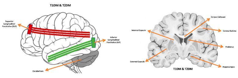
This medical illustration consists of two diagrams identifying brain regions frequently associated with microstructural tissue alterations in patients with Type 1 (T1DM) and Type 2 (T2DM) diabetes mellitus. The left diagram shows a lateral view of the human brain, highlighting two major white matter tracts: the Superior Longitudinal Fasciculus (SLF) in red and the Inferior Longitudinal Fasciculus (ILF) in green, with an orange arrow indicating the Cerebellum. The right diagram provides a coronal section of the brain with orange arrows labeling several key internal structures: the Corpus Callosum (superior midline), Corona Radiata, Thalamus (deep gray matter), Hippocampus (medial temporal lobe), and the Internal and External Capsules (white matter pathways). The diagrams serve an educational purpose by mapping common neuroanatomical sites of diabetic impact on brain integrity, relevant for clinical neurology and radiology students studying the long-term metabolic complications of diabetes on the central nervous system.
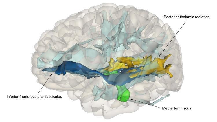
This diagnostic visualization features a 3D glass-brain model displaying white matter tracts reconstructed using diffusion MRI fiber tractography. The image highlights neuroanatomical structures within the translucent cortical shell to illustrate associations with polygenic scores for cognition. Three key tracts are color-coded based on nominal statistical significance: the inferior fronto-occipital fasciculus (IFOF) is shown in dark blue, extending horizontally from the frontal to the occipital pole; the medial lemniscus is highlighted in bright green at the level of the brainstem/pons; and the posterior thalamic radiation is depicted in yellow, branching through the posterior subcortical regions toward the visual cortex. Non-significant background tracts are rendered in a pale light-blue. This anatomical diagram serves to demonstrate the spatial distribution of white matter microstructural integrity and its correlation with genetic determinants of cognitive performance, providing a visual representation of how specific fiber pathways across different regions of the brain relate to genomic data in a population-based study.
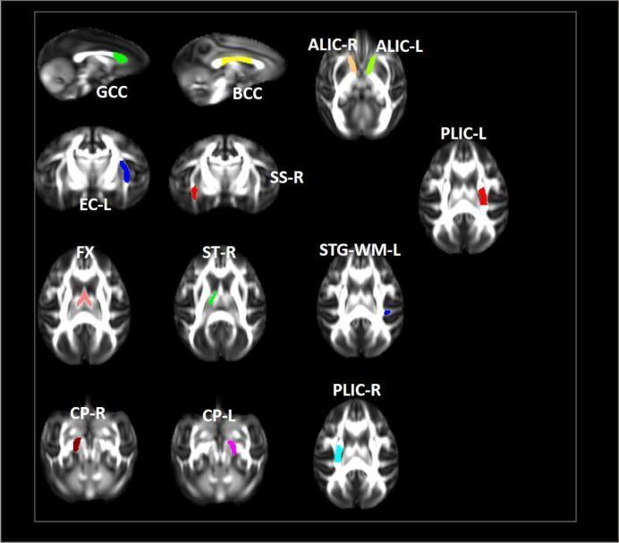
Anatomical diagram and diagnostic images of a non-human primate brain (rhesus macaque), specifically displaying fractional anisotropy (FA) maps derived from diffusion tensor imaging (DTI). The image shows multiple sagittal and axial brain slices with colored regions of interest (ROIs) identifying key white matter tracts. Midline structures include the genu of the corpus callosum (GCC, green), body of the corpus callosum (BCC, yellow), and fornix (FX, red). Lateralized tracts include the anterior limb of the internal capsule (ALIC-R/L, orange and green), posterior limb of the internal capsule (PLIC-L/R, red and cyan), external capsule (EC-L, blue), sagittal striatum (SS-R, red), stria terminalis (ST-R, green), superior temporal gyrus white matter (STG-WM-L, blue), and cerebral peduncles (CP-R/L, red and magenta). This educational visual illustrates the localization of neuroanatomical structures used to study microstructural white matter changes in a simian immunodeficiency virus (SIV) model of neuroHIV and the effects of antiretroviral therapy (cART).
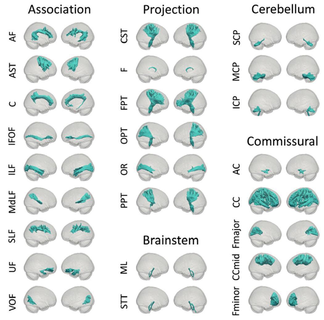
An anatomical diagram from the IIT Human Brain Atlas v.5.0 illustrating major white matter fiber bundles. The visualization is organized into a grid categorized by functional white matter types: Association, Projection, Cerebellum, Brainstem, and Commissural. Each specific tract is represented by teal-colored streamlines overlaid on a semi-transparent gray 3D brain model, shown from two perspective views to indicate spatial trajectory. Association fibers, such as the Arcuate Fasciculus (AF) and Cingulum (C), demonstrate curvilinear paths connecting cortical regions within the same hemisphere. Projection fibers, including the Corticospinal Tract (CST) and Frontopontine Tract (FPT), exhibit vertical orientations connecting the cortex to subcortical structures. Cerebellar and brainstem sections show localized pathways like the Superior Cerebellar Peduncle (SCP) and Medial Lemniscus (ML). Commissural tracts, such as the Corpus Callosum (CC) and Anterior Commissure (AC), show interhemispheric connectivity. This diagram serves as a neuroanatomical reference for white matter connectivity, tractography, and neuroimaging atlas interpretation.
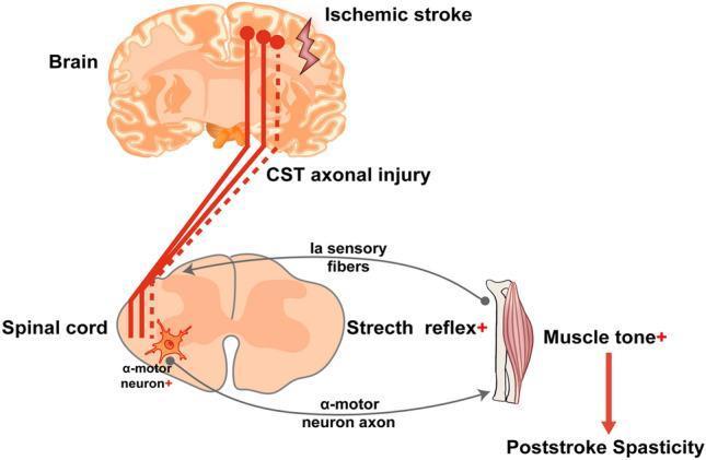
spinothalamic tract sensory pathway diagram
Tracts of the Brain
1. Projection Fibers
Internal Capsule
| Region | Location | Fiber content |
|---|---|---|
| Anterior limb | Between lenticular nucleus & head of caudate | Frontopontine fibers; frontal-thalamic connections |
| Genu | Junction of anterior & posterior limbs | Corticobulbar fibers |
| Posterior limb | Between lenticular nucleus & thalamus | Corticospinal fibers (largest component) |
| Retrolenticular limb | Posterior to lenticular nucleus | Optic radiation (lateral geniculate → primary visual cortex) |
| Sublenticular limb | Inferior to lenticular nucleus | Auditory radiation (medial geniculate → primary auditory cortex) |
2. Major Descending (Motor) Tracts
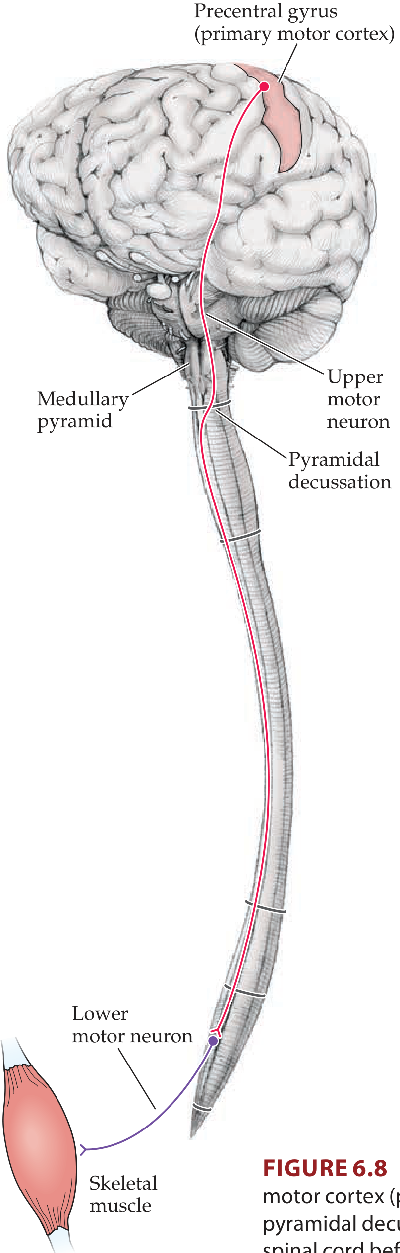
Corticospinal Tract (Pyramidal Tract)
- Origin: >50% from primary motor cortex (Brodmann area 4, precentral gyrus); remainder from premotor/supplementary motor areas (area 6) and parietal lobe (areas 3, 1, 2, 5, 7)
- Cells: Mostly cortical layer 5 pyramidal neurons; ~3% are giant Betz cells (largest neurons in the CNS)
- Course: Corona radiata → posterior limb of internal capsule → cerebral peduncles → ventral pons → medullary pyramids
- Decussation: ~90% cross at the pyramidal decussation (caudal medulla) → descend as lateral corticospinal tract in the lateral funiculus of the spinal cord → synapse on anterior horn cells (laminae IV-VII, IX)
- Remainder: ~10% do NOT cross → descend as ventral (anterior) corticospinal tract → cross later via ventral white commissure at cervical/upper thoracic levels
Corticobulbar Tract
- Travels with corticospinal fibers through the genu of the internal capsule
- Terminates on motor cranial nerve nuclei in the brainstem (CN V, VII, IX, X, XI, XII)
Rubrospinal Tract (Corticorubrospinal)
- Cortical areas 4, 6, 3, 1, 2 → ipsilateral red nucleus → axons decussate → descend in the lateral funiculus
- Functionally overlaps with the lateral corticospinal tract
Vestibulospinal Tract
- Lateral vestibulospinal tract: From lateral vestibular nucleus → entire length of spinal cord (anterior aspect of lateral funiculus) - facilitates extensor tone
- Medial vestibulospinal tract: From medial vestibular nucleus → cervical and upper thoracic cord (ventral funiculus) - controls head/neck position
Reticulospinal Tracts
- Lateral (medullary) reticulospinal: From medullary reticular formation → ventrolateral funiculus - mostly facilitates flexors, inhibits extensors
- Medial (pontine) reticulospinal: From pontine reticular formation → ipsilateral ventral funiculus - facilitates extensors
3. Major Ascending (Sensory) Tracts
Posterior Column - Medial Lemniscal Pathway
- 1st-order neuron: Enters dorsal root → ascends ipsilaterally in posterior column (fasciculus gracilis for legs; fasciculus cuneatus for arms) → synapses in gracile/cuneate nuclei in medulla
- 2nd-order neuron: Decussates in the medulla as internal arcuate fibers → forms the medial lemniscus → ascends to VPL of thalamus
- 3rd-order neuron: VPL → somatosensory cortex (postcentral gyrus, Brodmann areas 3, 1, 2)
- Lesion: Ipsilateral loss of fine touch and proprioception below the lesion
Spinothalamic Tract (Anterolateral System)
- 1st-order neuron: Enters dorsal root → synapses in dorsal horn (laminae I, V)
- 2nd-order neuron: Crosses via anterior commissure (2-3 segments above entry) → ascends in anterolateral white matter
- Passes through medulla (lateral), pontine tegmentum (just lateral to medial lemniscus), midbrain → VPL of thalamus
- 3rd-order neuron: VPL → somatosensory cortex (postcentral gyrus)
- Somatotopy: Legs lateral, arms medial in spinal cord (opposite of posterior columns)
- Lesion: Contralateral loss of pain and temperature a few segments below the lesion
Sub-components of the Anterolateral System:
| Tract | Termination | Function |
|---|---|---|
| Spinothalamic | VPL thalamus | Discriminative pain/temperature (location, intensity) |
| Spinoreticular | Medullary-pontine reticular formation → intralaminar thalamus | Emotional/arousal aspects of pain |
| Spinomesencephalic | Periaqueductal gray (PAG), midbrain | Pain modulation, analgesia |
Trigeminothalamic Tract
- Analogous to spinothalamic for the face
- Pain/temperature from face → trigeminal spinal nucleus → crosses → ascends to VPM of thalamus → somatosensory cortex
4. Commissural Fibers
Corpus Callosum
- Largest fiber bundle in the brain (~300 million axons)
- Parts: Rostrum → Genu → Body → Isthmus → Splenium
- Genu/Splenium: Prefrontal, temporal, parietal cortices (higher cognitive and sensory info)
- Body/Isthmus: Visual, auditory, somatosensory information
- Most cortical regions connected, except: hand area of motor/somatosensory cortex and primary visual cortex
Anterior Commissure
- Compact bundle anterior to the fornix
- Interconnects the two temporal lobes and anterior olfactory nuclei
Other Commissures
- Posterior commissure: Connects caudal diencephalon
- Hippocampal (fornical) commissure: Interconnects the two hippocampal formations
5. Association Fibers
Short Association Fibers
- Connect adjacent gyri ("U fibers")
Long Association Fasciculi
| Fasciculus | Connections | Clinical relevance |
|---|---|---|
| Superior longitudinal fasciculus (SLF) | Frontal - parietal - occipital (above insula) | Attention, spatial awareness |
| Arcuate fasciculus | Frontal lobe ↔ temporal lobe | Language (Broca's ↔ Wernicke's); conduction aphasia if damaged |
| Uncinate fasciculus | Orbital frontal ↔ anterior temporal | Emotion, memory |
| Inferior occipitofrontal fasciculus | Occipital ↔ frontal | Visual processing to frontal decision-making |
| Cingulum | Runs within the cingulate gyrus; frontal-parietal-temporal | Limbic functions, memory |
| Inferior longitudinal fasciculus (ILF) | Occipital ↔ temporal | Object recognition, face processing |
Summary Table
| Category | Tract | Modality/Function | Key Feature |
|---|---|---|---|
| Projection - Motor | Lateral corticospinal | Voluntary limb movement | Crosses at pyramidal decussation |
| Projection - Motor | Ventral corticospinal | Axial/proximal movement | Crosses at cord level |
| Projection - Motor | Corticobulbar | Voluntary cranial nerve motor | Genu of internal capsule |
| Projection - Motor | Rubrospinal | Limb movement | Via red nucleus |
| Projection - Motor | Vestibulospinal | Balance, extensor tone | Lateral vestibular nucleus |
| Projection - Motor | Reticulospinal | Posture, autonomic motor | Reticular formation |
| Projection - Sensory | Posterior columns/medial lemniscus | Fine touch, proprioception, vibration | Crosses in medulla |
| Projection - Sensory | Spinothalamic | Pain, temperature, crude touch | Crosses in spinal cord |
| Projection - Sensory | Optic radiation | Vision | Lateral geniculate → V1 |
| Projection - Sensory | Auditory radiation | Hearing | Medial geniculate → A1 |
| Commissural | Corpus callosum | Interhemispheric integration | Largest fiber bundle |
| Commissural | Anterior commissure | Temporal lobe, olfaction | Anterior to fornix |
| Association | Arcuate fasciculus | Language (Broca-Wernicke) | Conduction aphasia if cut |
| Association | Superior longitudinal fasciculus | Attention, spatial | Lateral hemisphere |
| Association | Uncinate fasciculus | Emotion, memory | Orbital frontal-temporal |
Leminiscus
medial lemniscus pathway brainstem diagram
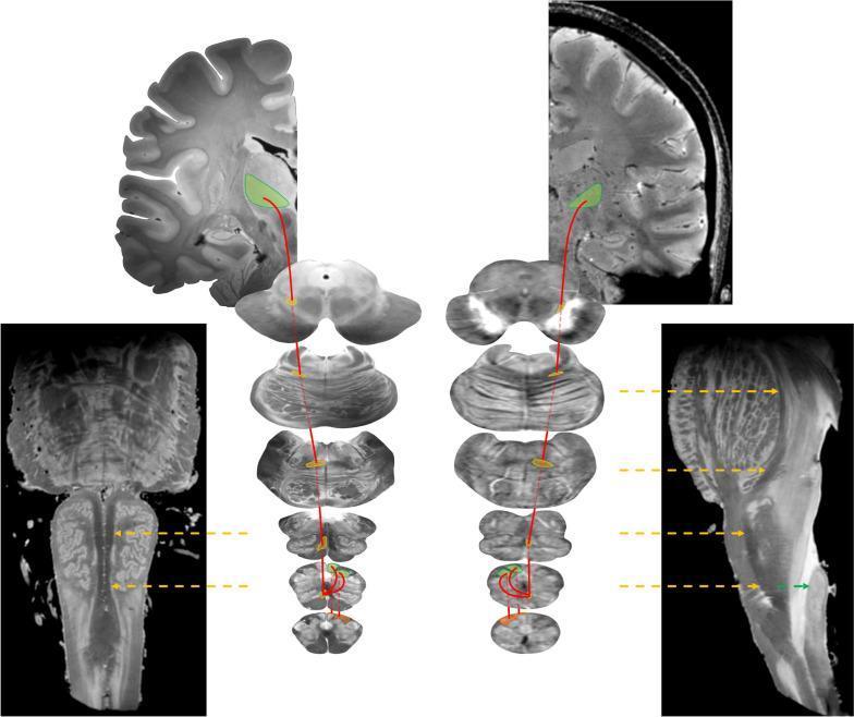
Educational neuroanatomical diagram and MRI composite illustrating the dorsal column-medial lemniscus (DCML) system. The visual uses a side-by-side comparison of ex vivo (left) and in vivo (right) high-resolution 7-T MR images to trace the somatosensory pathway. Red lines represent the trajectory of second-order neurons from the brainstem to the thalamus. Key structures are color-coded: the gracile and cuneate fasciculi are highlighted in orange at the caudal medulla; the gracile and cuneate nuclei are in green, marking the site of first-order synapse. The internal arcuate fibers are shown decussating before forming the medial lemniscus, which is highlighted in yellow across axial slices of the medulla, pons, and midbrain. Yellow arrows in the oblique sagittal and coronal brainstem views indicate the ascending trajectory of the medial lemniscus. The pathway terminates at the ventral posterolateral (VPL) nucleus of the thalamus, highlighted in green on coronal sections. This infographic serves as a diagnostic and anatomical reference for tracing epicritic sensation and conscious proprioception pathways.
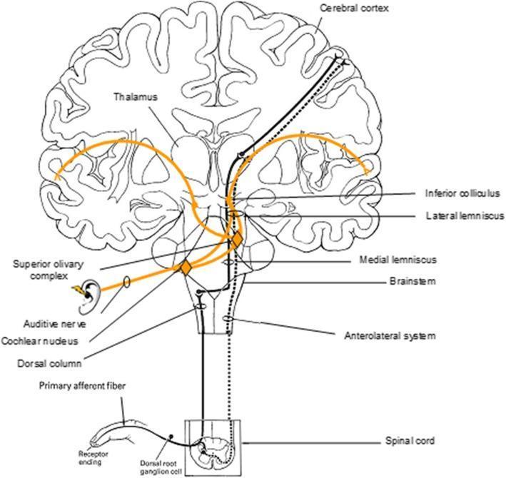
A pathophysiology diagram illustrating the ascending sensory pathways from the periphery to the cerebral cortex. The schematic uses a coronal section of the human brain, brainstem, and spinal cord to trace three major systems: the auditory-lateral lemniscal system (solid orange line), the dorsal column-medial lemniscus system (solid black line), and the anterolateral system (dashed black line). The auditory pathway is traced from the ear through the auditive nerve, cochlear nucleus, superior olivary complex, lateral lemniscus, and inferior colliculus, relaying through the medial geniculate nucleus of the thalamus to the auditory cortex. Somatosensory pathways are traced from receptor endings in the finger through the dorsal root ganglion to the spinal cord. The dorsal column system ascends ipsilaterally before decussating in the medulla (medial lemniscus) and relaying via the thalamus to the somatosensory cortex. The anterolateral system decussates at the spinal level before ascending. This visual summarizes the functional neuroanatomy required for sensory reactivity, commonly assessed in electroencephalography (EEG) clinical evaluations.
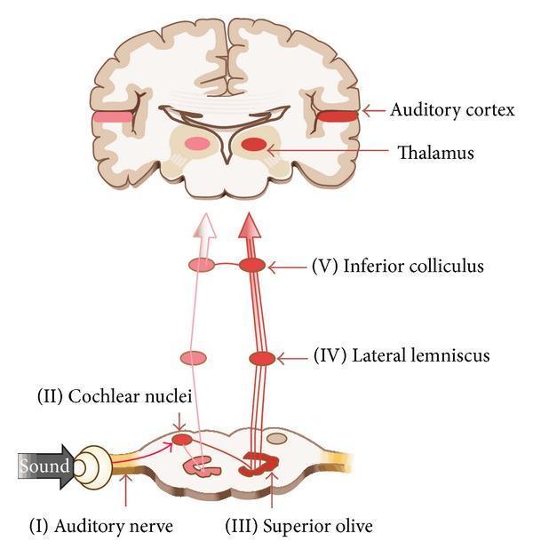
This medical illustration depicts the afferent auditory pathway and the neural generators of the auditory brainstem response (ABR). The diagram follows the sequential ascent of sound signals from the peripheral receptor to the cerebral cortex. The pathway initiates at the Auditory Nerve (I), proceeding to the Cochlear Nuclei (II) in the brainstem. The signal continues through the Superior Olive (III), ascends via the Lateral Lemniscus (IV), and reaches the Inferior Colliculus (V) in the midbrain. From the midbrain, the pathway is shown projecting to the Thalamus (medial geniculate nucleus) and finally to the primary Auditory Cortex located on the superior temporal gyrus. The top portion of the figure includes a coronal cross-section of the brain to contextualize the cortical and subcortical destinations. The diagram is designed for educational purposes in neurology and otolaryngology, illustrating the hierarchical processing stages involved in auditory perception and clinical diagnostic testing like ABR.
The Lemniscal System
- Medial Lemniscus (most important)
- Lateral Lemniscus
- Trigeminal Lemniscus
- Spinal Lemniscus
1. Medial Lemniscus
Pathway (3-Neuron Arc)
| Neuron | Location | Fiber | Notes |
|---|---|---|---|
| 1st order | Dorsal root ganglion | Enters medial dorsal root entry zone | Large-diameter, heavily myelinated (Aα, Aβ) |
| 2nd order | Posterior column nuclei (gracile/cuneate) in medulla | Internal arcuate fibers → medial lemniscus | Decussates in the medulla |
| 3rd order | VPL of thalamus | Thalamocortical radiation | → Postcentral gyrus (S1) |
Posterior Columns (1st order - ascending)
- Fasciculus gracilis (medial): carries legs and lower trunk (below T6)
- Fasciculus cuneatus (lateral): carries upper trunk (above T6), arms, neck
- Mnemonic: fibers add on laterally as they ascend - legs are always medial in the posterior columns
Formation of the Medial Lemniscus (2nd order)
- 1st-order neurons synapse in nucleus gracilis and nucleus cuneatus in the medulla
- Their axons curve anteriorly as internal arcuate fibers, cross the midline, and form the medial lemniscus on the opposite side
- Also receives input from the lateral cervical nucleus (LCN) and nucleus Z
Course of the Medial Lemniscus through the Brainstem
| Level | Position | Somatotopy |
|---|---|---|
| Medulla | Dorsal to pyramidal tract, near midline, vertical orientation | Gracile (legs) ventrolateral; Cuneate (arms) dorsomedial |
| Pons | Moves laterally, more inclined | Arms medial, legs lateral ("little person lies down") |
| Midbrain | Lateral in tegmentum, just lateral to red nucleus | Arms medial, legs lateral |
| Thalamus | Terminates in VPL (ventral posterolateral nucleus) | Full somatotopic map |
- Localization in Clinical Neurology, 8th ed., p. 45
Key somatotopy reversal: In the posterior columns, legs are medial; after crossing to form the medial lemniscus in pons/midbrain, legs become lateral.
From Thalamus to Cortex
- VPL → thalamocortical radiations → primary somatosensory cortex (S1) in the postcentral gyrus (Brodmann areas 3, 1, 2)
- Somatotopic map in S1: foot/leg medially, face laterally
Lesion Effects
| Site | Deficit |
|---|---|
| Posterior column (below decussation) | Ipsilateral loss of fine touch, vibration, proprioception below the lesion |
| Medial lemniscus (above decussation) | Contralateral loss of fine touch, vibration, proprioception |
| VPL thalamus | Contralateral hemisensory loss (all modalities) |
2. Lateral Lemniscus
Pathway
Nuclei of the Lateral Lemniscus
- Receives input from all major groups of ventral cochlear nucleus cells
- Responds predominantly to monaural input (contralateral ear)
- Neurons are inhibitory (glycinergic) and project to the inferior colliculus
- Thought to process the meaning/identity of sounds
- Receives input from lateral and medial superior olivary nuclei (binaural)
- Neurons are GABAergic and inhibitory
- Project to inferior colliculi bilaterally
- Generates persistent inhibition in the inferior colliculus → underlies the precedence effect (suppression of sound echoes for accurate sound localization)
3. Trigeminal Lemniscus
- Afferents from the face mediating joint position, vibration, and kinesthesia → principal (main sensory) trigeminal nucleus in the pons
- Post-synaptic neurons send axons across the brainstem as the trigeminal lemniscus
- Ascends in parallel with the medial lemniscus through the brainstem
- Terminates in the VPM (ventral posteromedial) nucleus of the thalamus (unlike the medial lemniscus which goes to VPL)
- From VPM → somatosensory cortex (S1), face region
Note: The trigeminothalamic tract (carrying pain and temperature from the face) travels separately but converges near the medial lemniscus before reaching VPM.
4. Spinal Lemniscus
Comparison Table: The Four Lemnisci
| Lemniscus | Modality | Origin | Thalamic Relay | Decussation |
|---|---|---|---|---|
| Medial | Fine touch, vibration, proprioception (body) | Nucleus gracilis/cuneatus (medulla) | VPL | Medulla (internal arcuate fibers) |
| Lateral | Auditory | Cochlear nuclei, superior olive | MGN (medial geniculate) | Multiple levels (bilateral) |
| Trigeminal | Fine touch, proprioception (face) | Principal trigeminal nucleus (pons) | VPM | Brainstem (crosses to opposite side) |
| Spinal | Pain, temperature, crude touch (body) | Dorsal horn (spinal cord) | VPL | Spinal cord (anterior commissure) |
Clinical Importance of the Medial Lemniscus
- Wallenberg syndrome (lateral medullary syndrome): Spares the medial lemniscus (it lies medially); causes loss of pain/temperature on ipsilateral face and contralateral body but fine touch and proprioception are preserved.
- Medial medullary syndrome: Damages the medial lemniscus and pyramid → contralateral loss of fine touch/proprioception + contralateral hemiplegia.
- Pure sensory stroke: Lesion in VPL thalamus disrupts both lemniscal and spinothalamic inputs → hemisensory loss of all modalities.