
Cardiomyopathies (DCM, HOCM, RCM)- definition,etiology, classification, symptoms, diagnosis, differential diagnosis treatment
Reading File
Finding Sources
Finding Sources
Finding Sources
Now let me read the key sources in parallel - Goldman-Cecil (comprehensive medicine), Robbins (pathology), Tintinalli (emergency/clinical), and search for RCM:
Finding Sources
Reading File
Reading File
Excellent content from Robbins Basic Pathology. Now let me read Goldman-Cecil (comprehensive clinical content) and Harrison's (RCM) and Tintinalli in parallel:
Reading File
Reading File
Reading File
Excellent. Now let me read the Goldman-Cecil content for HOCM and RCM, and the Robbins content for RCM:
Reading File
Reading File
Reading File
Now let me read the Goldman-Cecil HOCM section more fully, and get Robbins HOCM pathology + search for images:
Reading File
Reading File
Searching Images
dilated cardiomyopathy echocardiography gross pathology
Now let me search for HCM/HOCM and RCM images, and also get the clinical management content from Goldman-Cecil:
Searching Images
hypertrophic obstructive cardiomyopathy HOCM septal hypertrophy echocardiography
Searching Images
restrictive cardiomyopathy amyloidosis cardiac histology
Reading File
I now have comprehensive content from multiple authoritative sources. Let me also get the HOCM clinical management and treatment section and the DCM treatment section from Goldman-Cecil:
Reading File
I have comprehensive content from all three cardiomyopathies. Let me update todos and now compile the full response:# Cardiomyopathies: DCM, HOCM, and RCM
Overview & Classification
Cardiomyopathies are defined as diseases of the myocardium that are not explained by coronary artery disease, valvular disease, hypertension, or congenital heart disease. They are classified by their predominant functional/structural phenotype:
| Feature | DCM | HCM/HOCM | RCM |
|---|---|---|---|
| LV cavity | Dilated | Normal/small | Normal/small |
| Wall thickness | Normal/thin | Increased | Normal/increased |
| Systolic function (EF) | Reduced (<40%) | Preserved (50–80%) | Preserved or mildly reduced (25–50%) |
| Primary dysfunction | Systolic (pump failure) | Diastolic (poor relaxation) + LVOTO | Diastolic (impaired filling) |
- Robbins & Kumar Basic Pathology, Table 9.5
- Goldman-Cecil Medicine
1. DILATED CARDIOMYOPATHY (DCM)
Definition
DCM is characterized by progressive dilation and contractile (systolic) dysfunction of the left ventricle or both ventricles, usually with concurrent hypertrophy, in the absence of coronary artery disease, valvular abnormalities, or pericardial disease. It is the most common form of cardiomyopathy in children and the leading indication for cardiac transplantation.
- Prevalence: ~1 in 250 adults
- Males and females approximately equally affected (except X-linked forms, which are male-predominant)
Etiology & Classification
Genetic (20–50% of cases):
- Autosomal dominant (most common): mutations in cytoskeletal or sarcomere-linking genes
- TTN (titin) — most common, accounts for ~25% of familial and ~18% of sporadic cases
- MYH7 (β-myosin heavy chain), TNNT2 (troponin T)
- LMNA (lamin A/C) — causes arrhythmogenic DCM with progressive AV conduction disease
- FLNC (filamin C) — lethal ventricular arrhythmias in adolescents/young adults
- DSP (desmoplakin)
- X-linked (2–5%): mutations in dystrophin — associated with Duchenne/Becker/Emery-Dreifuss muscular dystrophies
- Key molecular mechanism: loss-of-function mutations in sarcomeric proteins (contrast with gain-of-function in HCM)
Acquired causes:
| Category | Examples |
|---|---|
| Infectious | Viral myocarditis (coxsackievirus B, parvovirus B19, HHV-6, HIV, EBV, CMV, Chagas disease) |
| Toxic | Alcohol (direct toxicity of ethanol + acetaldehyde), doxorubicin/anthracyclines, cyclophosphamide, cocaine |
| Metabolic | Iron overload (hemochromatosis), nutritional deficiencies (thiamine, selenium, carnitine) |
| Endocrine | Hypothyroidism, acromegaly, pheochromocytoma |
| Autoimmune/Inflammatory | SLE, sarcoidosis, giant cell myocarditis |
| Peripartum | Peripartum cardiomyopathy (last month of pregnancy to 5 months postpartum) |
| Tachycardia-induced | Tachycardiomyopathy (reversible once rate controlled) |
| Idiopathic | When no cause found |
Pathology
- Gross: Four-chamber dilation; thin, flabby walls; mural thrombus (especially LV apex); pale, dilated heart
- Microscopy: Myocyte hypertrophy, interstitial and replacement fibrosis (Masson trichrome: blue collagen), no specific changes
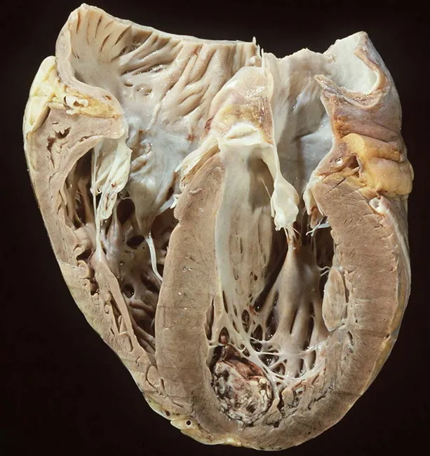
Gross pathology of DCM showing biventricular dilation, thin myocardial walls, and apical LV thrombus.
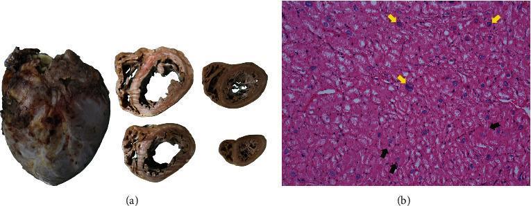
Gross (a) and H&E microscopy (b) of idiopathic DCM. Note biventricular dilation and myocyte hypertrophy (yellow arrows) with vacuolar degeneration (black arrows).
Clinical Features (Symptoms)
- Early: Reduced exercise tolerance, dyspnea on exertion
- Progressive: Dyspnea at rest, orthopnea, paroxysmal nocturnal dyspnea (PND), peripheral edema, ascites
- Arrhythmias: Palpitations, presyncope, syncope; AF, VT/VF
- Embolic events: Stroke, systemic embolism (from mural thrombus)
- Sudden cardiac death may be first presentation
- Low output signs: Sinus tachycardia, weak pulses, hypotension, fatigue, abdominal discomfort (mesenteric ischemia)
Examination:
- Displaced, diffuse apical impulse
- Elevated JVP; third heart sound (S3 gallop); ± fourth heart sound (S4)
- Pansystolic murmur of functional mitral regurgitation (apex → axilla)
- Basal lung crackles, hepatomegaly, peripheral edema
Diagnosis
ECG: Sinus tachycardia; nonspecific ST-T changes (inferior/lateral leads); atrial enlargement; LBBB/intraventricular conduction delay; AV block (LMNA/desmin mutations); VT
Chest X-ray: Cardiomegaly (cardiothoracic ratio >0.5); pulmonary vascular congestion; pleural effusions
Echocardiography (key diagnostic tool):
- LV end-diastolic dimension >2 SD above BSA-corrected mean (or >112% predicted)
- Fractional shortening <25%; EF <40%
- Functional MR and TR; diastolic dysfunction
- Mural thrombus at LV apex
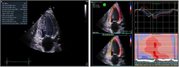
TTE of DCM: apical 4-chamber view showing severely dilated LV with markedly reduced EF (27–29%) and GLS analysis confirming global hypokinesia.
Labs: CBC, renal/thyroid/hepatic function, CK (elevated in dystrophin-linked DCM), troponins, BNP/NT-proBNP (prognostic), iron studies
Cardiac MRI: Areas of myocardial fibrosis (late gadolinium enhancement, LGE); characterizes etiology (non-ischemic pattern)
Cardiac catheterization: Rarely needed; to exclude significant CAD or provide hemodynamic data pre-transplantation
Endomyocardial biopsy: For suspected myocarditis, metabolic disorders; not routine
Genetic testing: Recommended in all; enables cascade screening of first-degree relatives
Differential Diagnosis
- Ischemic cardiomyopathy (CAD-related LV dysfunction — most common mimic)
- Hypertensive heart disease (with decompensation)
- Valvular heart disease (especially AR, MR causing volume overload)
- Myocarditis (acute/subacute)
- Takotsubo (stress) cardiomyopathy
- Congenital heart disease
- Peripartum cardiomyopathy
- Tachycardia-induced cardiomyopathy
Treatment
General/Supportive:
- Sodium and fluid restriction; alcohol avoidance; remove toxins
- Exercise (supervised cardiac rehabilitation for stable patients)
Pharmacotherapy (standard HFrEF therapy — "four pillars"):
| Drug Class | Examples | Notes |
|---|---|---|
| ACE inhibitor / ARB | Enalapril, ramipril; losartan | Reduce mortality; all tolerated patients |
| Beta-blocker | Carvedilol, metoprolol succinate, bisoprolol | Reduce mortality and sudden death |
| Mineralocorticoid antagonist | Spironolactone, eplerenone | Add if EF ≤35%, symptomatic HF |
| SGLT2 inhibitor | Dapagliflozin, empagliflozin | Reduce HF hospitalization and CV death |
| ARNI (sacubitril/valsartan) | Entresto | Superior to ACEi alone for HFrEF |
| Loop diuretics | Furosemide | Symptom relief (fluid overload); no mortality benefit |
| Hydralazine + nitrates | Alternative if ACEi/ARB intolerant; benefit in Black patients |
Device Therapy:
- ICD: EF ≤35% despite ≥3 months GDMT, for primary prevention of sudden cardiac death
- CRT: LBBB + QRS ≥150 ms + EF ≤35% — improves symptoms, EF, and mortality
- CRT-D: Combines CRT + ICD
Anticoagulation:
- Warfarin or NOAC for AF or documented LV thrombus
- Consider if EF very low + clinical risk factors
Advanced therapies:
-
Cardiac transplantation (end-stage refractory DCM)
-
LV assist device (LVAD) as bridge to transplant or destination therapy
-
Goldman-Cecil Medicine; Robbins Basic Pathology
2. HYPERTROPHIC (OBSTRUCTIVE) CARDIOMYOPATHY — HCM / HOCM
Definition
HCM is defined as unexplained LV hypertrophy in the absence of abnormal loading conditions (valve disease, hypertension, congenital heart defects) sufficient to explain the degree of hypertrophy. When this hypertrophy causes dynamic LV outflow tract obstruction (LVOTO), it is termed hypertrophic obstructive cardiomyopathy (HOCM) — present in ~1/3 of HCM patients at rest and ~2/3 with provocation.
- Prevalence: 0.2–0.5% (worldwide, all racial groups)
- Leading cause of sudden cardiac death in young athletes
Etiology & Genetics
- Autosomal dominant with variable penetrance and expressivity
- Gain-of-function mutations in sarcomeric proteins → myocyte hypercontractility, increased energy use, net negative energy balance
-
1,400 mutations identified; 50–60% of cases have identifiable sarcomeric mutation
- Most commonly mutated genes:
- MYH7 (β-myosin heavy chain): 30–40% of cases
- MYBPC3 (myosin-binding protein C): another major cause
- TNNT2 (troponin T), TNNI3 (troponin I)
- These three genes account for 70–80% of all HCM
- Other causes: Friedreich's ataxia, glycogen storage diseases (Pompe, Danon), Fabry disease, Noonan syndrome, infant of diabetic mother
Pathology
-
Gross: Massive myocardial hypertrophy without ventricular dilation; heavy heart
- Asymmetric septal hypertrophy (ASH) in ~90% — disproportionate IVS thickening
- In remaining 10%: concentric hypertrophy
- LV cavity compressed into "banana-like" shape on cross-section
- Endocardial plaque in LVOT (contact lesion from anterior mitral leaflet)
- Mitral leaflet thickening
-
Microscopy (pathognomonic):
- Myocyte disarray: chaotic, tangled arrangement replacing normal parallel orientation
- Myocyte hypertrophy
- Interstitial fibrosis (replacement fibrosis — substrate for arrhythmias)
- Abnormal intramural coronary arteries (medial hypertrophy, luminal narrowing)
Clinical Features (Symptoms)
- Many patients are asymptomatic (discovered incidentally or at family screening)
- Classic triad in symptomatic patients:
- Exertional dyspnea (diastolic dysfunction; impaired filling)
- Angina/chest pain (myocardial oxygen demand > supply; small vessel disease)
- Syncope/presyncope (LVOTO worsening with effort; arrhythmia; inadequate cardiac output)
- Palpitations (AF, VT)
- Sudden cardiac death — may be first manifestation (especially young athletes)
Key clinical feature of LVOTO: symptoms worsen with:
- Exercise (decreased afterload + increased contractility)
- Dehydration/hypovolemia
- Standing (decreased preload)
- Vasodilators, diuretics
- Tachycardia
Symptoms improve with:
- Lying down (increased preload)
- Squatting
- Handgrip (increased afterload)
Examination:
- Double or triple apical impulse (atrial kick + bifid LV contraction)
- Loud S4 gallop (stiff ventricle, forceful atrial contraction)
- Harsh crescendo-decrescendo systolic ejection murmur at LLSB/apex (LVOTO) — NOT radiating to neck (distinguishes from AS)
- Murmur increases with Valsalva, standing; decreases with squatting, handgrip
- Systolic anterior motion (SAM) of mitral valve → MR murmur (apex → axilla)
- Bisferiens carotid pulse (rare)
Diagnosis
ECG:
- LVH (voltage criteria)
- Deep, narrow Q waves (septal depolarization from disarray) — inferior/lateral leads
- T-wave inversion, ST changes
- AF, SVT
- Pre-excitation (WPW pattern — associated with glycogen storage diseases)
Echocardiography (gold standard):
- Asymmetric IVS thickness ≥15 mm (or ≥13 mm in first-degree relatives)
- IVS:posterior wall ratio >1.3–1.5
- Systolic anterior motion (SAM) of mitral valve (hallmark of LVOTO)
- LVOT gradient ≥30 mmHg at rest or ≥50 mmHg with provocation (Valsalva, amyl nitrite)
- Preserved EF (often hyperdynamic, 60–80%)
- Diastolic dysfunction
- Mitral regurgitation (posterior jet from SAM)
- Small LV cavity
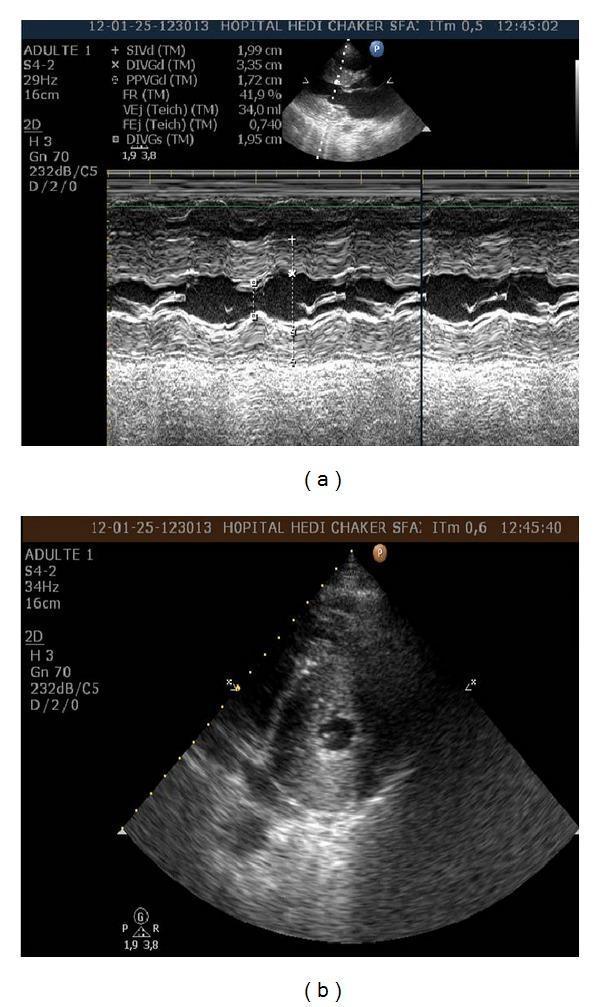
TTE of HOCM (a) parasternal long-axis with M-mode: IVS = 20 mm, SAM of mitral valve. (b) Short-axis: severe concentric hypertrophy with near-obliteration of LV cavity.
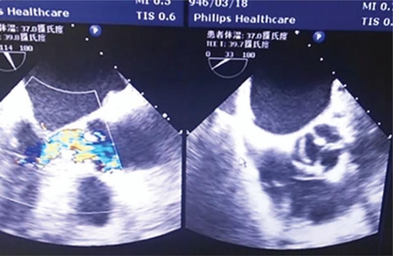
Color Doppler TEE in HOCM: turbulent, high-velocity flow through narrowed LVOT (left). Short-axis showing restricted aortic leaflet opening due to LVOT hemodynamics — not intrinsic AS (right).
Cardiac MRI: Quantifies hypertrophy distribution; detects LGE (fibrosis — prognostic for SCD); characterizes phenotype
Holter/ambulatory ECG: Assesses arrhythmia burden (AF, NSVT — SCD risk factor)
Exercise stress testing: Assess symptoms; abnormal BP response (failure to rise ≥20 mmHg or fall) = SCD risk factor; provocable LVOT gradient
Genetic testing: Recommended for all HCM patients; cascade family screening
Cardiac catheterization: Rarely needed; confirms LVOT gradient; Brockenbrough sign (post-PVC decrease in pulse pressure in HOCM vs increase in AS)
Differential Diagnosis
- Hypertensive heart disease (LVH from pressure overload — most common mimic)
- Aortic stenosis (calcific/bicuspid — fixed LVOTO; gradient does NOT change with Valsalva)
- Athlete's heart (physiological hypertrophy — wall thickness usually <13 mm; normal diastolic function; cavity not reduced; regresses with detraining)
- Cardiac amyloidosis (infiltrative — low-voltage ECG; sparkling echo pattern)
- Fabry disease (angiokeratomas, corneal deposits; reduced α-galactosidase A)
- Glycogen storage diseases (Pompe, Danon)
- Noonan syndrome (dysmorphic features)
- RCM overlap (some sarcomere mutations cause mixed phenotype)
Treatment
General Measures:
- Avoid dehydration, extreme exercise, vasodilators, diuretics (worsen obstruction)
- Avoid alcohol in excess
- Competitive sports restriction for patients with symptomatic HOCM (though recent data suggest vigorous exercise may be safe in carefully selected patients)
Pharmacotherapy:
| Drug | Mechanism | Use |
|---|---|---|
| Beta-blockers (first-line) | ↓ HR → ↑ diastolic filling time; ↓ contractility → ↓ LVOTO | Symptomatic HOCM; all symptomatic patients |
| Non-dihydropyridine CCBs (verapamil, diltiazem) | ↓ HR and contractility; improve diastolic relaxation | Alternative to beta-blockers |
| Disopyramide | Negative inotrope → ↓ LVOTO gradient | Add to beta-blocker for refractory obstruction |
| Mavacamten (novel) | Cardiac myosin inhibitor → directly reduces LVOT gradient | Approved for symptomatic HOCM (EXPLORER-HCM trial, 2020); significant gradient reduction |
| Anticoagulation | For AF (rate control preferred over rhythm) |
Avoid: Digoxin, dihydropyridine CCBs, nitrates, vasodilators, diuretics — worsen LVOTO
Septal Reduction Therapy (for patients with gradient ≥50 mmHg + severe symptoms refractory to medical therapy):
- Surgical septal myectomy (Morrow procedure) — gold standard; excises basal septal muscle; durable relief; preferred at expert centers
- Alcohol septal ablation — percutaneous injection of alcohol into first septal perforator → controlled septal infarction; less invasive; more LBBB/complete heart block risk; appropriate for older patients or poor surgical risk
ICD implantation (primary prevention of SCD) — indications:
- Massive hypertrophy (wall thickness ≥30 mm)
- History of VF/sustained VT
- Family history of SCD
- Unexplained syncope
- Abnormal BP response to exercise
- NSVT on Holter
Cardiac transplantation: End-stage HCM with refractory symptoms
- Goldman-Cecil Medicine; Robbins Basic Pathology; Olivotto et al. (mavacamten), Lancet 2020
3. RESTRICTIVE CARDIOMYOPATHY (RCM)
Definition
RCM is characterized by increased ventricular stiffness, impaired diastolic filling, elevated LV diastolic pressures, and reduced diastolic volume of the LV or RV, despite normal or near-normal systolic function and wall thickness. It is the least common of the three major cardiomyopathies.
Etiology & Classification
Primary (Idiopathic):
- ~30% have familial disease with sarcomere gene mutations (TNNI3, MYH7)
- Desmin (DES) mutations — RCM + skeletal myopathy + cardiac conduction abnormalities
- May overlap phenotypically with HCM
Secondary (most common — infiltrative/storage/fibrotic):
| Category | Examples |
|---|---|
| Infiltrative | Amyloidosis (AL and ATTR — most prevalent, increasing with age), Sarcoidosis |
| Storage disorders | Hemochromatosis, Fabry disease, Glycogen storage diseases, Mucopolysaccharidoses |
| Fibrotic/Toxic | Radiation-induced fibrosis, Scleroderma, Drugs (anthracyclines, serotonin, ergotamine) |
| Endomyocardial | Endomyocardial fibrosis (Africa/tropical — most common RCM worldwide), Löffler endomyocarditis (hypereosinophilia) |
| Metabolic | Carnitine deficiency, Fatty acid oxidation defects |
| Other | Carcinoid syndrome |
Wild-type (senile) ATTR amyloidosis is the most prevalent form of RCM and increases with age, especially in older males. Four percent of African Americans carry a specific TTR mutation with >4× risk.
Pathology
Endomyocardial fibrosis:
- Diffuse fibrosis of ventricular endocardium/subendocardium, often involving mitral/tricuspid valves
- Most common worldwide (tropical Africa)
- Linked to nutritional deficiencies and helminthic infections
Löffler endomyocarditis:
- Peripheral hypereosinophilia → eosinophil granule contents damage myocardium
- Endocardial fibrosis + large mural thrombi → thrombus organization
Cardiac amyloidosis:
- AL type (immunoglobulin light chains — multiple myeloma)
- ATTR type (transthyretin — hereditary variants or wild-type/senile)
- Extracellular deposition of β-pleated sheet protein fibrils in myocardial interstitium
Gross: Normal-sized or slightly enlarged ventricles; non-dilated cavities; firm myocardium; bilateral atrial dilation (hallmark — consequence of impaired ventricular filling); thrombus in atrial appendages
Microscopy: Variable interstitial fibrosis; myocyte disarray (less than HCM); cause-specific findings on biopsy (amyloid, granulomas, iron)
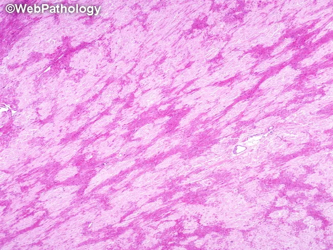
Histology of ATTR cardiac amyloidosis: pale eosinophilic amyloid deposits in myocardial interstitium, displacing and atrophying cardiomyocytes, producing restrictive physiology.
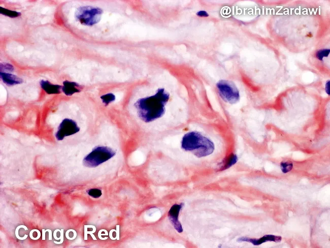
Congo red stain of cardiac amyloidosis: salmon-pink perimyocytic amyloid deposits around cardiomyocytes. Under polarized light, these show pathognomonic apple-green birefringence.
Clinical Features (Symptoms)
- Dyspnea on exertion → at rest (most common)
- Orthopnea, PND
- Recurrent respiratory infections
- Fatigue and weakness (low output)
- Right-sided predominance: Peripheral edema, ascites, hepatomegaly (prominent in severe RCM — due to impaired RV filling and tricuspid regurgitation)
- Chest pain, palpitations
- Supraventricular arrhythmias (AF, heart block) — common
- Sudden cardiac death (rare, usually first presentation)
Examination:
- Elevated JVP with prominent y descent
- Kussmaul's sign (JVP fails to fall or rises with inspiration) — also seen in constrictive pericarditis
- Loud P2 (if pulmonary hypertension)
- S3 and/or S4 gallop
- Peripheral edema, ascites, hepatomegaly
Diagnosis
ECG:
- P mitrale, P pulmonale
- Nonspecific ST-T changes; ST depression; T-wave inversion (inferolateral)
- Voltage criteria for LVH or RVH
- Low-voltage QRS — classic in cardiac amyloidosis (despite thick walls)
- Conduction abnormalities; abnormal Q waves; AV block
Chest X-ray: Prominent atrial silhouettes; mild cardiomegaly; pulmonary congestion
Echocardiography:
- Markedly dilated atria (can dwarf ventricles)
- Normal or mildly increased wall thickness; non-dilated LV cavity; preserved EF
- "Sparkling" granular appearance of myocardium (cardiac amyloidosis)
- Diastolic dysfunction: ↑ E/A ratio; ↓ E deceleration time; ↓ IVRT; Restrictive filling pattern
- Tissue Doppler: reduced diastolic annular velocities (e'↓); elevated E/e' ratio
- Pulmonary/hepatic vein Doppler: ↑ diastolic > systolic velocities
Cardiac MRI:
- LGE — diffuse subendocardial pattern (amyloidosis); patchy/midwall/epicardial (sarcoidosis)
- T1 mapping: elevated native T1 (amyloid infiltration)
- Concentric LV hypertrophy + bilateral atrial dilation
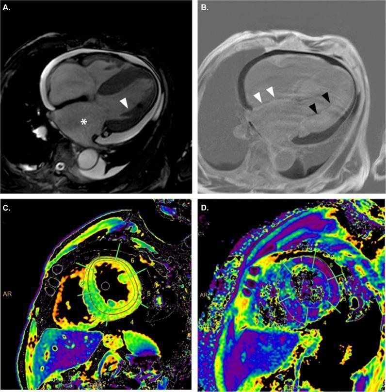
CMR panels of cardiac amyloidosis (RCM): (A) concentric LVH + bilateral atrial dilation; (B) diffuse subendocardial LGE; (C/D) T1 and T2 mapping showing diffuse infiltration.
Cardiac catheterization:
- "Dip-and-plateau" pattern (square root sign): rapid early diastolic pressure decline → rapid rise to plateau
- LVEDP, PCWP markedly elevated, usually 5 mmHg or more above right-sided pressures (distinguishes from constrictive pericarditis where pressures equalize)
Specific diagnostic tests:
- Amyloidosis: Serum/urine protein electrophoresis (AL type); TTR gene sequencing; Tc-99m pyrophosphate scintigraphy (ATTR amyloid — highly specific); endomyocardial biopsy (Congo red staining, apple-green birefringence)
- Hemochromatosis: Serum ferritin, transferrin saturation, HFE gene
- Sarcoidosis: ACE level, CT chest, FDG-PET, biopsy
- Fabry disease: Reduced α-galactosidase A (males); GLA gene; lyso-Gb3
Differential Diagnosis
Most critical distinction: RCM vs. Constrictive Pericarditis (CP)
| Feature | RCM | Constrictive Pericarditis |
|---|---|---|
| JVP | Elevated; y descent > x | Elevated; both x and y descent prominent |
| Kussmaul sign | Present | Present |
| Pericardium | Normal | Thickened/calcified |
| LVEDP vs RVEDP | LVEDP > RVEDP (≥5 mmHg) | LVEDP = RVEDP (equalization) |
| Respiratory variation (echo) | Minimal | Exaggerated (>25% mitral E variation) |
| Tissue Doppler e' | Reduced (<8 cm/s) | Normal or increased (>8 cm/s) — annulus paradoxus |
| CT/MRI | Normal pericardium | Pericardial thickening (>4 mm) ± calcification |
| Endomyocardial biopsy | Abnormal (cause-specific) | Normal |
| Surgery | Does not help | Pericardiectomy curative |
Also differentiate from:
- HCM (marked LVH; LVOTO; myocyte disarray; dynamic murmur)
- Hypertensive heart disease
- Acute decompensated heart failure from other causes
Treatment
General:
- Treat underlying cause when possible:
- ATTR amyloidosis: Tafamidis (TTR stabilizer) — approved; slows progression, reduces mortality and hospitalization; patisiran/inotersen (TTR gene silencers)
- AL amyloidosis: Treat underlying plasma cell dyscrasia (chemotherapy/stem cell transplant)
- Hemochromatosis: Phlebotomy, chelation
- Sarcoidosis: Corticosteroids
- Fabry disease: Enzyme replacement therapy (agalsidase)
Symptomatic/Supportive:
- Diuretics — cautious use (small preload-dependent ventricle; can precipitate low output)
- Rate control for AF (beta-blockers, verapamil); avoid digoxin in amyloidosis (binds amyloid fibrils → toxicity at therapeutic levels)
- Anticoagulation for AF or intracardiac thrombus
Device therapy:
- ICD — for sustained VT/VF or high SCD risk (sarcoidosis, giant cell myocarditis)
- Pacemaker — for symptomatic bradycardia/heart block
- CRT generally not beneficial (preserved EF)
Cardiac transplantation: For end-stage idiopathic RCM; generally not performed for systemic amyloidosis unless underlying disease also treated
- Goldman-Cecil Medicine; Robbins Basic Pathology; Harrison's Principles of Internal Medicine 22E
Comparative Summary Table
| Feature | DCM | HOCM | RCM |
|---|---|---|---|
| Definition | Dilation + systolic dysfunction | Unexplained LVH, ± LVOTO | Stiffened ventricle, impaired filling |
| EF | <40% (reduced) | 60–80% (hyperdynamic) | 25–50% (preserved-mildly reduced) |
| Key gene | TTN, LMNA (loss-of-function) | MYH7, MYBPC3 (gain-of-function) | TNNI3, DES; or secondary causes |
| Pathological hallmark | 4-chamber dilation, interstitial fibrosis | Myocyte disarray, ASH | Interstitial fibrosis ± infiltrate |
| Classic murmur | Functional MR (pansystolic) | Ejection murmur ↑ Valsalva | None specific; ± S3/S4 |
| Echo finding | Dilated, hypokinetic LV | ASH, SAM, LVOTO | Biatrial dilation, restrictive filling |
| Kussmaul sign | No | No | Yes |
| Key investigation | Echo + BNP + genetics | Echo + Holter + genetics | Echo + MRI + biopsy + amyloid studies |
| Specific treatment | GDMT (ACEi/ARB, BB, MRA, SGLT2i); ICD/CRT | Beta-blockers; mavacamten; septal myectomy/ablation | Treat cause (tafamidis, chelation, steroids) |
| SCD risk | Yes (ICD if EF ≤35%) | Yes (major — especially athletes) | Lower; highest in sarcoidosis |
| Transplant | Yes (end-stage) | Yes (refractory) | Selected cases (idiopathic) |
Sources: Goldman-Cecil Medicine (International Ed.) — Chs. 47, 62; Robbins & Kumar Basic Pathology — Ch. 9; Harrison's Principles of Internal Medicine 22E — Ch. 29; Tintinalli's Emergency Medicine; Olivotto I et al., mavacamten EXPLORER-HCM, Lancet 2020.This is a comprehensive review of all three major cardiomyopathies. Here's a quick orientation to the content:
What's covered for each (DCM, HOCM, RCM):
- Definition & epidemiology — prevalence, demographics
- Etiology & classification — genetic (specific genes/mutations) and acquired causes
- Pathology — gross and microscopic findings with images
- Symptoms — from early to advanced, plus classic examination findings
- Diagnosis — ECG, echo (with images), MRI, catheterization, labs, genetic testing
- Differential diagnosis — including the critical RCM vs. constrictive pericarditis table
- Treatment — pharmacotherapy, device therapy, surgical/interventional options, and disease-specific therapies
Key clinical pearls to remember:
- DCM murmur is functional MR (pansystolic); does not change with Valsalva
- HOCM murmur increases with Valsalva and decreases with squatting — opposite of AS
- RCM vs constrictive pericarditis: LVEDP > RVEDP by ≥5 mmHg in RCM (equalize in CP); annulus paradoxus (high e') is seen in CP not RCM
- Avoid in HOCM: vasodilators, diuretics, digoxin, dihydropyridine CCBs — all worsen LVOTO
- Avoid digoxin in amyloidosis: binds amyloid fibrils → toxicity even at "therapeutic" levels
- Mavacamten is the first cardiac myosin inhibitor approved specifically for symptomatic HOCM
This is a shared conversation. Sign in to Orris to start your own chat.