
Apathy
Reading File
The query "Apathy" is quite broad. Let me clarify before proceeding.
Asking for Preferences
What aspect of apathy are you interested in?
Finding Sources
Searching PubMed
apathy neurology dementia clinical assessment
Good - there's a dedicated "Apathy" section in Bradley and Daroff's Neurology. Let me read the core content.
Reading File
Finding Sources
Now let me read the dedicated "Apathy" section content starting at line 568 and 635:
Reading File
Finding Sources
Excellent - Stahl's has the ABC model of apathy with neurocircuitry. Let me read that section and also the Huntington's and FTD apathy sections:
Reading File
Reading File
Now let me fetch the Stahl's neurocircuitry figure and read the FTD/PSP apathy section:
Loading Image
Reading File
I now have enough high-quality content to give a thorough answer. Let me compile it.
Apathy - Clinical and Neurological Overview
Definition
Apathy is defined as diminished motivation not attributable to decreased level of consciousness, cognitive impairment, or emotional distress. At its core is a lack of motivation, manifested as three interrelated deficits:
- Lack of goal-directed behavior (spontaneous or reactive)
- Lack of goal-directed cognitive activity (loss of interest)
- Lack of spontaneous or reactive emotional expression (emotional blunting)
- Bradley and Daroff's Neurology in Clinical Practice
The ABC Model (Stahl's Framework)
The ABC (Affective/emotional - Behavioral - Cognitive) model categorizes three subtypes of apathy, each linked to deficits in distinct brain regions and their connections to reward centers in the basal ganglia:
| Type | Core Deficit | Brain Region Implicated |
|---|---|---|
| Affective | Loss of emotional expression/reactivity | Ventromedial prefrontal cortex (VMPFC), OFC |
| Behavioral | Lack of initiative, reduced activity | Dorsomedial prefrontal cortex (DMPFC) |
| Cognitive | Loss of interest, reduced cognitive engagement | Dorsolateral prefrontal cortex (DLPFC) |
All three types converge on disrupted connectivity with reward centers in the basal ganglia. Both dopaminergic and cholinergic neurotransmitter systems are involved.
- Stahl's Essential Psychopharmacology, p. 552
Apathy vs. Depression
A key clinical distinction:
| Feature | Apathy | Depression |
|---|---|---|
| Motivation | Absent | Reduced |
| Mood | Neutral/flat | Dysphoric |
| Guilt / Worthlessness / Hopelessness | Absent | Present |
| Emotional reactivity | Blunted | Reactive (sad) |
| Response to SSRIs | Poor | Often effective |
Apathy is not depression. Guilt, worthlessness, and hopelessness - the hallmarks of depression - are typically absent in apathy. Antidepressants that work for depression do not work well for apathy of dementia for this reason.
Prevalence Across Neurological Conditions
The chart below shows NPI (Neuropsychiatric Inventory) apathy vs. depression scores across dementia types - note apathy consistently exceeds depression in severity:
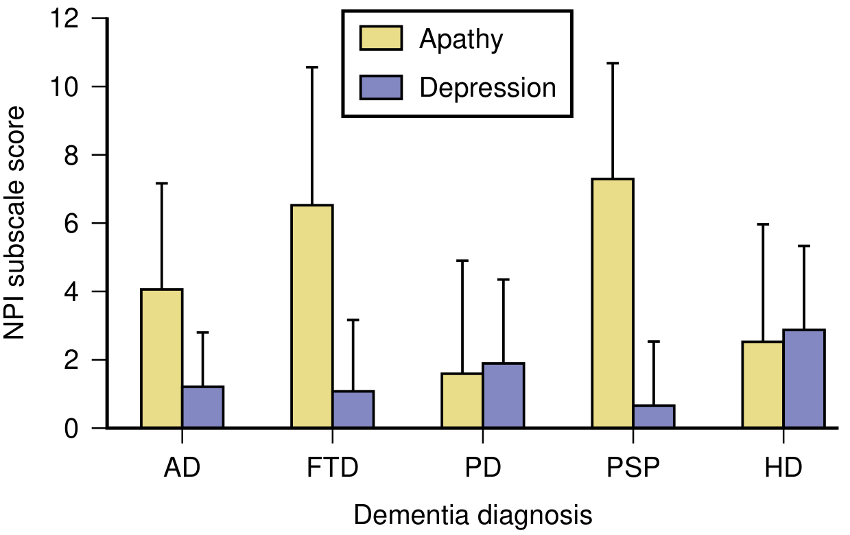
PSP shows the highest apathy burden, followed by FTD and AD.
Alzheimer's Disease (AD)
- One of the most common behavioral changes in AD
- Associated with bilateral gray matter loss in the anterior cingulate cortex, orbitofrontal cortex, dorsolateral prefrontal cortex, and putamen
- Linked to greater functional and cognitive impairment and lower quality of life
- Difficult to assess because inactivity may reflect inability rather than lack of motivation
Frontotemporal Dementia (FTD)
- Affective apathy is particularly prominent in the behavioral variant of FTD
- FTD patients may respond better to SSRIs (citalopram, escitalopram) or SNRIs than to dopaminergic agents
Parkinson's Disease (PD)
- Prevalence: 16.5% to 40%
- Associated with greater cognitive impairment
- Early signs in HD overlap: withdrawal from activities, decline in personal appearance, decreased spontaneous speech, constriction of emotional expression (often mistaken for depression)
Huntington's Disease (HD)
- Early HD apathy can be misidentified as depression
- Features: withdrawal, lack of behavioral initiation, decreased spontaneous speech
Neural Circuitry
Apathy in AD has been shown to correlate with bilateral reductions in gray matter volume in:
- Anterior cingulate cortex (ACC)
- Orbitofrontal cortex (OFC)
- Dorsolateral prefrontal cortex (DLPFC)
- Putamen
This is consistent with frontal-subcortical circuitry disruption, which underlies motivational behavior via reward pathways.
Assessment
- Neuropsychiatric Inventory (NPI) - widely used across dementia types
- Clinical challenge: distinguishing apathy from depression and from inability to perform activities
- Key diagnostic question: Is the patient unable to act, or unmotivated to act?
Treatment
Treatments are largely off-label and modestly effective:
Pharmacological
| Agent | Rationale |
|---|---|
| Dopamine agonists (first-line in PD) | Restore dopaminergic reward signaling |
| Methylphenidate / stimulants | Dopamine/norepinephrine reuptake inhibition |
| Bupropion | Dopaminergic/noradrenergic agonism |
| Levodopa | Dopamine precursor |
| Cholinesterase inhibitors | First-line in AD; better for prevention than treatment |
| SSRIs/SNRIs | Preferred in FTD (citalopram, escitalopram) |
| Modafinil | Reported benefit in PD apathy |
No agent is FDA-approved specifically for apathy. None is particularly robust in efficacy.
Non-Pharmacological (first-line)
- Structured daily routine and scheduled activities
- Environmental cuing and prompting from caregivers
- Behavioral activation strategies
- Involvement of neuropsychologist in a multidisciplinary team
Given risks of pharmacological treatments and their modest efficacy, non-pharmacological interventions should always be considered first.
Sources:
- Bradley and Daroff's Neurology in Clinical Practice (sections on AD, PD, HD, PSP apathy)
- Stahl's Essential Psychopharmacology, p. 552 (ABC model of apathy and treatment)
Menorragia and iron deficiency anemia
Menorrhagia (heavy menstrual bleeding) is one of the most common causes of iron deficiency anemia in women of reproductive age. The chronic blood loss depletes iron stores, reducing hemoglobin synthesis and leading to microcytic, hypochromic anemia.
Searching sources...
Finding Sources
Finding Sources
Finding Sources
Reading File
Reading File
Reading File
Reading File
Finding Sources
Reading File
Reading File
Finding Sources
Reading File
Reading File
Loading Image
I now have comprehensive content from multiple authoritative sources. Let me compile the full answer.
Menorrhagia and Iron Deficiency Anemia
The Link
Menorrhagia (heavy menstrual bleeding, defined as blood loss >80 mL/cycle or bleeding lasting >8 days) is one of the most common causes of iron deficiency anemia in women of reproductive age. Menstrual and GI blood loss are the two most common sources of chronic iron depletion. Because iron has no regulated excretion pathway, repeated blood loss steadily depletes both storage and functional iron pools.
Notably, average total body iron is only ~2.5 g in healthy young females vs. ~6 g in males, reflecting the ongoing loss from menstruation and increased demands during pregnancy. The storage pool (ferritin/hemosiderin) is therefore smaller and more easily exhausted.
- Robbins, Cotran & Kumar Pathologic Basis of Disease
Iron Metabolism: Key Concepts
Iron homeostasis is maintained by regulated absorption (there is no regulated excretion):
- Daily loss via shed epithelium: 1-2 mg/day
- Daily dietary requirement: 7-10 mg (men), 7-20 mg (women)
- Only 10-15% of ingested iron is absorbed; heme iron (meat) is far more bioavailable (~20%) than nonheme iron (~1-2%)
- Iron is transported in plasma bound to transferrin, delivered to erythroid precursors
- Stored as ferritin (in hepatocytes and macrophages) and hemosiderin (after lysosomal degradation of ferritin)
- Regulated by hepcidin (liver-derived peptide): inhibits ferroportin → traps iron in enterocytes and macrophages when stores are replete; falls with iron deficiency to facilitate absorption
In iron deficiency: low iron → low hepcidin → upregulated intestinal absorption (compensatory, but often insufficient with ongoing blood loss).
Stages of Iron Depletion
Iron deficiency progresses in sequential stages:
| Stage | Iron Stores | Serum Iron | Transferrin Sat. | Ferritin | Hb/Hct |
|---|---|---|---|---|---|
| 1 - Depletion | ↓↓ | Normal | Normal | ↓ | Normal |
| 2 - Iron-deficient erythropoiesis | Absent | ↓ | ↓ | Low | Normal |
| 3 - Iron deficiency anemia | Absent | ↓↓ | ↓↓ (<15%) | ↓↓ (<12 µg/L) | ↓↓ |
"Anemia appears only when iron stores are completely depleted." - Robbins, Cotran & Kumar
Laboratory Findings
| Test | Iron Deficiency Anemia | Anemia of Chronic Disease | Thalassemia |
|---|---|---|---|
| Ferritin | Low (<12 µg/L) | Increased | Normal/Increased |
| Serum Iron | Low | Low or Normal | Normal/Increased |
| TIBC | Increased | Decreased | Normal |
| Transferrin Sat. | Decreased (<10%) | Normal/Increased (>10%) | Normal/Increased |
| Serum Transferrin Receptor | Increased | Normal | Normal/Increased |
| Hepcidin | Decreased | Increased | Variable |
Note: Ferritin is an acute-phase reactant - it may be falsely normal or elevated in concurrent inflammation, so a normal ferritin does not completely exclude iron deficiency.
- Tietz Textbook of Laboratory Medicine, 7th Edition
Peripheral Blood Smear in IDA

Classic smear features:
- Microcytosis (small RBCs, low MCV)
- Hypochromia - central pallor exceeds 1/3 of cell diameter; hemoglobin visible only in a narrow peripheral rim
- Poikilocytosis - "pencil cells" (small, elongated red cells) are characteristic
- Bone marrow: absence of stainable iron on Prussian blue stain is diagnostically significant
Clinical Features Beyond Anemia
In severe, long-standing iron deficiency, depletion of iron-containing enzymes causes systemic manifestations:
- Koilonychia (spoon-shaped nails)
- Alopecia
- Atrophic glossitis (smooth, red tongue)
- Pica - craving for non-food substances (clay - geophagia) or ice (pagophagia)
- Intestinal malabsorption
- Plummer-Vinson syndrome (rare triad): microcytic hypochromic anemia + atrophic glossitis + esophageal webs
When to Investigate Beyond Menorrhagia
Clinical guidelines (Symptom to Diagnosis, Evidence-Based Guide) stratify investigation by age and menstrual status:
| Group | GI Evaluation Needed? |
|---|---|
| All men | Yes - always |
| Postmenopausal women | Yes - always (rule out GI cancer) |
| Women <40 with confirmed menorrhagia | Not routinely, unless GI symptoms or family history of early colon cancer |
| Women 40-50 with menorrhagia | Low threshold for colonoscopy (watch for celiac disease mimicking IBS) |
| Women of any age with menorrhagia | GI evaluation warranted after excluding menorrhagia as sufficient cause |
"To prematurely ascribe iron deficiency in such individuals to any other cause is to run the risk of missing a gastrointestinal cancer." - Robbins
Treatment
1. Iron Replacement (for IDA)
- Oral iron supplementation is first-line: produces reticulocyte response within 5-7 days, followed by steady Hb rise and normalization of red cell indices
- Factors enhancing absorption: ascorbic acid (vitamin C), citric acid, amino acids, sugars
- Factors inhibiting absorption: tannins (tea), carbonates, oxalates, phosphates
- IV iron for those who cannot tolerate oral therapy or have malabsorption
2. Managing the Menorrhagia (Cause)
| Treatment | Mechanism | Notes |
|---|---|---|
| LNG-IUS (Mirena) | Progestin-mediated endometrial atrophy | Most effective hormonal option; reduces bleeding 70-90% |
| Combined oral contraceptives | Reduce endometrial proliferation | Effective; caution in smokers/cardiovascular risk |
| Tranexamic acid | Antifibrinolytic - reduces plasminogen activation | 1.3 g TDS for 3-5 days during menses; reduces blood loss significantly |
| NSAIDs | Reduce prostaglandin-mediated vasodilation | Effective for idiopathic HMB; less effective with fibroids |
| GnRH agonists | Hypoestrogenic state; fibroid shrinkage 30-35% | Short-term use only (bone loss after 6 months); bridge to surgery |
| Progestins alone | Reduces endometrial proliferation | Regularizes bleeding but may not reduce days |
| Endometrial ablation | Destroys endometrium | Definitive non-surgical option; ~80% patient satisfaction |
| Hysterectomy | Definitive | For refractory cases |
- Berek & Novak's Gynecology
Key takeaway: Menorrhagia and IDA are treated in parallel - iron stores must be replenished while the underlying cause of blood loss is controlled. Failure to address the bleeding source will lead to recurrent anemia despite iron supplementation.
Sources:
- Robbins, Cotran & Kumar Pathologic Basis of Disease (iron metabolism, IDA pathogenesis, morphology, clinical features)
- Tietz Textbook of Laboratory Medicine, 7th Edition (lab findings table)
- Symptom to Diagnosis: An Evidence-Based Guide, 4th Edition (investigation algorithm)
- Berek & Novak's Gynecology (menorrhagia treatment)
Iron metabolism
Iron metabolism involves absorption in the duodenum (regulated by hepcidin), transport via transferrin, storage as ferritin/hemosiderin, and recycling from senescent RBCs by macrophages. Hepcidin is the master regulator - it degrades ferroportin to block iron export from enterocytes and macrophages when stores are replete.
Searching sources...
Finding Sources
Finding Sources
Excellent sources. Let me read them in parallel - Robbins (already read last session, great detail), Harrison's, Guyton, and Goodman & Gilman's:
Reading File
Reading File
Reading File
Excellent content. Let me now fetch key diagrams and read Harrison's hepcidin/regulation section more fully:
Loading Image
Loading Image
Reading File
Reading File
Reading File
Loading Image
Loading Image
I now have outstanding diagrams and comprehensive content from four authoritative sources. Let me compile the full answer.
Iron Metabolism
Body Iron Distribution
Total body iron averages 4-5 g in men and ~2.5 g in women (lower due to menstrual losses). It is distributed across three functional compartments:
| Compartment | Form | Amount (Men) | % of Total |
|---|---|---|---|
| Functional | Hemoglobin | ~2100 mg | ~65% |
| Myoglobin | ~300 mg | ~4% | |
| Iron enzymes (cytochromes, catalase, peroxidase) | ~50 mg | ~1% | |
| Transport | Transferrin-bound (plasma) | ~3 mg | <0.1% |
| Storage | Ferritin | ~1000 mg (men) / ~400 mg (women) | 15-30% |
| Hemosiderin | smaller fraction |
- Guyton and Hall Medical Physiology; Robbins, Cotran & Kumar
The Iron Cycle
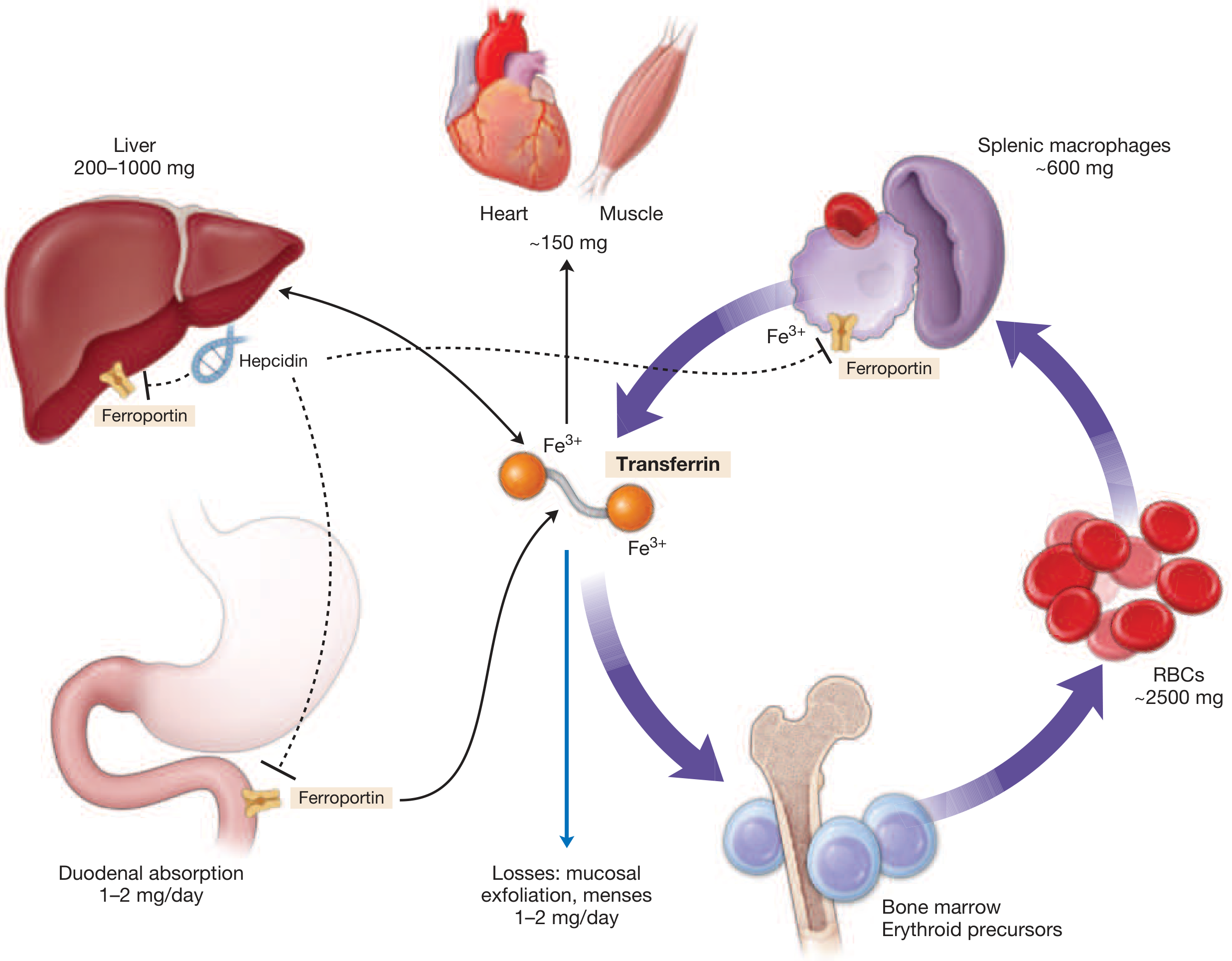
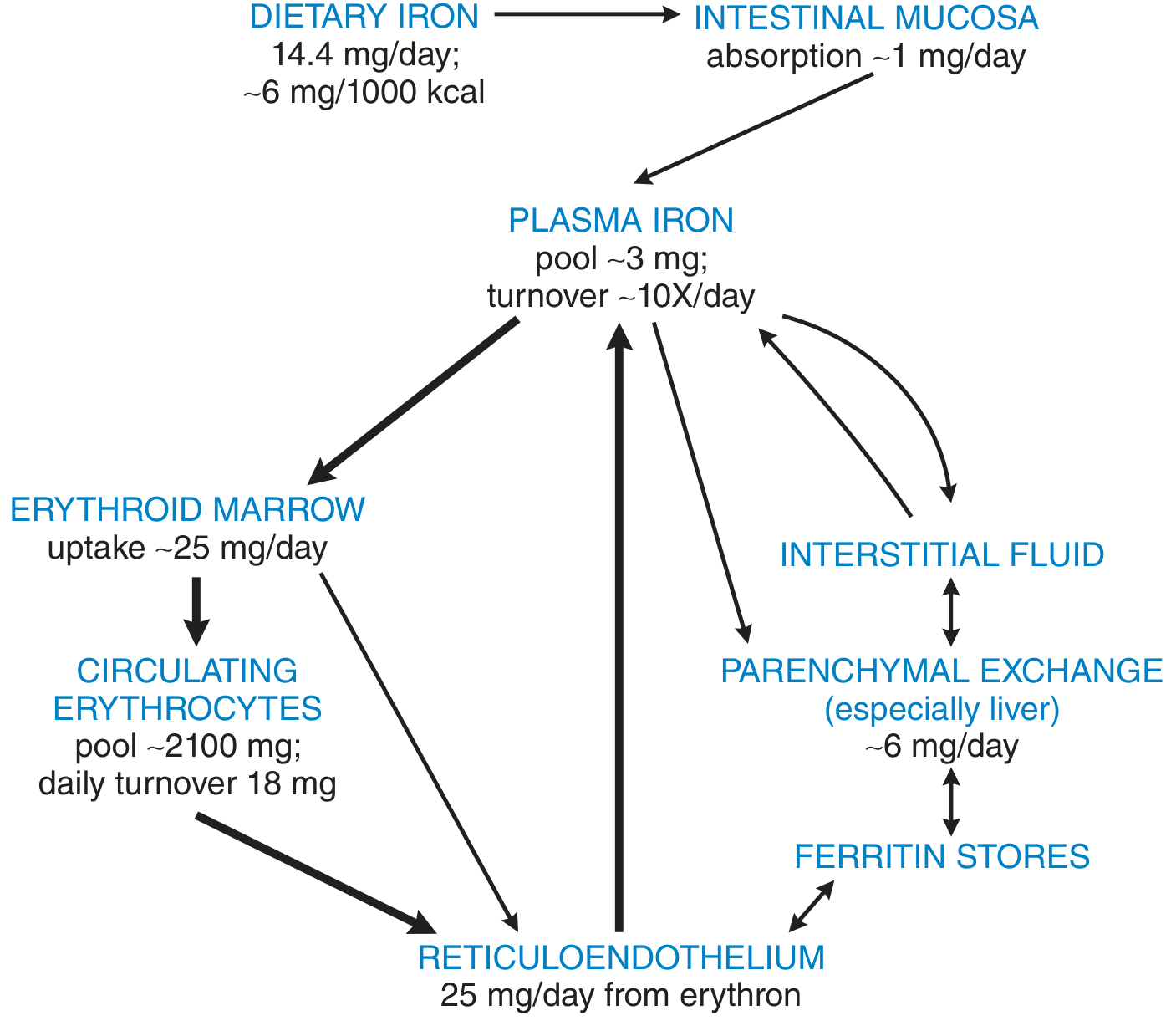
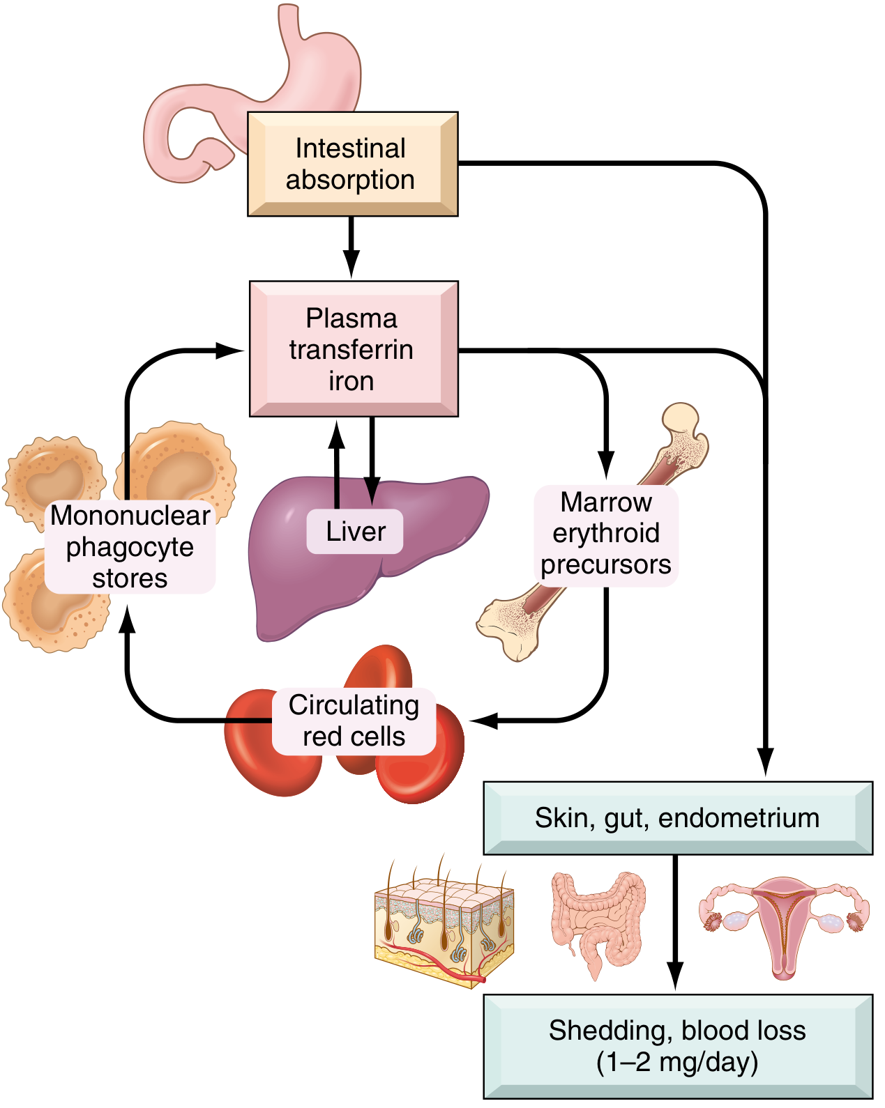
Key concept: Transferrin is central. Of the ~20-25 mg iron needed daily for erythropoiesis, only 1-2 mg comes from intestinal absorption - the rest (~80%) is recycled from senescent RBCs by macrophages (primarily in spleen, liver, and bone marrow). The plasma iron pool itself is tiny (~3 mg) but turns over ~10x per day.
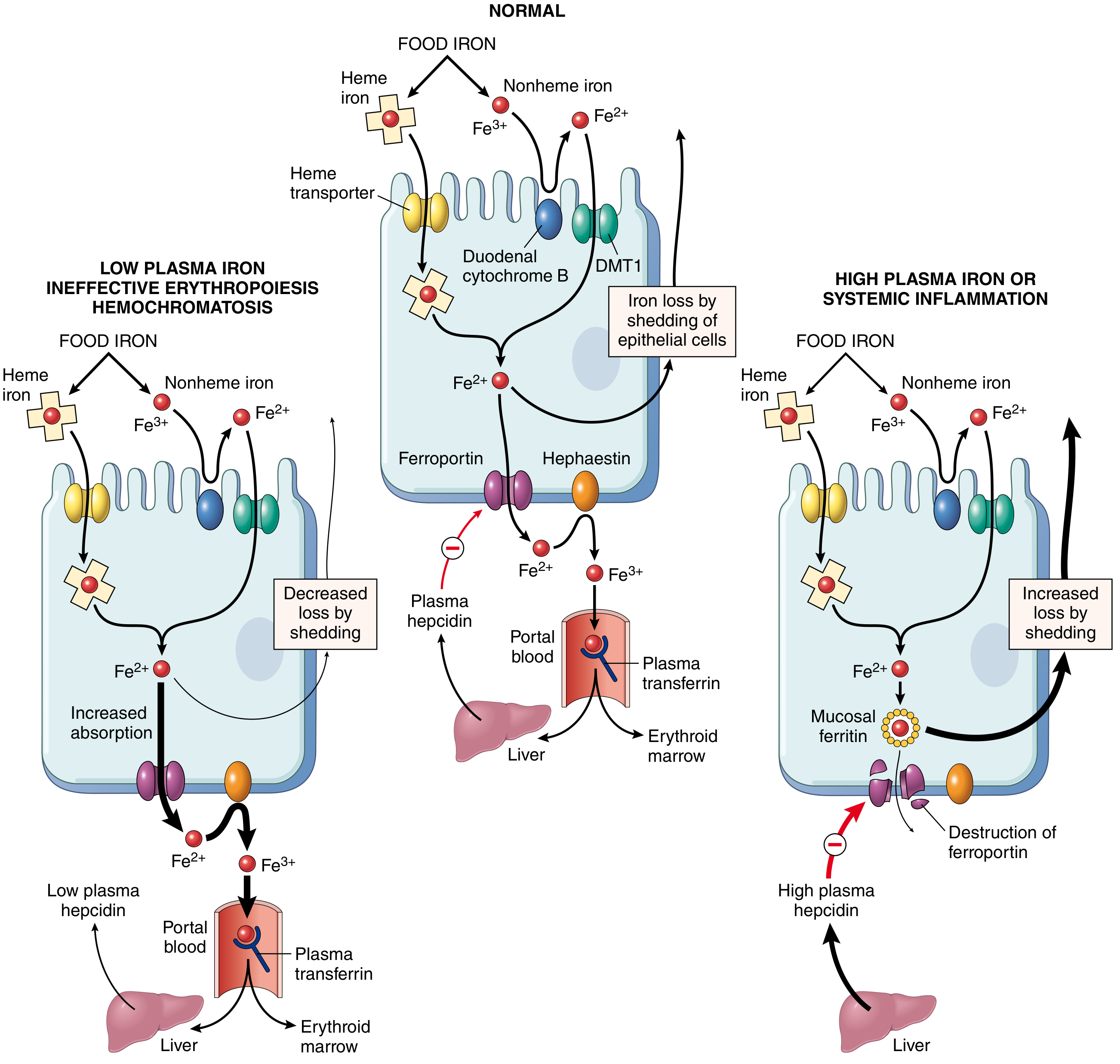
Step 1: Intestinal Absorption (Duodenum and Upper Jejunum)
This is the only regulated entry point for iron into the body.
Nonheme Iron (inorganic Fe³⁺)
- Reduction: Gastric acid + duodenal cytochrome B (DCYTB, a ferrireductase) reduces Fe³⁺ → Fe²⁺ (ferrous) at the brush border
- Apical uptake: DMT1 (Divalent Metal Transporter 1, SLC11A2) imports Fe²⁺ across the apical membrane
- Inside enterocyte: Fe²⁺ either:
- Stored as mucosal ferritin (lost when enterocyte sloughs - a safety mechanism)
- Transported to the basolateral membrane by ferroportin (FPN1, SLC40A1)
- Basolateral export: Ferroportin exports Fe²⁺; hephaestin (and ceruloplasmin) re-oxidizes it to Fe³⁺
- Plasma binding: Fe³⁺ rapidly binds to transferrin
Heme Iron (from meat/myoglobin)
- Absorbed via an incompletely characterized heme transporter at the apical membrane
- More bioavailable (~20%) vs. nonheme iron (<5%)
- Inside enterocyte, heme is metabolized by heme oxygenase to release Fe²⁺ into a common pool with nonheme iron
Step 2: Plasma Transport - Transferrin
- Transferrin: a 76-kDa bilobed glycoprotein synthesized by the liver; carries only 3-4 mg of iron at any time but has rapid turnover
- Each molecule has two iron-binding sites → monoferric or diferric transferrin
- Normal serum iron: ~120 µg/dL (males), ~100 µg/dL (females); transferrin normally one-third saturated
- Total iron-binding capacity (TIBC) reflects plasma transferrin concentration
Cellular Uptake (Receptor-Mediated Endocytosis)
- Fe³⁺-transferrin binds transferrin receptor (TfR1) on cell surface
- Complex internalized via clathrin-coated pits into endosomes
- Proton-pumping ATPase acidifies endosome to pH ~5.5 → iron dissociates from transferrin
- DMT1 transports Fe²⁺ out of endosome into cytoplasm
- Apotransferrin + TfR1 recycled back to cell surface; apotransferrin released into plasma
Erythroid precursors in bone marrow have the highest density of TfR1 because they require the most iron for hemoglobin synthesis (~20-25 mg/day for ~200 billion new erythrocytes daily).
- Goodman & Gilman's Pharmacological Basis of Therapeutics
Step 3: Storage - Ferritin and Hemosiderin
Ferritin:
- Shell protein: apoferritin (~450-460 kDa), 24 polypeptide subunits
- Stores up to 4000 iron atoms per molecule as polynuclear ferric oxide
- Located mainly in hepatocytes, splenic macrophages, and bone marrow macrophages
- Serum ferritin correlates with body iron stores: normal ~20-200 µg/L; <12 µg/L = iron deficiency; can reach ~5000 µg/L in iron overload
Hemosiderin:
- Aggregated, partially degraded ferritin in lysosomes
- Insoluble, less readily mobilized than ferritin
- Predominates when iron stores are overloaded
- Stains blue-black with Prussian blue (potassium ferrocyanide) - basis of Perls' stain
Step 4: Macrophage Recycling (The Dominant Iron Source)
Specialized macrophages in the spleen, liver (Kupffer cells), and bone marrow remove senescent RBCs (after ~120 days of circulation):
- Phagocytosis of aged RBCs
- Heme oxygenase degrades hemoglobin → liberates Fe²⁺
- Fe²⁺ either:
- Returned directly to plasma via ferroportin → binds transferrin
- Stored as ferritin for gradual release
- This recycling supplies ~25 mg/day - the vast majority of daily erythropoietic iron demand
In intravascular hemolysis, hepatocytes and macrophages recover iron via hemoglobin-haptoglobin and heme-hemopexin complexes.
The Master Regulator: Hepcidin
Hepcidin is a 25-amino acid peptide hormone produced by hepatocytes. It is the body's main systemic iron regulator.
Mechanism of Action
- Binds to ferroportin on enterocytes, macrophages, and hepatocytes
- Causes ferroportin internalization and lysosomal degradation
- Result: iron is trapped inside cells - enterocytes cannot export absorbed iron; macrophages cannot release recycled iron
Hepcidin Inducers (↑ hepcidin → ↓ iron release)
| Signal | Mediator | Effect |
|---|---|---|
| High iron stores | BMP6/BMP2 → BMP-SMAD pathway → HJV → hepcidin | Reduces absorption and macrophage release |
| Systemic inflammation | IL-6 → JAK-STAT3 pathway → hepcidin | Sequesters iron in macrophages (anemia of chronic disease) |
Hepcidin Suppressors (↓ hepcidin → ↑ iron release)
| Signal | Mediator |
|---|---|
| Iron deficiency / low stores | TMPRSS6 (matriptase-2) antagonizes BMP pathway |
| Increased erythropoietic demand (hemorrhage, hypoxia) | Erythroferrone (secreted by erythroblasts after EPO stimulation) sequesters BMPs |
| Testosterone | Adapts iron supply to male erythropoiesis |
Clinical correlates of hepcidin dysregulation:
| Condition | Hepcidin | Mechanism |
|---|---|---|
| Iron deficiency anemia | Low | Appropriately suppressed; increases absorption |
| Anemia of chronic disease | High | IL-6 drives hepcidin; iron trapped in macrophages |
| Hereditary hemochromatosis | Low (inappropriately) | Mutations in HFE, HJV, hepcidin gene, or TfR2 → unregulated absorption |
| IRIDA (iron-refractory IDA) | High (inappropriately) | TMPRSS6 mutations → cannot suppress hepcidin |
| β-thalassemia / ineffective erythropoiesis | Low | Excess erythroferrone from expanded erythroid mass |
Cellular Iron Regulation: IRP-IRE System
At the cellular level, iron homeostasis is fine-tuned post-transcriptionally:
- IRP1 and IRP2 (Iron Regulatory Proteins): cytosolic RNA-binding proteins
- When cellular iron is low: IRPs bind to iron-responsive elements (IREs) in mRNA
- 5' IRE of ferritin mRNA → blocks translation (less storage)
- 3' IRE of TfR1 mRNA → stabilizes transcript (more uptake)
- When cellular iron is high: IRPs release IREs → opposite effect (more ferritin, less TfR1)
- IRPs also regulate HIF-2α, erythropoietin, and ALA synthase 2 (first enzyme of heme biosynthesis)
Iron Losses
There is no regulated iron excretion pathway. Daily losses are small and obligatory:
| Source | Amount |
|---|---|
| GI tract (bile, exfoliated mucosa) | ~0.6 mg/day (men) |
| Skin desquamation | small |
| Urine | trace |
| Menstruation (average) | +0.5 mg/day (~1.3 mg/day total in women) |
| Menstruation (10th percentile) | >2 mg/day |
| Full-term pregnancy (total) | ~1190 mg |
Dietary Bioavailability
| Factor | Effect on Iron Absorption |
|---|---|
| Heme iron (meat) | High bioavailability (~20%) |
| Nonheme iron | Low (<5%) |
| Ascorbic acid (vitamin C) | Enhances (reduces Fe³⁺ → Fe²⁺) |
| Citric acid, amino acids, sugars | Enhance |
| Tannins (tea), oxalates, carbonates, phytates | Inhibit (stabilize Fe³⁺) |
| Gastric acidity | Essential for nonheme absorption |
| Gastrectomy | Reduces absorption (less acidity, faster transit) |
Sources:
- Robbins, Cotran & Kumar Pathologic Basis of Disease (Figs 14.20, 14.21 - iron cycle and absorption regulation)
- Harrison's Principles of Internal Medicine 22E (Fig. 102-1 - the iron cycle with organ-level quantities)
- Guyton and Hall Textbook of Medical Physiology (iron distribution and transport)
- Goodman & Gilman's The Pharmacological Basis of Therapeutics (Fig. 45-4 - quantitative iron flow; IRP-IRE system; pregnancy requirements)
This is a shared conversation. Sign in to Orris to start your own chat.