
Acute kidney Injury
Reading File
Now let me search the library and PubMed in parallel for comprehensive AKI coverage.
Finding Sources
Searching PubMed
"acute kidney injury"[MeSH Terms] AND management
Good. The main AKI chapter is in block15. Let me read it comprehensively in parallel chunks.
Reading File
Reading File
Finding Sources
Now let me read the dedicated AKI chapter from the NKF Primer and the KDIGO staging table, plus the pathophysiology section.
Reading File
Reading File
Finding Sources
Excellent content. Now let me read the NKF Primer AKI definition continuation, the Goldman-Cecil AKI section, and the Frameworks for Internal Medicine section on AKI causes.
Reading File
Reading File
Reading File
Now let me read the Goldman-Cecil management and epidemiology sections, plus the AKI causes and management from the Goldman-Cecil and NKF Primer.
Reading File
Reading File
Now let me fetch the key AKI diagrams and read the management/treatment sections.
Reading File
Loading Image
Loading Image
Now let me get the management/treatment and AKI complications sections.
Reading File
Finding Sources
Reading File
Reading File
Loading Image
Now I have comprehensive material to write a thorough, well-structured AKI review. Let me now compile the full answer.
Acute Kidney Injury (AKI)
Definition
Acute kidney injury is a clinical syndrome defined by a rapid decline in the kidney's ability to clear waste products, occurring within approximately 1 week of an injurious stimulus. The 2012 KDIGO (Kidney Disease: Improving Global Outcomes) criteria are the current gold standard:
AKI is defined by ANY ONE of the following:
- Rise in serum creatinine (sCr) by ≥0.3 mg/dL within 48 hours, OR
- Rise in sCr to ≥1.5 times baseline within 7 days, OR
- Urine output <0.5 mL/kg/hour for ≥6 hours
The term "Acute Kidney Disease" (AKD) was introduced by KDIGO to capture cases where creatinine rises evolve over periods longer than 7 days and thus do not meet the formal AKI definition. - NKF Primer on Kidney Diseases, 8e
KDIGO Staging
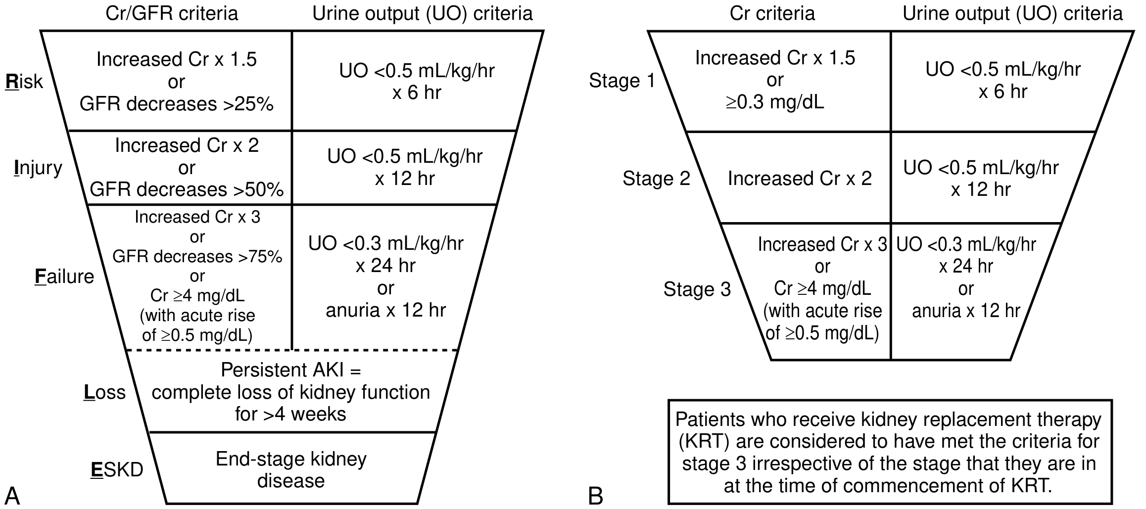
| Stage | Serum Creatinine | Urine Output |
|---|---|---|
| 1 | 1.5-1.9x baseline OR ≥0.3 mg/dL increase | <0.5 mL/kg/h for 6-12 hrs |
| 2 | 2.0-2.9x baseline | <0.5 mL/kg/h for ≥12 hrs |
| 3 | ≥3.0x baseline OR sCr ≥4.0 mg/dL OR initiation of KRT | <0.3 mL/kg/h for ≥24 hrs OR anuria ≥12 hrs |
Patients receiving kidney replacement therapy (KRT) are considered to have met Stage 3 criteria regardless of the creatinine level at the time of KRT initiation. - Goldman-Cecil Medicine, Table 106-1
The earlier RIFLE classification used a funnel-like staging system (Risk, Injury, Failure, Loss, ESKD) that preceded KDIGO but applied similar creatinine and urine output thresholds.
Epidemiology
-
~20-25% of hospitalized adults have an elevated serum creatinine
-
Up to 45% of ED admissions present with elevated sCr
-
55-65% of ICU patients develop AKI
-
Community-acquired AKI: annual incidence <1%
-
Prerenal azotemia accounts for 40% of hospital-acquired and 60-70% of community-acquired AKI cases
-
Intrinsic AKI is present in 5-6% of ED admissions and up to 60% of ICU patients
-
Intrinsic AKI requiring dialysis has increased substantially over the past three decades
-
Goldman-Cecil Medicine, p. 1241
Three Anatomic Categories
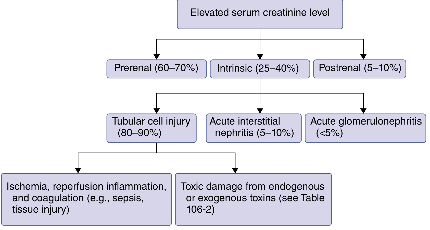
1. Prerenal AKI (60-70% of cases)
Mechanism: Decreased renal blood flow/perfusion without structural kidney damage.
Causes:
- True volume depletion: Limited oral intake, fever/sweating, burns, diarrhea, diuretics, hemorrhage
- Effective volume depletion: Heart failure, liver failure/cirrhosis, nephrotic syndrome
- Medications reducing GFR: NSAIDs, ACE inhibitors, angiotensin receptor blockers, cyclosporine, radiocontrast agents
Key features:
- BUN:Creatinine ratio >20:1 (increased urea reabsorption due to low effective arterial volume)
- FENa <1% in oliguric patients (intact tubular sodium reabsorption)
- Rapidly reversible with volume repletion within hours
- Urinary biomarkers of tubular injury not particularly elevated
FENa can be influenced by diuretics; in such cases, FEurea <35% is more reliable. - NKF Primer, p. 346
2. Intrinsic AKI (25-40% of cases)
An abrupt, sustained decline in function due to structural kidney damage - cannot be readily reversed for days to weeks.
Subtypes and frequencies:
- Tubular cell injury (80-90% of intrinsic AKI):
- Ischemic/ATN: Ischemia, reperfusion, inflammation, coagulation (e.g., sepsis, major surgery, cardiogenic shock)
- Toxic ATN: Endogenous toxins (myoglobin, hemoglobin, uric acid crystals, calcium oxalate) or exogenous (radiocontrast, aminoglycosides, vancomycin, amphotericin B, cisplatin, tacrolimus, cyclosporine)
- Acute Interstitial Nephritis (5-10%): Drug-induced (sulfonamides, penicillins, cephalosporins, NSAIDs, PPIs, checkpoint inhibitors), infections (Legionella, leptospirosis, SARS-CoV-2), autoimmune (SLE, Sjögren's)
- Acute Glomerulonephritis (<5%): Rapidly progressive GN, vasculitis
Molecular pathobiology: Activation of inflammatory, ischemic, and coagulation pathways; slow resolution due to vasoconstriction, tubular damage, cell death, and interstitial edema. Urinary biomarkers (NGAL, IL-18, KIM-1, TIMP-2, IGFBP7) are prominently elevated. - Goldman-Cecil Medicine, p. 1242
3. Postrenal AKI (5-10% of cases)
Mechanism: Obstruction of urine flow from kidneys to urethra.
Causes:
- Men: Benign prostatic hypertrophy (most common), bladder outlet obstruction
- Both sexes: Nephrolithiasis, pelvic malignancy, retroperitoneal fibrosis, neurogenic bladder
- Children: Congenital anatomic abnormalities
Relieving the obstruction can restore kidney function, but prolonged obstruction leads to tubular damage and may not be fully reversible.
Pathophysiology of AKI (ATN)
The proximal tubule and medullary thick ascending limb are most vulnerable due to high metabolic activity and limited oxygen supply (the medullary-cortical oxygen gradient). Following ischemia:
- Depletion of ATP → loss of cell polarity → detachment of tubular epithelial cells
- Cells form casts obstructing tubular flow → back-leak of filtrate
- Intense afferent arteriolar vasoconstriction (tubuloglomerular feedback via macula densa)
- Endothelial activation → inflammation, neutrophil and monocyte infiltration
- Reparative phase involves upregulation of genes for cell-cycle regulation, growth factors (KIM-1, NGAL), and cytokines
Diagnosis and Evaluation
Serum Creatinine - Limitations
sCr is widely available but has important limitations:
- Reflects GFR only at steady-state; there is a 24-72 hour lag before sCr stabilizes after an acute injury
- Influenced by age, sex, muscle mass, and catabolic state
- Volume resuscitation dilutes sCr, masking true AKI severity (the FACTT trial demonstrated fluid-corrected sCr identified more AKI cases)
- In the elderly and malnourished, lower creatinine generation means sCr rises more slowly even with severe injury
Urine Studies
| Test | Prerenal | ATN |
|---|---|---|
| FENa | <1% | >2% |
| FEUrea | <35% | >50% |
| Urine Na | <10 mEq/L | >40 mEq/L |
| Urine Osmolality | >500 mOsm/kg | <350 mOsm/kg |
| Urine sediment | Normal/hyaline casts | Muddy brown granular casts, tubular cells |
| BUN:Cr ratio | >20:1 | ~10-15:1 |
Urinary electrolytes should not be used in isolation - they are influenced by diuretics, sepsis, cirrhosis, and the phase of AKI when obtained. - NKF Primer, p. 347
Novel Biomarkers
| Biomarker | Source | Use |
|---|---|---|
| NGAL (Neutrophil Gelatinase-Associated Lipocalin) | Distal tubule | Early ATN detection (rises within hours) |
| KIM-1 (Kidney Injury Molecule-1) | Proximal tubule | Ischemic/toxic tubular injury |
| IL-18 | Proximal tubule | Predicts AKI progression and long-term mortality; AUC ~0.74-0.76 in cardiac surgery |
| TIMP-2 × IGFBP7 | Tubular cells | Cell-cycle arrest markers; FDA-cleared for AKI risk prediction |
| Cystatin C | All nucleated cells, freely filtered | Earlier GFR marker than creatinine |
| α1-Microglobulin | Proximal tubule reabsorption | AUC ~0.86 for predicting need for RRT |
Brenner and Rector's The Kidney, 2-Volume Set
Management
General Principles (Goldman-Cecil Medicine, p. 1243)
"Treatments involve removal of offending stimuli, correcting any obstruction, and supportive care to address volume depletion and electrolyte disturbances."
1. Identify and Treat the Cause
- Discontinue nephrotoxins (NSAIDs, aminoglycosides, contrast, ACE inhibitors if prerenal)
- Relieve urinary obstruction (Foley catheter, ureteric stent, percutaneous nephrostomy)
- Treat sepsis aggressively (source control, antibiotics, hemodynamic support)
- Treat AIN with steroids when drug-induced and offending agent removed
2. Fluid and Hemodynamic Management
- Prerenal AKI: IV fluids (isotonic crystalloids preferred); target euvolemia
- Avoid fluid overload - associated with worse outcomes in AKI
- Vasopressors in septic shock to maintain MAP ≥65 mmHg
- Balanced crystalloids (e.g., lactated Ringer's) are preferred over normal saline to reduce hyperchloremic acidosis and AKI risk
3. Electrolyte Management
- Hyperkalemia: Dietary restriction, sodium bicarbonate, calcium gluconate (membrane stabilization), insulin/dextrose, kayexalate, dialysis if severe
- Metabolic acidosis: Sodium bicarbonate if pH <7.2 or HCO3 <15
- Hyperphosphatemia: Dietary restriction, phosphate binders
- Hyponatremia/hypervolemia: Fluid restriction
4. Nutritional Support
- Standard caloric needs (20-25 kcal/kg/day non-catabolic; 25-30 in hypercatabolic)
- Protein: 0.8-1.0 g/kg/day (non-dialysis); 1.0-1.5 g/kg/day if on RRT
5. Drug Dosing Adjustments
- Renally cleared drugs must be dose-adjusted (antibiotics, anticoagulants, antivirals)
- Avoid NSAIDs, contrast, aminoglycosides when possible
6. Renal Replacement Therapy (RRT)
Indications (AEIOU):
- Acidosis refractory to medical management
- Electrolyte disturbance (hyperkalemia) not responding to treatment
- Ingestion/intoxication
- Overload (fluid overload refractory to diuretics)
- Uremic symptoms (encephalopathy, pericarditis, coagulopathy, nausea/vomiting)
Modalities:
- Intermittent Hemodialysis (IHD): Hemodynamically stable patients; more efficient solute removal
- Continuous RRT (CRRT): Hemodynamically unstable patients; preferred in ICU
- Peritoneal dialysis: Alternative when vascular access not available
Timing of RRT: Multiple RCTs have shown that in patients with KDIGO Stage 3 AKI, delayed initiation (watchful waiting) showed no increase in mortality compared to early initiation, and patients with delayed dialysis often had earlier diuresis and fewer adverse events. - Goldman-Cecil Medicine, p. 1349
Complications of AKI
| Complication | Mechanism |
|---|---|
| Hyperkalemia | Reduced K+ excretion + tissue breakdown |
| Metabolic acidosis | Reduced H+ excretion, reduced HCO3 reabsorption |
| Hyperphosphatemia | Reduced phosphate excretion |
| Volume overload/pulmonary edema | Reduced urine output |
| Uremia | Accumulation of urea and other uremic toxins |
| Hyponatremia | Water retention exceeding Na retention |
| Anemia | Reduced EPO production |
| Infection | Immune dysregulation, indwelling catheters |
| AKI-CKD progression | ~25-50% of severe AKI survivors develop CKD |
Prognosis and AKI-to-CKD Transition
- About 1/3 of hospitalized patients with AKI resolve within 24 hours, ~60% by 48 hours, and ~70% by 72 hours
- ATN carries a higher risk of CKD progression than non-ATN AKI
- Severe AKI requiring dialysis carries ~40-60% in-hospital mortality in ICU settings
- Each episode of AKI independently increases the risk of CKD, ESRD, cardiovascular events, and all-cause mortality
- Long-term follow-up with nephrology is essential after hospitalized AKI
Prevention
- Identify high-risk patients early (CKD, elderly, diabetic, volume-depleted, septic)
- Minimize nephrotoxin exposure; use N-acetylcysteine + hydration for contrast nephropathy
- Optimize hemodynamics before major surgery
- Use balanced crystalloids rather than normal saline for resuscitation
- Biomarker-guided (TIMP-2 × IGFBP7) protocols may allow preventive care bundles in high-risk surgical patients
Key Sources:
- Goldman-Cecil Medicine International Edition, Ch. 106 - Acute Kidney Injury, p. 1241-1243
- NKF Primer on Kidney Diseases, 8e, Ch. 31 - Acute Kidney Injury Definition, p. 346-348
- Brenner and Rector's The Kidney, 2-Volume Set - AKI Biomarkers and Pathophysiology chapters
- Sabiston Textbook of Surgery, 14e - KDIGO Criteria Table 26.4, p. 449
- Frameworks for Internal Medicine - AKI Categories, p. 448
cause of aki in detail with rational
AKI causes are classified into prerenal, intrinsic (renal), and postrenal. Prerenal involves reduced perfusion without structural damage; intrinsic involves actual kidney parenchymal damage (tubular, glomerular, interstitial, vascular); postrenal involves obstruction. Each has a distinct pathophysiologic rationale.
Searching sources...
Finding Sources
Finding Sources
Finding Sources
Reading File
Finding Sources
Finding Sources
Reading File
Reading File
Reading File
Reading File
Reading File
Loading Image
Loading Image
Excellent - superb diagrams and detailed text obtained. Now let me get the specific sections on intrinsic AKI causes - tubular toxins, contrast nephropathy, rhabdomyolysis, glomerulonephritis, and vascular causes.
Reading File
Finding Sources
Finding Sources
Reading File
Reading File
I now have all the material needed for a detailed, well-structured, rational account of AKI causes. Let me compose the full answer.
Causes of AKI - In Detail with Rationale
AKI causes are divided into three anatomic categories, each with a distinct pathophysiologic basis:
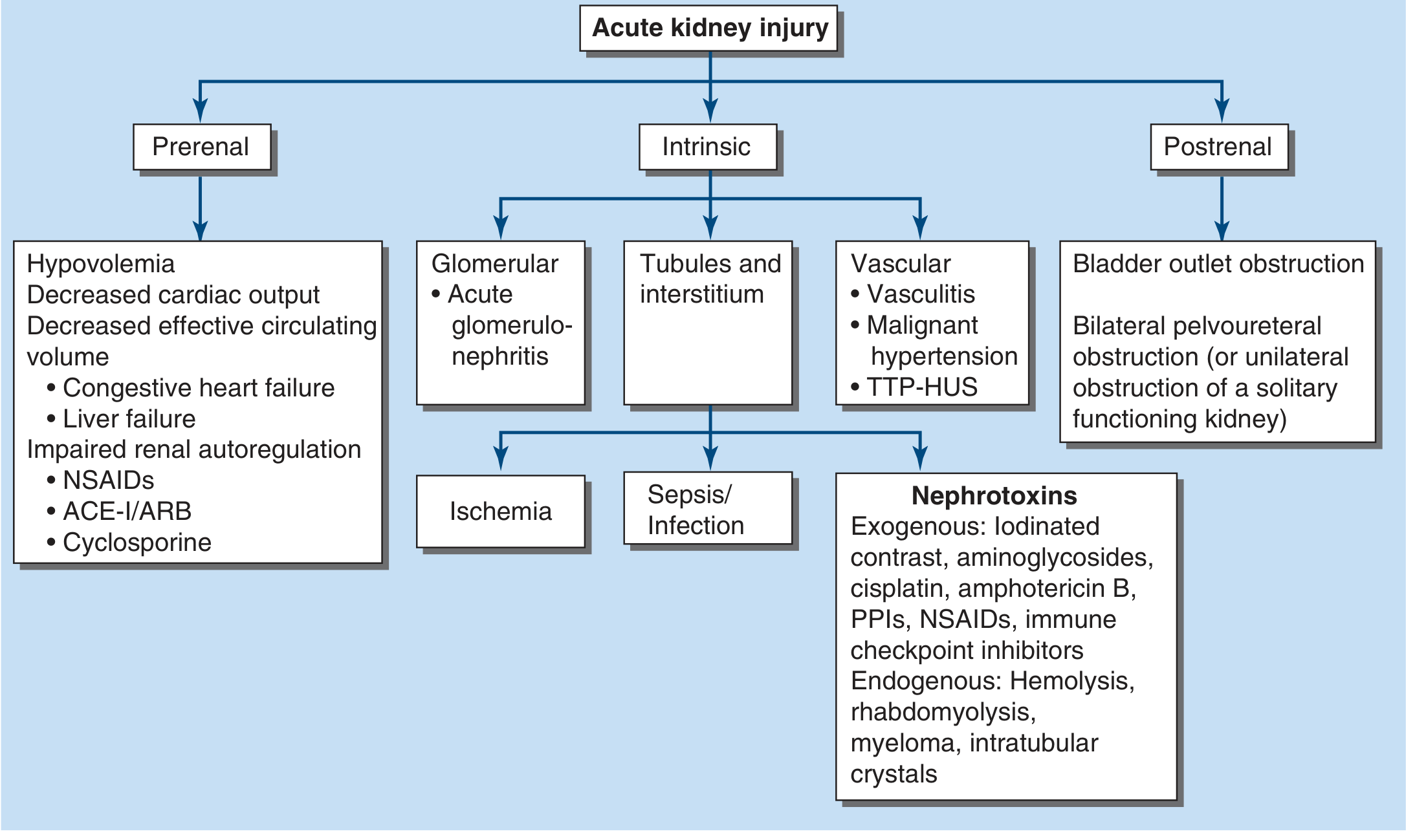
CATEGORY 1: PRERENAL AKI (40-80% of all AKI)
Core Rationale
Prerenal AKI arises from inadequate renal plasma flow and intraglomerular hydrostatic pressure to support normal glomerular filtration - without any structural damage to the kidney itself. The kidney parenchyma is intact; the problem is upstream. Because no tissue is destroyed, it is fully reversible within hours of restoring perfusion. - Harrison's Principles of Internal Medicine, 22e
Normal Autoregulatory Defense (Why Prerenal AKI Has a Threshold Effect)
The kidney defends GFR through three autoregulatory mechanisms:
- Myogenic reflex: Afferent arteriole dilates in response to reduced perfusion pressure
- Prostaglandins (PGI2, PGE2): Vasodilate the afferent arteriole under hypoperfusion
- Angiotensin II: Preferentially vasoconstricts the efferent arteriole to maintain glomerular capillary pressure
- Tubuloglomerular feedback: Macula densa senses low solute delivery → afferent arteriole dilates → maintains GFR
These compensatory responses maintain GFR until mean arterial pressure falls below ~80 mmHg, at which point GFR drops steeply and AKI ensues.
Causes of Prerenal AKI with Rationale
A. True Hypovolemia (Reduced Circulating Blood Volume)
| Cause | Mechanism |
|---|---|
| GI losses (vomiting, diarrhea, NGT drainage) | Fluid lost from the gut → reduced intravascular volume → low renal perfusion pressure |
| Hemorrhage | Direct loss of blood volume → reduced MAP → reduced afferent arteriolar pressure |
| Burns | Massive plasma leak into burn wound (third-space loss) → reduced effective circulating volume |
| Diuretic overuse | Forced sodium and water excretion → reduced intravascular volume |
| Insensible losses (fever, sweating) | Increased free water loss not replaced → hypovolemia |
| Pancreatitis/Peritonitis | Third-space fluid sequestration into inflamed cavity → decreased effective circulating volume |
Rationale: All of these reduce the effective blood volume reaching the kidneys. The reduced afferent arteriolar pressure triggers the renin-angiotensin-aldosterone system (RAAS) and sympathetic nervous system. While angiotensin II initially helps by constricting the efferent arteriole, once perfusion falls below the autoregulatory threshold, GFR falls.
B. Reduced Cardiac Output (Effective Arterial Hypovolemia)
| Cause | Mechanism |
|---|---|
| Myocardial infarction | Pump failure → low cardiac output → low renal perfusion pressure |
| Cardiomyopathy / Congestive Heart Failure | Despite fluid overload, the effective arterial volume (volume perceived as filling the vascular tree) is low → RAAS and SNS activation → renal vasoconstriction |
| Valvular disease (severe aortic stenosis, mitral stenosis) | Low forward flow → reduced MAP and renal perfusion |
| Cardiac tamponade/massive PE | Obstructive shock → reduced cardiac output |
Rationale: In heart failure, the kidneys "see" underfilling of the arterial tree (low effective arterial volume) despite total body sodium and water excess. This triggers intense RAAS activation and ADH release. The kidneys retain sodium and water (worsening edema) while GFR falls - a vicious cycle. The term "cardiorenal syndrome" describes this bidirectional interaction. - Rosen's Emergency Medicine
C. Decreased Effective Arterial Volume (Distributive States)
| Cause | Mechanism |
|---|---|
| Liver failure / Cirrhosis | Splanchnic vasodilation (from nitric oxide) → underfilling of systemic circulation → compensatory RAAS/SNS activation → renal vasoconstriction → hepatorenal syndrome |
| Nephrotic syndrome | Profound hypoalbuminemia → low oncotic pressure → fluid escapes to interstitium → reduced effective circulating volume |
| Sepsis (early/distributive) | Massive peripheral vasodilation → profound drop in SVR → decreased effective perfusion pressure despite high cardiac output |
| Anaphylaxis | Extreme vasodilation and capillary leak → acute distributive shock → reduced renal perfusion |
Special note - Hepatorenal Syndrome: This represents the extreme end of cirrhotic prerenal physiology. Intense splanchnic vasodilation causes compensatory renal vasoconstriction via RAAS, ADH, and endothelin. The kidney itself is normal - when transplanted into a healthy recipient, it functions perfectly. It is defined by urine sodium <10 mEq/L, absence of proteinuria, and failure to respond to volume expansion.
D. Impaired Renal Autoregulation (Drug-Induced Prerenal AKI)
This is one of the most clinically important and avoidable causes:
NSAIDs (including COX-2 inhibitors)
- Normal state: Renal prostaglandins (PGI2, PGE2) play a minor role in renal blood flow
- Hypoperfused state: Prostaglandins become critically important for maintaining afferent arteriolar dilation
- NSAIDs block COX-1/COX-2 → prostaglandin synthesis inhibited → afferent vasoconstriction → GFR falls
- Risk is highest in elderly, CKD, CHF, cirrhosis, or volume-depleted patients
ACE Inhibitors / ARBs
- Angiotensin II normally maintains GFR under hypoperfusion by constricting the efferent arteriole
- ACE-I/ARBs block this → efferent arteriole dilates → glomerular capillary hydrostatic pressure drops → GFR falls
- Critical scenario: Bilateral renal artery stenosis - GFR is entirely dependent on efferent vasoconstriction; ACE-I/ARB causes precipitous GFR loss
- Also important: combined NSAID + ACE-I use ("triple whammy" with diuretic) dramatically increases AKI risk
Cyclosporine / Tacrolimus (Calcineurin inhibitors)
- Cause intense afferent arteriolar vasoconstriction → reduced GFR
- This is a hemodynamic (prerenal-type) effect, distinct from their chronic nephrotoxicity
SGLT-2 inhibitors lower intraglomerular pressure by reducing proximal tubule sodium reabsorption, but recent studies show a protective effect against AKI - probably through reduction in hyperfiltration injury and anti-inflammatory effects. - Harrison's Principles, 22e
CATEGORY 2: INTRINSIC (RENAL) AKI
Core Rationale
Intrinsic AKI involves structural damage to the kidney parenchyma - tubules, interstitium, glomeruli, or blood vessels. Unlike prerenal AKI, restoring perfusion does NOT immediately reverse function because cells have been destroyed. Recovery requires cellular regeneration, which takes days to weeks.
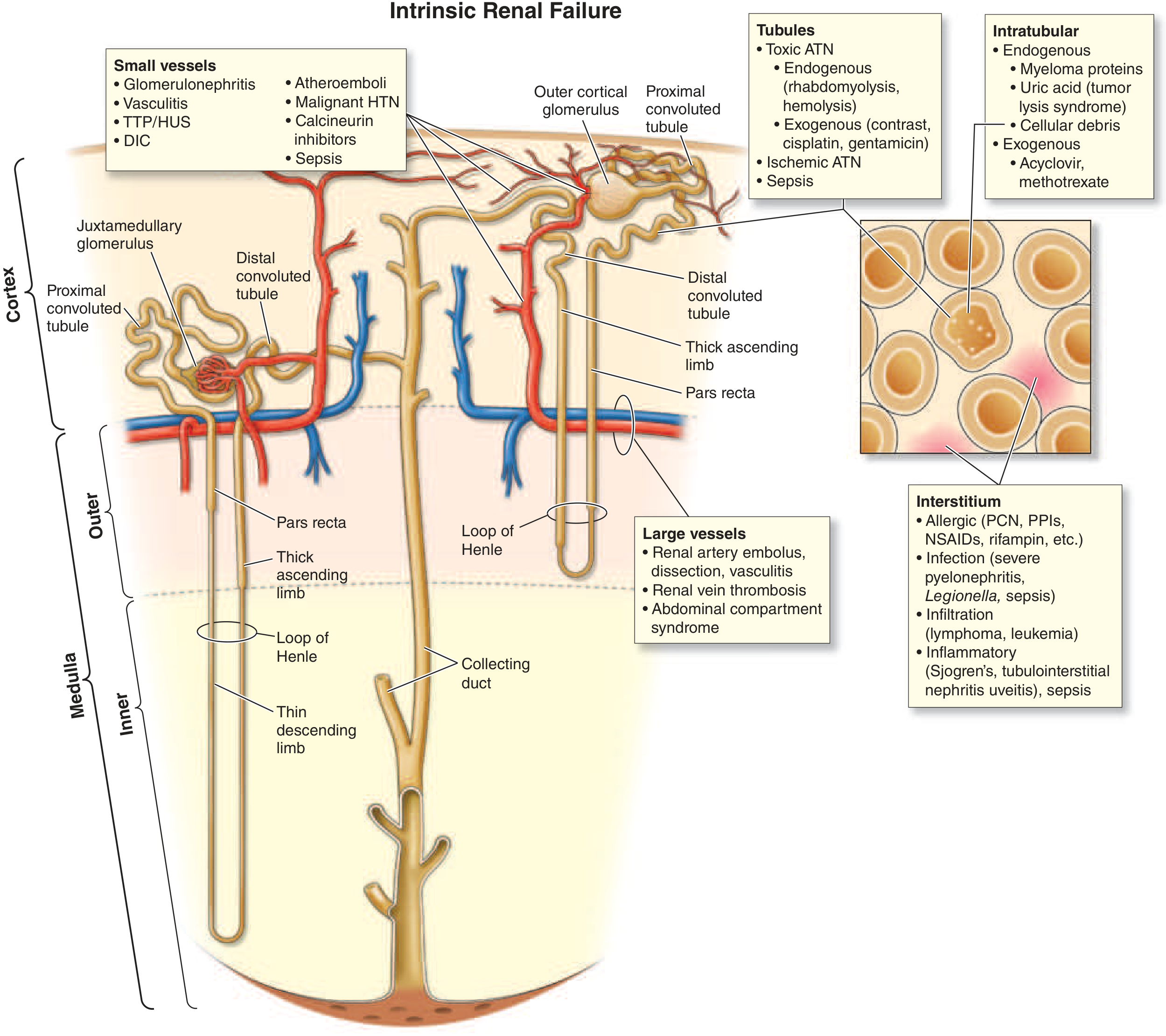
2A: TUBULAR INJURY - Acute Tubular Necrosis (ATN) - 80-90% of Intrinsic AKI
The S3 segment of the proximal tubule (pars recta) and the medullary thick ascending limb are the most vulnerable because:
- They have the highest metabolic demand (ATP-intensive active transport)
- They exist in the most hypoxic zone of the kidney (renal medulla has a naturally low pO2)
- They cannot switch to anaerobic glycolysis under ischemia
i. Ischemic ATN
Causes: Prolonged prerenal azotemia, cardiogenic shock, septic shock, aortic cross-clamping, major surgery, hemorrhagic shock, burns
Pathophysiology - Step by Step:
- ATP depletion → failure of Na-K-ATPase → cells swell, lose polarity
- Cytoskeletal disruption → loss of brush border → tubular cells detach into lumen
- Detached cells + cellular debris form casts → tubular obstruction → raised intratubular pressure → backpressure on glomerulus → GFR falls
- Backleak of filtrate across denuded, leaky tubular epithelium → net fluid absorption is impaired
- Afferent arteriolar vasoconstriction (from tubuloglomerular feedback - macula densa senses increased distal solute delivery due to failed proximal reabsorption) → further reduces GFR
- Inflammatory cascade: Neutrophils and monocytes infiltrate the interstitium → cytokine release (TNF-α, IL-1, IL-6) → endothelial injury → microvascular thrombosis → worsens medullary ischemia
- Reactive oxygen species (ROS) from mitochondrial dysfunction cause lipid peroxidation, cell membrane damage, and apoptosis
"Transient ischemia alone in a normal kidney is usually not sufficient to cause severe AKI, as evidenced by the relatively low risk of severe AKI even after total interruption of renal blood flow during suprarenal aortic clamping or cardiac arrest." Prerenal azotemia and ischemic ATN represent a continuum. - Harrison's Principles, 22e
ii. Sepsis-Associated AKI (most common cause of ATN in ICU)
Pathophysiology:
- Renal vasoconstriction: Activation of RAAS, sympathetic nervous system, vasopressin, and endothelin
- Paradox: In early sepsis, renal blood flow may actually be normal or even high, yet GFR falls - suggesting altered intrarenal microvascular distribution (shunting away from medullary tubules)
- Endothelial damage → increased leukocyte adhesion and migration → microvascular thrombosis → tubular ischemia
- Cytokine storm: TNF-α, IL-1, IL-6, IL-8 → direct tubular toxicity, increased permeability
- Reactive oxygen species from activated neutrophils and macrophages → oxidative tubular damage
- Sepsis may also cause AIN (see below)
iii. Exogenous Nephrotoxins
Radiocontrast Agents (Contrast-Induced AKI)
- Mechanism 1 - Renal medullary ischemia: Contrast causes intense intrarenal vasoconstriction via endothelin, adenosine, and reduced nitric oxide → medullary hypoxia (S3 segment and thick ascending limb most affected)
- Mechanism 2 - Direct tubular cytotoxicity: Contrast agents are directly toxic to tubular epithelial cells via reactive oxygen species and apoptosis
- Timeline: SCr rises at 24-48 hours, peaks at 3-5 days, typically resolves within 1 week
- Risk factors: CKD, diabetes, volume depletion, high contrast volume, heart failure, NSAID/ACE-I use
- Prevention: IV fluid hydration with isotonic saline before and after, minimize contrast dose, use iso-osmolar agents
Aminoglycosides (Gentamicin, Tobramycin, Amikacin)
- Freely filtered at glomerulus → concentrated in proximal tubular cells (via megalin receptor)
- Accumulate in lysosomes → phospholipidosis → lysosomal rupture → mitochondrial dysfunction → cell death
- Non-oliguric AKI in 10-30% of courses, even at therapeutic levels
- Onset: typically 5-7 days into therapy (can appear even after stopping)
- Causes hypomagnesemia (tubular Mg wasting) as an early sign
- Once-daily dosing reduces nephrotoxicity compared to multiple daily doses
Amphotericin B
- Binds ergosterol (and cholesterol) in tubular cell membranes → pores form → tubular cell lysis
- Also causes intense afferent arteriolar vasoconstriction → ischemia
- Causes distal RTA, hypokalemia, hypomagnesemia, and nephrotoxic ATN
Vancomycin
- Tubular injury, especially when trough levels are high
- Can crystallize in tubules causing intratubular obstruction
- Synergistically nephrotoxic with aminoglycosides and piperacillin-tazobactam
Cisplatin and other Platinum Chemotherapeutics
- Enter proximal tubular cells via organic cation transporters
- Form platinum-DNA adducts in tubular cell nuclei → apoptosis and necrosis
- Non-oliguric AKI; also causes hypomagnesemia (tubular Mg wasting), hypokalemia
iv. Endogenous Nephrotoxins
Myoglobin (Rhabdomyolysis)
Causes: Crush injury, prolonged immobilization, seizures, statin toxicity, extreme exertion, heat stroke, drugs (cocaine, ecstasy), hypokalemia, hypothyroidism
Triple mechanism of renal damage:
- Cast formation: Myoglobin precipitates with Tamm-Horsfall protein in acidic, concentrated urine → obstructive casts in distal tubules → tubular obstruction
- Direct proximal tubular cytotoxicity: At pH ≤5.6, myoglobin dissociates into ferriheme + free iron → ferriheme causes lipid peroxidation → oxidative cell death in proximal tubules
- Intrarenal vasoconstriction: Myoglobin scavenges nitric oxide (a potent renal vasodilator) → net vasoconstrictive effect; also activates endothelin-1, thromboxane A2, and RAAS
All three mechanisms are worsened by volume depletion and aciduria - hence the rationale for aggressive isotonic saline (± bicarbonate to alkalinize urine) as treatment. - Rosen's Emergency Medicine
Hemoglobin (Hemolysis - Pigment Nephropathy)
- Mechanism is analogous to myoglobin-induced ATN
- Causes: Transfusion reactions, G6PD deficiency, TTP/HUS, malaria, snake envenomation
- Hemoglobin cast formation in distal tubules + proximal tubular direct toxicity via heme iron
Uric Acid Crystals (Acute Urate Nephropathy)
- Context: Tumor Lysis Syndrome (after cytotoxic therapy for high-burden tumors - lymphoma, leukemia) or spontaneous (Burkitt's, ALL)
- Massive cell death → huge purine load → uric acid overproduction → uric acid precipitates in collecting ducts and distal tubules (poorly soluble at acidic pH)
- Causes: tubular obstruction → anuria; also intrarenal vasoconstriction
- Treatment: rasburicase, allopurinol, IV hydration, urine alkalinization
Myeloma Cast Nephropathy ("Myeloma Kidney")
- Light chains (Bence Jones proteins) filtered at glomerulus → co-precipitate with Tamm-Horsfall protein in distal tubules → obstruct tubules
- Light chains also directly toxic to proximal tubule cells (endocytosis → lysosomal injury)
- Volume depletion, contrast, hypercalcemia, and NSAIDs dramatically worsen myeloma AKI
Calcium Oxalate Crystals
- Ethylene glycol ingestion → oxalic acid → calcium oxalate crystals → tubular obstruction
- Also in primary oxaluria, after jejunoileal bypass
2B: ACUTE INTERSTITIAL NEPHRITIS (AIN) - 5-10% of Intrinsic AKI
Core Rationale: An immune-mediated inflammatory reaction in the renal interstitium, not primarily the tubules. Pathologically shows interstitial edema and inflammatory infiltrate (T lymphocytes, macrophages, ± eosinophils) with relative preservation of glomeruli.
Causes:
1. Drug-Induced (most common ~75%)
- Mechanism: Drugs act as haptens or directly trigger a T-cell-mediated delayed hypersensitivity reaction against tubular antigens
- Key drugs:
| Drug Class | Examples |
|---|---|
| Beta-lactam antibiotics | Penicillin, methicillin, cephalosporins |
| Sulfonamides | TMP-SMX |
| Fluoroquinolones | Ciprofloxacin |
| Rifampin | - |
| NSAIDs | Any NSAID (can also cause nephrotic syndrome simultaneously) |
| Proton pump inhibitors | Omeprazole, pantoprazole (now a leading cause) |
| Diuretics | Furosemide, thiazides |
| Checkpoint inhibitors | Ipilimumab, nivolumab, pembrolizumab (~5% of treated patients) |
| Allopurinol | - |
Classic triad (present in only ~1/3 of cases): Fever + rash + eosinophilia - absence does NOT exclude AIN.
2. Infection-Associated AIN
- Mechanism: Direct infection or immune-complex deposition
- Legionella pneumophila, Leptospira, Streptococcus, Mycobacterium tuberculosis, CMV, EBV, SARS-CoV-2, Hantavirus
3. Autoimmune / Systemic Disease
- Sjogren's syndrome, SLE, sarcoidosis (granulomatous AIN), tubulointerstitial nephritis-uveitis (TINU) syndrome, IgG4-related disease
2C: ACUTE GLOMERULONEPHRITIS - <5% of Intrinsic AKI
Core Rationale: Immune-mediated inflammation directly in the glomerulus → reduced filtration surface area + increased glomerular capillary permeability. GFR falls because glomerular inflammation causes:
- Proliferation of mesangial, endothelial, or epithelial cells → physically narrows the capillary lumen
- Fibrin deposition → occludes capillary loops
- Inflammatory cells → release cytokines/ROS damaging the filtration barrier
Rapidly Progressive GN (RPGN) - most severe form:
| Type | Mechanism | Cause |
|---|---|---|
| Type I - Anti-GBM | Antibodies vs. collagen IV in GBM → linear IgG deposits | Goodpasture's syndrome (lung + kidney) |
| Type II - Immune complex | Immune complex deposition → complement activation → inflammation | Post-streptococcal GN, lupus nephritis, IgA nephropathy (cresentic), endocarditis |
| Type III - Pauci-immune (ANCA) | ANCA-mediated neutrophil activation → vessel wall destruction | GPA (Wegener's), MPA, EGPA (Churg-Strauss) |
Clinical clues: Hematuria with red cell casts, proteinuria (often 1-3 g/day), hypertension, edema. Absence of red cell casts, hematuria, and significant proteinuria effectively excludes GN.
2D: VASCULAR CAUSES - Large and Small Vessel Disease
Large Vessel Disease
| Cause | Mechanism |
|---|---|
| Renal artery occlusion (thromboembolism, dissection, vasculitis) | Abrupt cessation of arterial inflow → cortical ischemia/infarction |
| Renal vein thrombosis | Venous congestion → reduced net filtration pressure → AKI (particularly in nephrotic syndrome, hypercoagulable states) |
| Abdominal compartment syndrome | Raised intra-abdominal pressure → renal vein and inferior vena cava compression → venous hypertension → reduced perfusion pressure |
| Aortic dissection | Involvement of renal artery ostia → ischemic AKI |
Small Vessel and Microvascular Disease
| Cause | Mechanism |
|---|---|
| TTP (Thrombotic Thrombocytopenic Purpura) | ADAMTS13 deficiency → uncleaved vWF multimers → platelet aggregation → microthrombi in glomerular capillaries → microangiopathic hemolytic anemia + AKI |
| HUS (Hemolytic Uremic Syndrome) | Shiga toxin (E. coli O157:H7) → endothelial injury in renal microvasculature → microthrombi in glomeruli → triad: hemolytic anemia + thrombocytopenia + AKI (predominant in children) |
| Malignant Hypertension | Extremely high BP → fibrinoid necrosis of arterioles → ischemic glomerular injury + tubular ischemia |
| Atheroembolic AKI | Cholesterol crystals embolize (after aortic catheterization or anticoagulation) → lodge in small renal arteries → local inflammation + ischemia; subacute onset; livedo reticularis, eosinophilia |
| Scleroderma Renal Crisis | Intense arteriolar spasm + endothelial proliferation → critical renal ischemia → malignant HTN + AKI |
| HELLP Syndrome / Preeclampsia | Endothelial dysfunction + microangiopathy in pregnancy → glomerular endotheliosis + platelet thrombi |
| Vasculitis (ANCA, PAN) | Necrotizing inflammation of arterioles → fibrinoid necrosis → ischemic glomerular injury |
CATEGORY 3: POSTRENAL AKI (5-10% of all AKI)
Core Rationale
Obstruction at any level of the urinary tract causes a retrograde rise in intratubular pressure that opposes glomerular filtration. Initially afferent arteriole dilates (hyperemia), but within hours, intrarenal vasoconstriction from angiotensin II, thromboxane A2, and vasopressin causes GFR to fall. The key insight: obstruction must be bilateral (or unilateral in a solitary functioning kidney) to cause AKI.
Pathophysiology of Obstruction
- Acute obstruction → ↑ intratubular pressure → opposes net filtration pressure → GFR falls
- Phase 1 (0-2 hrs): Compensatory afferent arteriolar dilation (prostaglandin-mediated) → increased renal blood flow
- Phase 2 (2-5 hrs): Efferent arteriolar vasodilation → reduced filtration pressure
- Phase 3 (>5 hrs): Afferent arteriolar vasoconstriction (angiotensin II, thromboxane A2, endothelin) → marked reduction in RBF and GFR
- Prolonged obstruction → tubular atrophy + interstitial fibrosis → permanent CKD
Causes by Level
Intrarenal (Collecting Duct Level)
- Crystal precipitation: Uric acid (tumor lysis), calcium oxalate (ethylene glycol), phosphate (bowel prep), acyclovir, sulfonamides, methotrexate, indinavir
- Myeloma proteins (see above)
Ureteral (Bilateral or Solitary Kidney)
- Nephrolithiasis (most common cause of bilateral ureteral obstruction in young patients)
- Retroperitoneal fibrosis (idiopathic or secondary to drugs - methysergide, beta-blockers; associated with AAA)
- Pelvic/retroperitoneal malignancy (cervical, bladder, prostate, lymphoma, metastases)
- Sloughed renal papillae (diabetes, analgesic nephropathy, sickle cell disease)
- Surgical ligation of ureter
- Blood clots
Bladder
- Benign prostatic hypertrophy - most common cause of postrenal AKI in older men
- Prostate cancer
- Neurogenic bladder (diabetic autonomic neuropathy, spinal cord injury, MS)
- Anticholinergic drugs (tricyclics, antihistamines, antipsychotics) → bladder atony
- Bladder carcinoma
- Blood clots, bladder stones
Urethra
- Urethral stricture (post-infectious, post-traumatic)
- Phimosis (in children)
- Obstructed urinary catheter (most preventable cause in hospital)
"Obstruction is an eminently reversible cause of AKI and should be considered in every patient with newly discovered azotemia." - Rosen's Emergency Medicine
A post-obstructive diuresis can occur after relief - the kidneys excrete retained salt, water, and urea; patients need careful IV fluid replacement to avoid volume depletion worsening AKI.
Summary Table: Distinguishing the Three Categories
| Feature | Prerenal | Intrinsic (ATN) | Postrenal |
|---|---|---|---|
| BUN:Cr | >20:1 | ~10-15:1 | Variable |
| FENa | <1% | >2% | Variable |
| Urine Na | <10 mEq/L | >40 mEq/L | Variable |
| Urine osmolality | >500 | <350 | Variable |
| Urine sediment | Normal / hyaline casts | Muddy brown granular casts, tubular cells | Normal or RBC casts if stone |
| Response to fluids | Rapid reversal | No immediate reversal | No reversal until obstruction relieved |
| Kidney size on US | Normal | Normal | Bilateral hydronephrosis |
| Biomarkers (NGAL, KIM-1) | Low | Very elevated | Low-moderate |
Key Sources:
- Harrison's Principles of Internal Medicine, 22e (2025) - Chapter 321, Prerenal Azotemia and Ischemia/Nephrotoxin-Associated AKI
- Rosen's Emergency Medicine - Chapters 83 and 116 (Rhabdomyolysis)
- Goldman-Cecil Medicine International Edition - Chapter 106, Intrinsic AKI
- Frameworks for Internal Medicine - Chapter 33, AKI Categories
- Smith and Tanagho's General Urology, 19e - Postrenal Kidney Injury
explain RIFILE
RIFLE is the original consensus classification system for AKI (2004), standing for Risk, Injury, Failure, Loss, End-stage kidney disease. It uses serum creatinine/GFR changes and urine output to stage severity. It preceded AKIN and KDIGO but established the framework both later built upon.
Searching sources...
Finding Sources
Finding Sources
Reading File
Reading File
Reading File
Reading File
Finding Sources
Loading Image
Reading File
Reading File
Loading Image
Finding Sources
I now have all the material for a thorough, well-illustrated explanation of RIFLE. Here is the full answer:
The RIFLE Classification System
Background and Origin
RIFLE was proposed in 2002 by the Acute Dialysis Quality Initiative (ADQI) working group - the first international consensus effort to standardize the definition and staging of AKI. Before RIFLE, there were over 35 different definitions of "acute renal failure" in the literature, making it impossible to compare incidence, outcomes, or treatments across studies. RIFLE solved this by creating a uniform, tiered staging system based on two functional parameters: serum creatinine (or GFR) and urine output. - Current Surgical Therapy 14e; Brenner and Rector's The Kidney
The RIFLE Acronym - What Each Letter Means
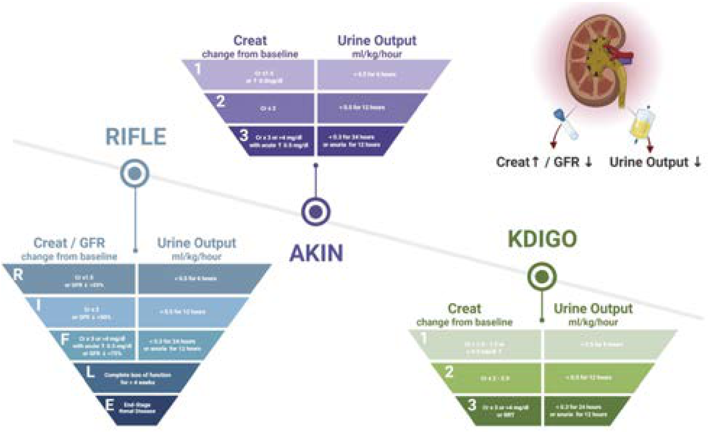
RIFLE has three severity classes (R, I, F) and two outcome stages (L, E):
Full RIFLE Criteria Table
| Stage | Serum Creatinine / GFR Criteria | Urine Output Criteria |
|---|---|---|
| R - Risk | Cr ×1.5 from baseline OR GFR decrease >25% | <0.5 mL/kg/h for >6 hours |
| I - Injury | Cr ×2 from baseline OR GFR decrease >50% | <0.5 mL/kg/h for >12 hours |
| F - Failure | Cr ×3 from baseline OR GFR decrease >75% OR sCr ≥354 µmol/L (≥4 mg/dL) with acute rise ≥44 µmol/L (0.5 mg/dL) | <0.3 mL/kg/h for >24 hours OR anuria for >12 hours |
| L - Loss | Complete loss of kidney function requiring RRT for >4 weeks | - |
| E - End-stage kidney disease | Complete loss of kidney function requiring RRT for >3 months | - |
Explaining Each Stage in Detail
R = Risk
What it means: The patient is at risk of kidney injury - there is an early, mild decline in GFR. No structural damage has necessarily occurred yet.
Creatinine rationale: A ×1.5 rise (i.e., 50% increase) over less than 7 days represents a meaningful GFR decline (~33% reduction in actual GFR, since creatinine and GFR have an inverse relationship). This is the most sensitive threshold - it identifies the largest number of patients, including those with mild AKI who may benefit from early intervention.
GFR rationale: A >25% drop in GFR further captures patients where creatinine has not yet visibly risen much but actual filtration has already meaningfully fallen.
Urine output rationale: <0.5 mL/kg/h for >6 hours = oliguria. This is the minimum threshold recognized as clinically significant oliguria. Six hours is chosen because shorter periods of low urine output are common physiologically (e.g., during sleep) and not necessarily pathological.
"The first stratum provides the greatest sensitivity for diagnosing AKI." - Brenner and Rector's The Kidney
I = Injury
What it means: The kidney has now sustained significant injury. GFR has fallen by more than half.
Creatinine rationale: A ×2 rise means the GFR has fallen by approximately 50%. At this point, the kidney is struggling to maintain homeostasis - electrolyte disturbances and fluid retention begin to appear.
Urine output rationale: <0.5 mL/kg/h for >12 hours represents sustained oliguria - the kidney has now been unable to maintain adequate urine output for half a day, indicating persistent rather than transient dysfunction.
Clinical significance: At the Injury stage, the patient requires active monitoring, nephrology input, and investigation for treatable causes. The "Injury" class in the original ADQI analysis was noted to represent a heterogeneous population - some were recovering, others progressing - which later prompted refinement in AKIN and KDIGO.
F = Failure
What it means: Severe, life-threatening kidney failure. This is the most specific threshold - virtually all patients identified here truly have serious AKI requiring intervention.
Creatinine rationale:
- ×3 rise = GFR has fallen by ~75% or more - the kidney retains barely a quarter of its filtration capacity
- sCr ≥4 mg/dL with acute rise ≥0.5 mg/dL - this absolute threshold catches patients with CKD who may not show a ×3 rise from their (already elevated) baseline but still have acute-on-chronic severe failure
Urine output rationale:
- <0.3 mL/kg/h for >24 hours = severe oliguria (almost anuric over a full day)
- Anuria for >12 hours = complete cessation of urine production, which in the absence of obstruction indicates either severe tubular necrosis, renal cortical necrosis, or vascular occlusion
Clinical significance: Patients in "F" class generally meet criteria for dialysis if they do not respond to medical management. Attributable mortality for RIFLE Failure = 26.3% vs. 5.5% in matched patients without AKI. - Current Surgical Therapy 14e
L = Loss
What it means: An outcome stage, not a severity stage. The patient has suffered complete, persistent loss of kidney function requiring renal replacement therapy (RRT) for more than 4 weeks.
Rationale: This distinguishes patients with ATN who recover (most within 1-4 weeks) from those with more severe, prolonged injury (e.g., cortical necrosis, severe bilateral ischemia) who do not recover within a month. The 4-week cutoff was chosen because most reversible causes of AKI (including severe ATN) are expected to recover within this window. If they have not, the prognosis for renal recovery is poor but not yet hopeless.
Clinical importance: "Loss" patients require decisions about long-term dialysis access, transition to peritoneal dialysis, or transplant listing.
E = End-Stage Kidney Disease (ESKD)
What it means: The patient requires RRT for >3 months - signifying permanent, irreversible kidney failure.
Rationale: Three months is the threshold because:
- KDIGO defines CKD as ≥3 months of impaired function/structure
- Beyond 3 months of RRT dependence after an AKI event, spontaneous renal recovery is extremely unlikely
- This marks the transition from acute/subacute to chronic kidney disease requiring indefinite renal replacement (dialysis or transplantation)
The Funnel Design - Why It's Shaped That Way
The RIFLE classification is intentionally displayed as an inverted funnel (pyramid):
- Wide at the top (Risk): Large number of patients qualify → high sensitivity, lower specificity → good for early detection and population-level surveillance
- Narrow at the bottom (Failure): Fewer patients qualify → lower sensitivity, very high specificity → good for identifying the most severe cases reliably
This design reflects the epidemiology of AKI: many patients have mild rises in creatinine (Risk), fewer progress to Injury, and fewer still to Failure.
"As proposed, the first stratum provides the greatest sensitivity for diagnosing AKI, whereas higher strata provide increasing specificity of diagnosis." - Brenner and Rector's The Kidney, 2-Volume Set
Mortality Correlation with RIFLE Grade
One of RIFLE's greatest validations is its correlation with outcomes:
| RIFLE Stage | Attributable In-Hospital Mortality |
|---|---|
| No AKI | 5.5% |
| Risk (R) | 8.8% |
| Injury (I) | 11.4% |
| Failure (F) | 26.3% |
In critically ill patients (ICU), mortality is even higher - rising from 49% at RIFLE Risk to 62.3% at Injury to 86.8% at Failure. - Brenner and Rector's The Kidney, 2-Volume Set
Key Diagnostic Rule - "Worst Criteria" Principle
A patient is classified by whichever criterion - creatinine OR urine output - places them in the highest (most severe) RIFLE stage. You do not need both criteria to be met; the worst single criterion determines staging.
What RIFLE Does NOT Include - Its Limitations
| Limitation | Explanation |
|---|---|
| No absolute creatinine threshold for early diagnosis | Only uses relative changes; misses early AKI in patients with low baseline muscle mass (elderly, malnourished) where creatinine rises slowly |
| Requires a known baseline creatinine | Difficult in emergency settings; ADQI recommended back-calculating using MDRD equation assuming eGFR of 75 mL/min/1.73m² if no baseline available |
| Does not capture sub-48-hour changes | The creatinine criterion uses a 7-day window; small but clinically important acute rises (e.g., 0.3 mg/dL over 48 hours) are not classified as AKI in RIFLE |
| GFR included | GFR is rarely measured directly in clinical practice - estimated GFR (eGFR) equations are not accurate in AKI (non-steady state); this was removed in AKIN and KDIGO |
| No biomarker component | Functional criteria only; does not identify subclinical structural tubular injury |
| L and E stages defined by RRT duration, not biology | No pathological or biomarker basis - purely operational definitions |
How RIFLE Led to AKIN and KDIGO
RIFLE's limitations drove two further refinements:
| Feature | RIFLE (2002) | AKIN (2007) | KDIGO (2012) |
|---|---|---|---|
| Minimum diagnosis | Cr ×1.5 over 7 days | Cr ×1.5 OR +0.3 mg/dL over 48 hrs | Both 48-hr absolute AND 7-day relative criteria |
| GFR criterion | Included | Removed | Removed |
| Time window for Cr | <7 days | <48 hours | 48 hrs (absolute) / 7 days (relative) |
| RRT = Stage 3 | Not explicit | Explicit | Explicit |
| Baseline unknown | Back-calculate MDRD | Requires known baseline | Addresses unknown baseline |
| Pediatric criteria | Not addressed | Not addressed | eGFR <35 mL/min/1.73m² for Stage 3 |
| L and E stages | Present | Removed | Removed |
"KDIGO further refined the AKIN criteria and developed a method to address unknown baseline sCr values." - Current Surgical Therapy 14e
The key advancement of AKIN was adding the absolute 0.3 mg/dL rise criterion - which captures early AKI that RIFLE would miss because patients with low muscle mass (elderly women, malnourished) may have a 0.3 mg/dL rise that is clinically very significant but does not constitute a ×1.5 increase from a very low baseline.
Pediatric RIFLE (pRIFLE)
A modified version, pRIFLE, was validated for children and uses estimated creatinine clearance (eCrCl) rather than absolute creatinine values, because normal creatinine is age- and size-dependent in children:
| pRIFLE Stage | eCrCl Change | Urine Output |
|---|---|---|
| Risk | eCrCl decrease 25% | <0.5 mL/kg/h for 8 hrs |
| Injury | eCrCl decrease 50% | <0.5 mL/kg/h for 16 hrs |
| Failure | eCrCl decrease 75% OR eCrCl <35 mL/min/1.73m² | <0.3 mL/kg/h for 24 hrs or anuria for 12 hrs |
| Loss | Persistent failure >4 weeks | - |
| End-stage | Failure >3 months | - |
Summary: Why RIFLE Matters
- Standardization - replaced 35+ competing definitions with one consensus framework
- Severity stratification - allows comparison of AKI severity across patients, studies, and populations
- Outcome prediction - each RIFLE stage correlates with mortality, length of ICU stay, and need for dialysis
- Early detection - the "Risk" stage was explicitly designed to enable preventive intervention before irreversible injury occurs
- Historical foundation - RIFLE is the ancestor of both AKIN and KDIGO, which are now the clinical standards
Key Sources:
- Brenner and Rector's The Kidney, 2-Volume Set - Table 29.2 and staging criteria
- Current Surgical Therapy 14e - ADQI, RIFLE, AKIN, KDIGO section with mortality data
- Morgan and Mikhail's Clinical Anesthesiology, 7e - Table 31-3 (RIFLE/AKIN/KDIGO comparison)
- Miller's Anesthesia, 10e - RIFLE Classification Scheme Table
- NKF Primer on Kidney Diseases, 8e - Fig. 31.1 RIFLE/AKIN/KDIGO systems
causes of AKI
You already have a very detailed breakdown of AKI causes from earlier in this conversation (the second response). Here is a clean, concise summary organized for quick reference:
Causes of AKI
AKI causes are divided into 3 anatomic categories:
1. PRERENAL (60-70%)
Reduced renal perfusion - no structural damage - reversible with fluid/hemodynamic correction
| Subcategory | Examples |
|---|---|
| True hypovolemia | Vomiting, diarrhea, hemorrhage, burns, diuretics, NGT drainage |
| Low cardiac output | MI, cardiomyopathy, CCF, cardiac tamponade, valvular disease |
| Effective arterial hypovolemia | Cirrhosis/hepatorenal syndrome, nephrotic syndrome, sepsis (distributive), anaphylaxis |
| Impaired renal autoregulation | NSAIDs (block prostaglandins → afferent vasoconstriction), ACE-I/ARBs (block efferent constriction), cyclosporine/tacrolimus |
| Renal artery stenosis | Bilateral RAS + ACE-I/ARB → critical GFR drop |
2. INTRINSIC RENAL (25-40%)
Structural damage to kidney parenchyma - not immediately reversible
A. Tubular Injury / ATN (80-90% of intrinsic)
| Type | Causes |
|---|---|
| Ischemic ATN | Prolonged prerenal azotemia, cardiogenic shock, septic shock, major surgery, aortic cross-clamping |
| Sepsis-associated | Most common ATN cause in ICU - cytokines, microvascular thrombosis, endothelial injury |
| Exogenous nephrotoxins | Aminoglycosides, vancomycin, amphotericin B, contrast agents, cisplatin, methotrexate |
| Endogenous toxins | Myoglobin (rhabdomyolysis), hemoglobin (hemolysis), uric acid crystals (tumor lysis syndrome), myeloma light chains |
B. Acute Interstitial Nephritis / AIN (5-10%)
| Type | Causes |
|---|---|
| Drug-induced | Penicillins, cephalosporins, NSAIDs, PPIs, sulfonamides, rifampin, checkpoint inhibitors |
| Infection | Legionella, Leptospira, SARS-CoV-2, EBV, Streptococcus, TB |
| Autoimmune | SLE, Sjögren's, sarcoidosis, IgG4-related disease |
C. Glomerulonephritis (<5%)
| Type | Mechanism | Cause |
|---|---|---|
| Anti-GBM (Type I RPGN) | Antibodies vs. GBM | Goodpasture's syndrome |
| Immune complex (Type II) | IC deposition | Post-strep GN, lupus, IgA nephropathy |
| Pauci-immune/ANCA (Type III) | Neutrophil-mediated necrotizing vasculitis | GPA (Wegener's), MPA, EGPA |
D. Vascular Causes
| Large vessels | Small vessels / Microvasculature |
|---|---|
| Renal artery occlusion/embolism/dissection | TTP (ADAMTS13 deficiency) |
| Renal vein thrombosis | HUS (Shiga toxin - E. coli O157:H7) |
| Abdominal aortic dissection | Malignant hypertension |
| Abdominal compartment syndrome | Atheroembolic disease |
| Scleroderma renal crisis | |
| HELLP syndrome / preeclampsia |
3. POSTRENAL (5-10%)
Obstruction of urine outflow - must be bilateral (or single functioning kidney)
| Level | Causes |
|---|---|
| Intrarenal / Collecting duct | Crystal precipitation: uric acid (tumor lysis), oxalate (ethylene glycol), acyclovir, sulfonamides, methotrexate, myeloma proteins |
| Ureteral (bilateral) | Nephrolithiasis, retroperitoneal fibrosis, pelvic/retroperitoneal malignancy, sloughed papillae, surgical ligation, blood clots |
| Bladder | BPH (most common in older men), prostate cancer, neurogenic bladder, anticholinergic drugs, bladder carcinoma |
| Urethra | Urethral stricture, phimosis, obstructed Foley catheter |
Quick Comparison: How to Distinguish Them
| Feature | Prerenal | Intrinsic (ATN) | Postrenal |
|---|---|---|---|
| FENa | <1% | >2% | Variable |
| Urine Na | <10 mEq/L | >40 mEq/L | Variable |
| BUN:Cr | >20:1 | ~10:1 | Variable |
| Urine osmolality | >500 mOsm/kg | <350 mOsm/kg | Variable |
| Urine sediment | Normal / hyaline casts | Muddy brown granular casts | Normal / RBCs if stone |
| Ultrasound | Normal kidneys | Normal kidneys | Bilateral hydronephrosis |
| Response to fluids | Rapid recovery | No immediate reversal | No reversal until obstruction relieved |
For the full mechanistic rationale of each cause (with detailed pathophysiology), refer to the earlier detailed response in this conversation above. Let me know if you want any specific cause explained further.
more details with rational
The detailed causes with rationale were already covered extensively in the second response of this conversation. Let me pull additional depth from textbooks on the areas not yet fully covered - specifically the vascular causes, specific toxin mechanisms, and GN subtypes.
Searching sources...
Finding Sources
Finding Sources
Finding Sources
Reading File
Reading File
Reading File
Reading File
Finding Sources
Finding Sources
Reading File
Reading File
Reading File
Loading Image
Reading File
Reading File
Now I have comprehensive additional detail on every category. Let me compile the complete, maximally detailed answer with rationale for every cause.
Causes of AKI - Complete Detail with Rationale
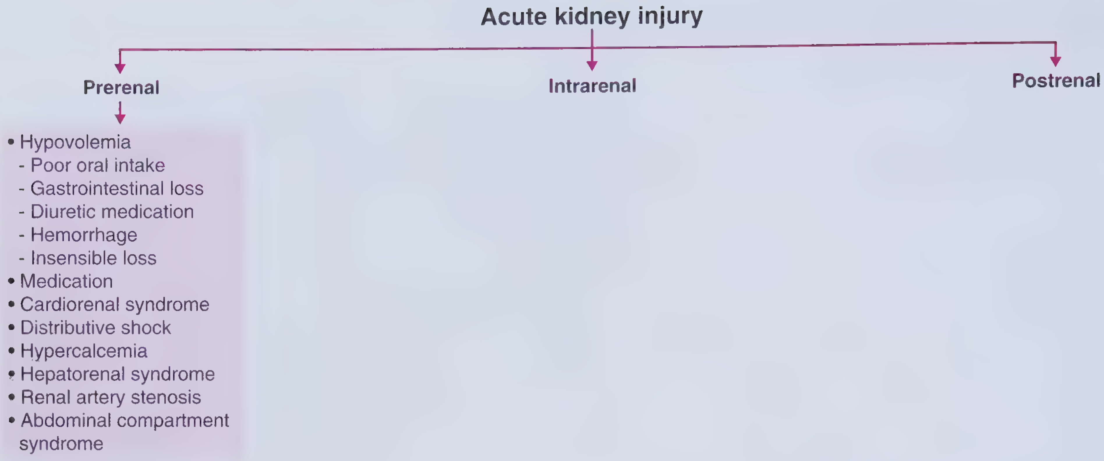
CATEGORY 1: PRERENAL AKI (60-70%)
Core Pathophysiologic Rationale
The kidney receives 20% of cardiac output and is exquisitely sensitive to perfusion. Under normal conditions, renal autoregulation maintains GFR across a wide MAP range (80-180 mmHg) through three mechanisms:
- Myogenic reflex - afferent arteriole dilates when wall tension falls (low perfusion pressure)
- Prostaglandins (PGI₂, PGE₂) - synthesized locally, keep afferent arteriole dilated under hypoperfusion
- Angiotensin II - constricts the efferent arteriole, preserving glomerular capillary hydrostatic pressure when perfusion is low
When MAP falls below ~80 mmHg, these defenses are overwhelmed → GFR drops steeply. No structural damage occurs initially - the kidney is functionally impaired but anatomically intact.
1A. TRUE HYPOVOLEMIA
Gastrointestinal Losses
Vomiting, diarrhea, NGT drainage, fistulas
Rationale: GI fluid losses reduce intravascular volume → low venous return → low cardiac output → reduced MAP → kidney senses underperfusion → RAAS + SNS activation → renal vasoconstriction → GFR falls.
- Diarrhea also causes hyperchloremic metabolic acidosis which, combined with hypovolemia, synergistically worsens renal hypoperfusion
- Vomiting causes metabolic alkalosis + hypochloremia → paradoxically, the kidney reabsorbs HCO₃⁻ avidly and excretes Na⁺ - an exception where FENa may be high despite true hypovolemia
Hemorrhage
Rationale: Acute blood loss → drop in circulating blood volume → baroreceptor activation → sympathetic discharge → peripheral vasoconstriction (maintain MAP to vital organs) → renal vasoconstriction → oliguria. Severe hemorrhage to Class III/IV (>30-40% blood loss) causes near-complete anuria. If hypotension persists >30-40 minutes, ischemic ATN supervenes.
Burns
Rationale: Burns cause massive third-space losses - plasma leaks into the burn wound because injured capillaries become hyperpermeable. Using the Parkland formula: 4 mL × kg × %TBSA burned of Ringer's lactate is needed in the first 24 hours to replace this. Without aggressive replacement, severe prerenal AKI (progressing to ATN) develops. Additionally, myoglobin from thermally injured muscle contributes toxic intrinsic injury.
Diuretic Overuse
Rationale: Loop diuretics block Na-K-2Cl in the thick ascending limb → massive natriuresis and water loss → volume depletion → prerenal AKI.
- Risk is highest in patients with CHF or cirrhosis given diuretics - these patients' kidneys depend on maximal RAAS-mediated sodium retention to maintain any effective circulating volume; diuretics undermine this
- Metolazone combined with furosemide can cause precipitous volume depletion
Insensible Losses
Rationale: Fever increases insensible water loss by ~250-500 mL/day per °C above 37°C. Patients unable to compensate by drinking (elderly, encephalopathic, post-operative) develop hypovolemia. Sweating during heat exposure or vigorous exercise can lose 1-2 L/hour. The kidney concentrates urine maximally (osmolality >500 mOsm/kg) until tubular damage supervenes.
Third-Space Sequestration
Pancreatitis, Peritonitis, Trauma, Sepsis
Rationale: Massive capillary leak and release of inflammatory mediators (IL-1, IL-6, TNF-α) → fluid redistributes into the interstitial "third space" → effective circulating volume falls despite normal or increased total body water. In pancreatitis, up to 4-6 L of fluid can sequester in the retroperitoneum and inflamed peripancreatic tissue within hours.
1B. REDUCED CARDIAC OUTPUT (Cardiorenal Syndrome)
Myocardial Infarction / Cardiomyopathy / Heart Failure
Rationale - The Cardiorenal Syndrome Paradox:
Despite total body Na⁺ and water excess (edema, ascites), the kidney detects low effective arterial blood volume - the volume perceived to be filling the arterial tree. This triggers:
- RAAS activation → aldosterone → sodium and water retention → worsens edema
- ADH release → free water retention → dilutional hyponatremia
- Sympathetic activation → renal vasoconstriction → GFR falls
- Reduced renal blood flow → ischemic tubular injury if prolonged
Pattern: High JVP, high BNP, low CO, high SVR - the "cardiorenal syndrome"
- Diuretics given to relieve congestion can worsen renal perfusion, raising creatinine - the clinical dilemma of "cardiorenal syndrome type 1"
- Every 10 mmHg drop in MAP from cardiac failure causes ~10% reduction in GFR
Cardiac Tamponade / Massive Pulmonary Embolism
Rationale: Obstructive shock → impaired cardiac filling or outflow → low CO → prerenal AKI. Beck's triad (hypotension + JVD + muffled heart sounds) in tamponade; massive PE causes acute right heart failure with septal shift compressing the LV.
1C. DECREASED EFFECTIVE ARTERIAL VOLUME (Distributive)
Hepatorenal Syndrome (HRS)
The archetypal "effective hypovolemia with total body overload" state
Pathophysiology - Step by Step:
- Portal hypertension (from cirrhosis, acute liver failure) → splanchnic vascular bed vasodilates
- This splanchnic vasodilation is driven by excess nitric oxide production by endothelial cells in splanchnic vessels
- Systemic circulation becomes "underfilled" (low effective arterial volume) despite total body volume excess
- The kidney "reads" this as severe hypovolemia → maximal RAAS + SNS + ADH activation → intense renal vasoconstriction (endothelin, angiotensin II, vasopressin)
- Renal blood flow falls dramatically → GFR → near zero
- Crucially: the kidneys are structurally normal - if transplanted into a healthy recipient, they function perfectly. If a normal liver is transplanted into an HRS patient, the kidneys recover.
Two types:
| HRS Type 1 (Now: AKI-HRS) | HRS Type 2 (Now: CKD-HRS) | |
|---|---|---|
| Onset | Rapid (<2 weeks) | Gradual |
| Trigger | Spontaneous bacterial peritonitis, GI bleed, large paracentesis | None (spontaneous) |
| Prognosis | Median survival 2 weeks without treatment | Weeks to months |
| Treatment | Terlipressin + albumin; bridge to transplant | Diuretic-resistant ascites management |
"Hepatorenal syndrome is a form of renal failure occurring in individuals with liver failure in whom there is no intrinsic morphologic or functional cause for kidney dysfunction." - Robbins Pathologic Basis of Disease
Nephrotic Syndrome
Rationale: Severe hypoalbuminemia (<2 g/dL) → reduced plasma oncotic pressure → fluid leaks from capillaries into interstitium → reduced effective circulating volume → RAAS activation → prerenal AKI. This occurs despite massive total body edema.
Sepsis (Distributive Shock)
Rationale: Bacterial endotoxins + cytokines → massive peripheral vasodilation → SVR drops → MAP falls → effective arterial underfilling → prerenal AKI. Note: In early sepsis, cardiac output is actually high (hyperdynamic state), yet GFR falls because SVR is so low that renal perfusion pressure is inadequate. Pattern: Low JVP, high CO, low SVR.
Hypercalcemia (Cancer, Hyperparathyroidism)
Rationale: Hypercalcemia → direct afferent arteriolar vasoconstriction (calcium-mediated smooth muscle contraction in afferent arterioles) → reduced glomerular perfusion → GFR falls. Additionally, hypercalcemia causes renal salt-wasting via direct tubular effects → volume depletion → worsens prerenal component. Common in multiple myeloma (osteoclast-activating factor), bony metastases, and primary hyperparathyroidism.
1D. DRUG-INDUCED IMPAIRMENT OF RENAL AUTOREGULATION
NSAIDs (Including COX-2 Inhibitors)
Full Mechanistic Rationale:
- Under normal conditions: renal prostaglandins have minimal effect on GFR
- Under hypoperfusion (CHF, cirrhosis, volume depletion, CKD, elderly): PGI₂ and PGE₂ are critically upregulated to maintain afferent arteriolar dilation against intense angiotensin II-mediated vasoconstriction
- NSAIDs inhibit COX-1 and COX-2 → prostaglandin synthesis blocked → unopposed afferent arteriolar vasoconstriction → GFR collapses
- "High-risk triad": NSAID + ACE-I + diuretic = "Triple whammy" → very high AKI risk
Other NSAID-related renal syndromes:
- Acute interstitial nephritis (especially with nephrotic syndrome and minimal change disease simultaneously)
- Papillary necrosis (chronic use)
- Salt and water retention → worsens hypertension
ACE Inhibitors / ARBs
Mechanistic Rationale:
- Under normal perfusion: angiotensin II plays a minor role in GFR maintenance
- Under hypoperfusion: efferent arteriolar constriction by angiotensin II is the LAST LINE OF DEFENSE to maintain glomerular capillary hydrostatic pressure and GFR
- ACE-I/ARBs block this → efferent arteriole dilates → glomerular capillary pressure drops → GFR falls
Critical scenario - Bilateral Renal Artery Stenosis:
- Both kidneys depend entirely on efferent constriction to maintain any GFR
- ACE-I or ARB → precipitous renal failure → presents as flash pulmonary edema
- Diagnosis: renal artery duplex ultrasound, CTA, or MRA
Synergistic risk: ACE-I + NSAID = doubled AKI risk; adding a diuretic = "triple whammy"
Cyclosporine / Tacrolimus (Calcineurin Inhibitors)
Rationale: Cause intense afferent arteriolar vasoconstriction by:
- Increasing endothelin-1 production
- Reducing prostaglandin and nitric oxide synthesis
- Activating sympathetic nervous system
This is a hemodynamic/functional effect (reversible with dose reduction) distinct from their chronic nephrotoxicity (structural interstitial fibrosis with "striped" pattern on biopsy). Both prerenal (hemodynamic) and intrinsic (structural) damage can co-exist.
Contrast Agents (CI-AKI)
Dual mechanism:
- Medullary ischemia: Contrast causes endothelin release + adenosine A1 receptor activation + reduced NO → intense intrarenal vasoconstriction particularly in the outer medulla → the S3 segment and thick ascending limb (highest O₂ demand, lowest O₂ supply) suffer ischemic injury
- Direct tubular cytotoxicity: ROS generation from contrast → apoptosis and necrosis of proximal tubular cells
Risk factors: CKD (especially diabetic), volume depletion, CHF, large contrast volume, iso-osmolar > high-osmolar agents
CATEGORY 2: INTRINSIC (RENAL) AKI (25-40%)
2A. ACUTE TUBULAR NECROSIS (ATN) - 80-90% of Intrinsic
Why the Proximal Tubule (S3/Pars Recta) and Medullary Thick Ascending Limb Are Vulnerable
The kidney lives in a constant state of physiological hypoxia in the medulla:
- Cortical pO₂: ~50 mmHg
- Medullary pO₂: ~10-15 mmHg (nearly ischemic at baseline)
Why? Because the countercurrent arrangement of vasa recta loses O₂ by diffusion before reaching the medulla. The thick ascending limb and S3 segment sit in this hypoxic zone AND have extremely high metabolic demands (active Na-K-2Cl transport). They cannot survive prolonged ischemia.
Ischemic ATN - Step-by-Step Molecular Mechanism
Step 1 - ATP depletion:
- Ischemia → oxidative phosphorylation fails → ATP depleted within minutes
- Na-K-ATPase fails → intracellular Na⁺ rises → cell swells
- Loss of cellular polarity: Na-K-ATPase redistributes from basolateral to apical membrane → tubule can no longer maintain Na⁺ gradient → more solute reaches macula densa
Step 2 - Cytoskeletal breakdown:
- Actin depolymerization → loss of brush border (seen as "muddy brown" casts in urine)
- Tight junction disruption → paracellular back-leak of filtrate
- Cell-cell adhesion molecules lost → tubular cells detach from basement membrane
Step 3 - Tubular obstruction:
- Detached cells + cellular debris + Tamm-Horsfall protein → form intratubular casts
- Cast obstruction → back-pressure on Bowman's capsule → glomerular filtration opposes its own pressure → GFR falls
Step 4 - Tubuloglomerular feedback amplification:
- Injured proximal tubules fail to reabsorb Na⁺ → increased Na⁺ delivery to macula densa → tubuloglomerular feedback → afferent arteriolar vasoconstriction → further reduces GFR (a vicious self-amplifying cycle)
Step 5 - Microvascular inflammation:
- Endothelial activation → increased ICAM-1, P-selectin, E-selectin → neutrophil adhesion and transmigration
- Neutrophils release: elastase, myeloperoxidase, superoxide → amplifies tubular injury
- Microvascular thrombosis (mediated by PAI-1 upregulation) → worsens medullary ischemia
Step 6 - Reperfusion injury (paradoxically worsens damage):
- Re-oxygenation → burst of reactive oxygen species from xanthine oxidase and NADPH oxidase
- ROS causes lipid peroxidation, DNA strand breaks, mitochondrial permeability transition → cell death
- Complement activation → MAC (membrane attack complex) formation on tubular cells
Phase of recovery:
- Surviving cells dedifferentiate → proliferate → redifferentiate → restore tubular integrity
- KIM-1, NGAL upregulated as repair signals
- Recovery typically 7-21 days for mild ATN; longer for severe
Sepsis-Associated ATN (Most Common ATN in ICU)
Mechanism - More Complex than Pure Ischemia:
- Early sepsis is a "high-flow, low-resistance" state - renal blood flow may be normal or elevated, yet GFR falls. This suggests intrarenal maldistribution of blood flow (shunting from medulla to cortex)
- Cytokines (TNF-α, IL-1β, IL-6, IL-8): direct tubular toxicity, endothelial activation, impaired autoregulation
- LPS (lipopolysaccharide) binds TLR4 on tubular cells → NF-κB activation → inflammatory gene expression → tubular injury independent of ischemia
- Microvascular coagulopathy: fibrin microthrombi in glomerular capillaries + peritubular capillaries → ischemia
- Reactive nitrogen species (peroxynitrite from iNOS) → tubular cell mitochondrial damage
- Glycocalyx shedding from endothelium → increased microvascular permeability + coagulation activation
2B. EXOGENOUS NEPHROTOXINS - Detailed Mechanisms
Aminoglycosides (Gentamicin, Tobramycin, Amikacin, Streptomycin)
Mechanism:
- Filtered at glomerulus → binds to megalin receptor and anionic phospholipids on the brush border of S1 and S2 proximal tubular cells
- Endocytosed into lysosomes → inhibits lysosomal phospholipases → phospholipidosis (lipid accumulation in lysosomes)
- Lysosomal rupture → release of cathepsins → mitochondrial damage → impaired oxidative phosphorylation → ATP depletion → cell death
- ROS generation from iron chelation
Clinical features:
- Onset: 5-7 days (drug accumulates in cortex; cortical concentration can exceed plasma by 10-100x)
- Non-oliguric AKI (preserved urine output despite GFR decline - distal tubule function partially preserved)
- Hypomagnesemia (early sign - Mg wasting from tubular injury)
- Can occur even after stopping drug (cortical accumulation persists)
- Risk reduced with: once-daily dosing, drug level monitoring, adequate hydration
Vancomycin
Mechanism:
- Direct tubular toxicity: enters proximal tubular cells via endocytosis → mitochondrial dysfunction → ROS generation → apoptosis
- Crystal deposition in tubular lumens → obstruction
- In high doses: endosomal accumulation → lysosomal membrane disruption
- Synergistic toxicity with aminoglycosides (additive tubular injury) and piperacillin-tazobactam (mechanism unclear but well-documented clinically)
- AKI risk correlates with: trough levels >15 mg/L, prolonged duration, concurrent nephrotoxins, prior CKD
Amphotericin B
Mechanism (Dual):
- Hemodynamic: Binds ergosterol (and cholesterol) in tubular cell membranes → pore formation → depolarizes cells → triggers tubuloglomerular feedback → intense afferent arteriolar vasoconstriction → medullary ischemia
- Direct cytotoxicity: Pore formation in tubular cell membranes → uncontrolled ion fluxes → cell lysis
Clinical syndrome:
- Distal renal tubular acidosis (type 1) - impaired H⁺ secretion
- Hypokalemia (tubular K⁺ wasting)
- Hypomagnesemia
- Nephrogenic diabetes insipidus
- Progressive non-oliguric AKI
Liposomal amphotericin B has significantly lower nephrotoxicity because it does not bind tubular membrane cholesterol as avidly.
Cisplatin / Carboplatin
Mechanism:
- Taken up into proximal tubular cells (S2 and S3 segments) via organic cation transporters (OCT2)
- Inside cell: forms platinum-DNA adducts → DNA strand breaks → p53 activation → apoptosis
- Also accumulates in mitochondria → inhibits oxidative phosphorylation → generates ROS → impairs ATP generation
- Causes tubular injury to both proximal and distal nephrons
Clinical syndrome:
- Non-oliguric AKI
- Hypomagnesemia (magnesium wasting - often persists long-term)
- Hypokalemia
- Hypocalcemia
- Dose-limiting toxicity: nephrotoxicity in ~30% without prophylaxis; intensive hydration (3-4 L/day) reduces but does not eliminate risk
Checkpoint Inhibitors (PD-1, PD-L1, CTLA-4 Inhibitors)
Mechanism:
- Remove normal T-cell suppression → hyperactivated T cells attack tubular cells
- AKI typically manifests as acute interstitial nephritis in ~3-5% of patients
- Average onset: 3-4 months after initiation (much later than classic drug-induced AIN)
- Risk factors: lower baseline GFR, concurrent PPI use, extrarenal immune-related adverse events
- Treatment: withdraw checkpoint inhibitor + high-dose corticosteroids; most (but not all) recover
Ifosfamide
Mechanism:
- Produces nephrotoxic metabolite chloroacetaldehyde → tubular injury
- Specifically damages the proximal tubule → Fanconi syndrome:
- Glycosuria (without hyperglycemia)
- Aminoaciduria
- Phosphaturia → hypophosphatemia
- Type 2 (proximal) RTA
- Polyuria
- Also causes hemorrhagic cystitis (mesna co-administration prevents this)
2C. ENDOGENOUS NEPHROTOXINS - Detailed Mechanisms
Myoglobin - Rhabdomyolysis
Causes of Rhabdomyolysis:
Crush injury, prolonged immobilization (found down), seizures, statin toxicity (especially with CYP3A4 inhibitors), cocaine, ecstasy (MDMA), alcohol, extreme exertion, heat stroke, malignant hyperthermia, hypokalemia, hypophosphatemia, hypothyroidism, inflammatory myopathies, compartment syndrome
Triple Mechanism of Kidney Injury:
1. Tubular Cast Obstruction (Distal Tubule)
- Myoglobin filtered at glomerulus → at urine pH ≤5.6, myoglobin co-precipitates with Tamm-Horsfall protein → dense, obstructive casts in distal convoluted tubules
- Volume depletion concentrates the urine and acidifies it → ideal conditions for cast formation
- Rationale for treatment: IV isotonic saline dilutes myoglobin, and bicarbonate alkalinizes urine (pH >6.5) to prevent precipitation
2. Direct Proximal Tubular Cytotoxicity
- Myoglobin dissociates at acidic pH → ferriheme + free iron + globin
- Ferriheme: undergoes lipid peroxidation → lipid peroxides damage cell membranes
- Free iron (Fe²⁺): participates in Fenton reaction: Fe²⁺ + H₂O₂ → Fe³⁺ + OH⁻ + •OH (hydroxyl radical) → intense oxidative stress → tubular cell death
3. Intrarenal Vasoconstriction
- Myoglobin scavenges nitric oxide (NO is a potent renal vasodilator) → unopposed endothelin-1, thromboxane A₂ activity → afferent vasoconstriction → ischemic overlay
- RAAS activation from volume depletion → further vasoconstriction
- Locally stimulated TNF-α, F2-isoprostanes → oxidant injury + vasoconstriction
Diagnosis: Urine dipstick positive for blood but microscopy shows NO red blood cells (positive dipstick = hemoglobin or myoglobin, not cells). Urine appears brown/tea-colored.
Hemoglobin - Intravascular Hemolysis
Causes: ABO incompatible transfusion, G6PD deficiency + oxidant stress, TTP/HUS, malaria (blackwater fever), snake envenomation, mechanical heart valves, PNH
Mechanism - Analogous to Myoglobin:
- Free hemoglobin (plasma) → filtered at glomerulus → distal tubular casts
- Heme iron → Fenton reaction → ROS → proximal tubular oxidative injury
- Hemoglobin scavenges NO → renal vasoconstriction
- Hemoglobin dimers bind avidly to haptoglobin (rapidly saturated) → free hemoglobin in plasma → crosses glomerular filtration barrier
Blackwater fever (severe P. falciparum malaria): Massive hemolysis → hemoglobinuria → pigment nephropathy + malaria-induced endothelial injury → AKI with high mortality
Uric Acid - Tumor Lysis Syndrome
Causes: After cytotoxic chemotherapy for high-burden hematologic malignancies (Burkitt's lymphoma, ALL, AML), occasionally solid tumors. Spontaneous TLS in rapidly growing tumors.
Mechanism:
- Massive cell death → release of purines (hypoxanthine, xanthine) → metabolized by xanthine oxidase → uric acid
- Uric acid supersaturation in renal tubules (especially collecting duct, distal tubule) → crystal precipitation
- At urine pH <5.5: uric acid is mostly un-ionized (pKa = 5.4) → highly insoluble → crystallizes
- Crystal obstruction → tubular inflammation → ATN
Also occurs simultaneously:
- Hyperphosphatemia → calcium phosphate crystals in tubules and microvasculature (at pH >7.0)
- Hypocalcemia from calcium-phosphate binding → tetany, seizures, arrhythmia
- Hyperkalemia from cell lysis → can be immediately life-threatening
- Crystal-independent uric acid toxicity: renal vasoconstriction, oxidative injury via ROS
Prevention: rasburicase (converts uric acid to allantoin, which is highly soluble), allopurinol, aggressive IV hydration, urinary alkalinization (pH 6.5-7.0)
Myeloma Cast Nephropathy ("Myeloma Kidney")
Mechanism (Two components, both required):
1. Direct light-chain tubular toxicity:
- Freely filtered Bence-Jones proteins → reabsorbed by proximal tubular cells via megalin → endosomal processing → released into cytoplasm
- Some κ and λ light chains have intrinsic physicochemical properties that directly injure lysosomes → lysosomal rupture → proximal tubular cell death
- Produces: Fanconi syndrome (glycosuria, phosphaturia, aminoaciduria, bicarbonaturia)
2. Cast formation (Distal tubule):
- Light chains reach the distal tubule at high concentrations → bind to Tamm-Horsfall protein under acidic, concentrated conditions → large, dense, fractured casts form
- Casts obstruct distal tubular lumens → retrograde pressure → GFR falls
- Inflammatory reaction: giant cells (activated macrophages) surround and engulf casts → local interstitial inflammation → additional injury
- Histology: pathognomonic fractured, fractured casts with pericast giant cell reaction
Aggravating factors: Volume depletion (NSAID, diuretics), hypercalcemia (common in myeloma), contrast agents, NSAIDs, infections → all worsen precipitation
Three additional mechanisms of AKI in myeloma:
- Amyloidosis (AL type, λ chains): glomerular deposit → nephrotic syndrome → gradually failing GFR
- Light-chain deposition disease (κ chains): non-fibrillar deposits in GBM and mesangium → glomerulopathy
- Hypercalcemia-induced: afferent vasoconstriction + volume depletion + direct tubular toxicity
2D. ACUTE INTERSTITIAL NEPHRITIS (AIN) - Detailed Mechanism
Immunopathogenesis
Two pathways operate simultaneously:
Type IV (T-cell-mediated) - Dominant pathway:
- Drug binds to tubular basement membrane or tubular cell surface antigen → acts as hapten
- Antigen presented by MHC class II on macrophages to CD4⁺ T helper cells
- Th1 cells: release IFN-γ → activate macrophages → granuloma formation (seen with rifampin, methicillin, thiazides)
- Cytotoxic CD8⁺ T cells: directly kill tubular cells bearing drug-modified antigens (tubulitis on biopsy)
- Tubulitis: lymphocytes migrating into and disrupting tubular epithelium - pathognomonic
Type I (IgE-mediated) - Secondary pathway:
- Drug acts as allergen → IgE production → mast cell + basophil degranulation → histamine + leukotrienes → eosinophil recruitment
- Explains: eosinophilia, rash, fever, elevated serum IgE
"Clinical evidence of hypersensitivity includes the latent period, eosinophilia and rash, the fact that the onset of nephropathy is not dose-related, and recurrence of clinical and pathologic manifestations after re-exposure to the same or a chemically related drug." - Robbins Pathologic Basis of Disease
Key drugs and their timing:
| Drug | Onset After Starting | Special Features |
|---|---|---|
| Methicillin/Penicillins | 2-40 days | Classic AIN description; granulomas if tubules rupture |
| NSAIDs | Months | Concurrent minimal-change nephrotic syndrome in ~50% |
| PPIs (omeprazole, pantoprazole) | 1-18 months (average 4 months) | Most common cause today; often missed |
| Checkpoint inhibitors | 3-6 months | T-cell hyperactivation; steroid-responsive |
| Rifampin | Days-weeks | Classic "flu-like" hypersensitivity reaction |
| Sulfonamides | 1-3 weeks | Eosinophil-heavy infiltrate |
Clinical presentation:
- Classic triad (fever + rash + eosinophilia) present in only 10-30% of cases
- More common: unexplained AKI + sterile pyuria + WBC casts + mild proteinuria (<1 g/day)
- Flank pain (from renal capsule distension) in ~1/3
- Absence of classic triad does NOT exclude AIN
- Definitive diagnosis: renal biopsy showing interstitial edema + mononuclear infiltrate + tubulitis
Treatment: Withdraw offending drug → ~60-70% recover. If no recovery in 3-5 days, or biopsy confirms AIN: prednisone 1 mg/kg/day for 4-6 weeks accelerates recovery. About 1/3 require dialysis acutely.
2E. GLOMERULONEPHRITIS - Detailed Mechanisms
Type I RPGN - Anti-GBM Disease (Goodpasture's Syndrome)
Mechanism:
- Autoantibodies against the α3 chain of type IV collagen in the glomerular basement membrane (and alveolar basement membrane)
- Antibodies bind → fix complement → recruit neutrophils → necrotizing injury to glomerular capillary walls
- Bowman's epithelial cells proliferate → form crescents (the histologic hallmark) → compress the glomerular tuft → GFR rapidly falls
- Linear IgG deposits on GBM (immunofluorescence)
- Pulmonary hemorrhage occurs when lung capillary basement membrane is also targeted (classic Goodpasture's)
Type II RPGN - Immune Complex
| Disease | Mechanism |
|---|---|
| Post-streptococcal GN | Streptococcal antigens → immune complex formation → complement activation → endocapillary proliferation → hump-shaped subepithelial deposits ("lumpy-bumpy" IF) |
| Lupus nephritis (Class III/IV) | Anti-dsDNA antibodies → immune complex deposition in mesangium + subendothelial space → complement activation → neutrophil influx → necrotizing GN |
| IgA nephropathy (crescentic) | Galactose-deficient IgA1 → mesangial IgA deposits → complement activation |
| Infective endocarditis | Circulating immune complexes + complement → diffuse proliferative GN |
Type III RPGN - ANCA-Associated Vasculitis (Pauci-Immune)
Mechanism:
- ANCA (anti-neutrophil cytoplasmic antibodies) bind surface-expressed PR3 (c-ANCA) or MPO (p-ANCA) on primed neutrophils
- ANCA-activated neutrophils adhere to endothelium → release proteases (elastase, proteinase 3) + ROS → necrotizing vasculitis of glomerular capillaries
- Fibrinoid necrosis of arterioles → capillary wall rupture → fibrin/cell crescent formation
- No or scanty immune deposits (pauci-immune = "few immune complexes") - distinguishing feature
| Disease | ANCA | Clinical |
|---|---|---|
| GPA (Wegener's) | c-ANCA (anti-PR3) 90% | Granulomatous: sinusitis, lung nodules, saddle-nose deformity, AKI |
| MPA | p-ANCA (anti-MPO) 60% | No granulomas; pulmonary hemorrhage + AKI |
| EGPA (Churg-Strauss) | p-ANCA 40% | Asthma + eosinophilia + mononeuritis multiplex + AKI |
2F. VASCULAR CAUSES - Detailed Mechanisms
TTP (Thrombotic Thrombocytopenic Purpura)
Mechanism:
- ADAMTS13 (a metalloprotease) normally cleaves ultra-large von Willebrand factor (UL-vWF) multimers
- Deficiency of ADAMTS13 (congenital: Upshaw-Schülman syndrome; or acquired: autoantibody, usually IgG)
- UL-vWF multimers accumulate → spontaneously bind platelets under shear stress → platelet-rich microthrombi in arterioles and capillaries (especially brain and kidney)
- Red blood cells sheared by these microthrombi → schistocytes (fragmented RBCs)
- Classic pentad: MAHA + thrombocytopenia + neurological symptoms + fever + renal failure
"TTP is now defined as MAHA associated with ADAMTS13 activity of <5-10%." - Harrison's Principles, 22e
Treatment: Plasma exchange (replaces ADAMTS13, removes autoantibody); caplacizumab (anti-vWF nanobody); steroids + rituximab for acquired TTP. Untreated mortality >90%.
HUS (Hemolytic Uremic Syndrome)
Mechanism:
- Shiga toxin (produced by E. coli O157:H7, Shigella dysenteriae) → binds Gb3 receptor on glomerular endothelial cells (highly expressed in children) → enters cell → inhibits protein synthesis → endothelial cell death
- Exposed subendothelial matrix → platelet activation + fibrin deposition → microvascular thrombosis predominantly in kidneys
- Triad: hemolytic anemia + thrombocytopenia + AKI (predominant, vs. TTP where neurological symptoms dominate)
- Bloody diarrhea prodrome (5-10 days before AKI) is the clinical hallmark
Atypical HUS (complement-mediated):
- Mutations in complement regulatory proteins (Factor H, Factor I, MCP/CD46) → uncontrolled complement activation on renal endothelium → TMA without Shiga toxin or ADAMTS13 deficiency
- Treatment: Eculizumab (anti-C5 monoclonal antibody)
Malignant Hypertension
Mechanism:
- Extreme BP (usually >180/120 mmHg) → fibrinoid necrosis of arterioles: smooth muscle cells destroyed, plasma proteins (including fibrin) infiltrate the arterial wall
- Onion-skin proliferation of intima in larger vessels → stenosis
- Ischemic injury to glomeruli → rapidly progressive AKI
- RBC fragmentation from high shear in damaged vessels → microangiopathic hemolytic anemia (similar to TMA)
Atheroembolic AKI
Mechanism:
- Cholesterol crystals embolize from atheromatous plaques in the aorta → lodge in renal interlobar arteries and arcuate arteries
- Triggers: aortic catheterization, angiography, aortic surgery, thrombolysis, anticoagulation (destabilizes plaques)
- Biconvex "needle-shaped" cholesterol crystal clefts on renal biopsy (crystals dissolve during processing, leaving empty clefts)
- Crystal deposition → eosinophilic granulomatous inflammation → intimal fibrosis → progressive ischemic nephropathy
- Subacute course (weeks-months) distinguishes it from acute embolic occlusion
- Systemic clues: livedo reticularis, blue toe syndrome, eosinophilia, hypocomplementemia, elevated ESR/CRP
CATEGORY 3: POSTRENAL AKI - Detailed Rationale
Pathophysiology of Obstruction - Phase Analysis
Phase 1 (0-2 hours): Acute intratubular pressure rise → renal blood flow transiently increases (afferent arteriolar dilation mediated by prostaglandins and NO) → "autoregulatory vasodilation"
Phase 2 (2-5 hours): Efferent arteriolar dilation (NO-mediated) → glomerular capillary pressure falls despite normal RBF → GFR begins to fall
Phase 3 (>5 hours - persistent): Afferent arteriolar vasoconstriction (angiotensin II, thromboxane A₂, endothelin-1) → RBF and GFR both fall markedly → frank oliguria/anuria
Phase 4 (prolonged - weeks/months): Tubular atrophy + interstitial fibrosis (TGF-β mediated) → permanent structural damage → CKD even after obstruction relieved
Why must obstruction be bilateral? One kidney can fully compensate for the loss of the other (compensatory hypertrophy and hyperfiltration). Unilateral obstruction causes pain and hydronephrosis but NOT AKI unless the other kidney is already non-functional.
Crystal Nephropathy (Collecting Duct Level)
| Crystal | Cause | Conditions |
|---|---|---|
| Uric acid | Tumor lysis, myeloproliferative disease | Acidic urine (pH <5.5), volume depletion |
| Calcium oxalate | Ethylene glycol poisoning, primary oxaluria, jejunoileal bypass | Hyperoxaluria |
| Calcium phosphate | Tumor lysis (hyperphosphatemia), bowel preps (oral sodium phosphate) | Alkaline urine |
| Acyclovir | High-dose IV acyclovir in bolus + volume depletion | Insoluble in tubular urine |
| Sulfonamides | TMP-SMX, sulfadiazine | Acidic urine |
| Methotrexate | High-dose IV methotrexate | Acidic urine (alkalinization prevents) |
| Indinavir | HIV antiretroviral therapy | Visible on plain X-ray (radiopaque) |
BPH - Why It Is the Leading Cause of Postrenal AKI in Older Men
Rationale: Prostatic enlargement → compresses the urethra/bladder neck → progressive outlet obstruction → bladder hypertrophy and trabeculation → detrusor muscle fails ("decompensated bladder") → urinary retention. Bilateral hydroureteronephrosis develops gradually, often without dramatic symptoms. Patients may present with azotemia discovered incidentally. Foley catheterization is both diagnostic and therapeutic.
Post-obstructive diuresis: After bladder catheterization and decompression of bilateral hydronephrosis, a massive diuresis occurs (sometimes >200-500 mL/hour) because:
- Retained urea acts as osmotic diuretic
- Natriuretic peptides accumulated during obstruction drive natriuresis
- Tubular concentrating ability impaired by prolonged hydronephrosis
- Patients require careful IV fluid replacement to prevent volume depletion (match urine output ml-for-ml with 0.45% saline)
MASTER TABLE: Rational Distinction of All Three Categories
| Feature | Prerenal | Intrinsic (ATN) | Postrenal |
|---|---|---|---|
| FENa | <1% | >2% | Variable (can be <1% early) |
| FEUrea | <35% | >50% | Variable |
| Urine Na | <10 mEq/L | >40 mEq/L | Variable |
| Urine osmolality | >500 mOsm/kg | <350 mOsm/kg | <350 if prolonged |
| BUN:Cr ratio | >20:1 | ~10:1 | Variable |
| Urine sediment | Normal / hyaline casts | Muddy brown granular casts, renal tubular epithelial cells | Normal, or RBCs/crystals if stone |
| Biomarkers (NGAL, KIM-1) | Low/normal | Very elevated | Low-moderate |
| Ultrasound | Normal kidneys | Normal kidneys | Bilateral hydronephrosis (unless early) |
| Response to fluids | Rapid reversal (hours) | No immediate reversal | No reversal until obstruction relieved |
| Structural damage | None | Yes | Develops if prolonged |
| Pathogenesis | Hemodynamic | Cellular/molecular injury | Pressure-induced |
Key Sources:
- Harrison's Principles of Internal Medicine, 22e (2025) - Chapter 321 (Prerenal, Ischemic, Nephrotoxin AKI, TMA)
- Robbins, Cotran & Kumar Pathologic Basis of Disease - Ch. 20 (Drug-induced AIN, Myeloma Kidney, Hepatorenal)
- Comprehensive Clinical Nephrology, 7th Edition - Rhabdomyolysis, TLS, AIN, HRS
- Rosen's Emergency Medicine - Prerenal causes, Myoglobin mechanisms
- Frameworks for Internal Medicine - Prerenal causes and hemodynamic patterns
This is a shared conversation. Sign in to Orris to start your own chat.