

Explain
CT scan paranasal sinuses sinusitis maxillary ethmoidal
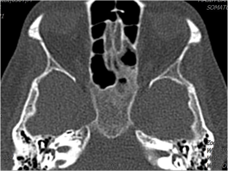
An axial non-contrast computed tomography (CT) scan of the paranasal sinuses at the level of the ethmoidal and maxillary regions. The image demonstrates significant bilateral mucosal thickening and opacification involving the maxillary sinuses and the ethmoidal air cells. These regions exhibit soft tissue density (intermediate gray) replacing the normal dark, air-filled spaces. The opacification appears diffuse, with notable involvement of the posterior ethmoid cells. The surrounding bony architecture, including the orbital walls and nasal septum, remains visible. Clinically, such findings in the context of a diabetic or immunocompromised patient are suggestive of severe inflammatory or infectious processes, such as acute rhinosinusitis or invasive fungal sinusitis (e.g., aspergillosis). This diagnostic image is essential for evaluating the extent of sinus disease and assessing for potential bony erosion or orbital extension in ENT and radiology education.
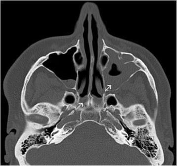
This diagnostic image is a non-contrast axial Computed Tomography (CT) scan of the paranasal sinuses. The scan demonstrates significant pathology in the left paranasal sinuses compared to the right. There is subtotal opacity of the left maxillary sinus, characterized by soft tissue density almost completely filling the sinus cavity, indicative of chronic rhinosinusitis or fluid accumulation. Additionally, the image shows evidence of ethmoidal sinusitis with mucosal thickening and opacification of the ethmoid air cells. In contrast, the right maxillary sinus remains primarily air-filled with minor peripheral mucosal thickening. White arrows indicate areas of significant inflammation and narrowing of the nasal passages. The surrounding bony structures of the sinus walls appear intact, and the mastoid air cells are visible inferiorly. This clinical imaging is essential for evaluating chronic sinus disease, which may be associated with systemic conditions such as Yellow Nail Syndrome (YNS).
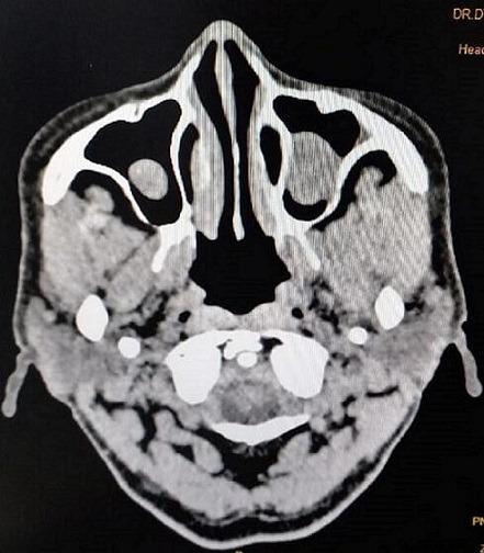
This diagnostic image is an axial non-contrast Computed Tomography (CT) scan of the paranasal sinuses. The scan demonstrates bilateral abnormalities within the maxillary and ethmoidal sinuses. Specifically, there is prominent mucosal thickening characterized by ill-defined, heterogeneous soft tissue signal intensity that partially opacifies the normally air-filled sinus cavities. The thickening is visible along the walls of both the right and left maxillary sinuses, appearing denser than air but less dense than cortical bone. The ethmoidal air cells also show evidence of soft tissue density, indicating inflammatory or infectious processes such as sinusitis. These findings are clinically significant in the context of paranasal sinus disease and are used to evaluate the extent of sinonasal involvement prior to interventions like Functional Endoscopic Sinus Surgery (FESS). The image serves as an educational example of radiologic manifestations of chronic or acute inflammatory sinus pathology.
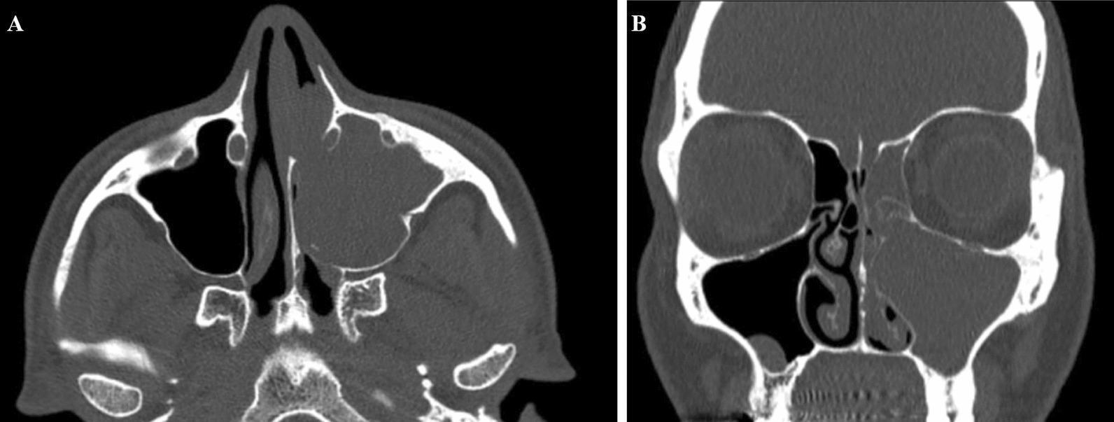
A head computed tomography (CT) scan displaying axial (A) and coronal (B) views of the paranasal sinuses. The imaging reveals a large, soft-tissue density mass measuring approximately 24 mm x 28 mm x 26 mm, which completely obliterates the left maxillary sinus. In both views, the lesion demonstrates expansion into the adjacent nasal cavity and causes total obstruction of the left anterior osteomeatal unit. Secondary findings include mucosal thickening and opacification of the left ethmoid air cells, consistent with secondary ethmoidal sinusitis. The right maxillary and ethmoid sinuses appear clear and well-aerated. This diagnostic image illustrates the radiological presentation of a sinonasal mass, such as an inverted papilloma or other neoplasm, highlighting the resulting regional obstruction and secondary inflammatory changes in the paranasal sinus system. It is an educational example of sinus pathology and the importance of evaluating the osteomeatal complex for obstructive complications.
CT Scan of Paranasal Sinuses - Report Explained
What is a CT Scan of the Paranasal Sinuses?
What the Report Found - Finding by Finding
- Sinusitis is inflammation of the mucosal lining of the sinuses, most commonly from infection or allergy.
- "Minimal" means the thickening is mild - not a full opacity or pus collection, just early/mild inflammatory changes.
- The sphenoid sinus is normal - a good sign, as sphenoid sinusitis can have serious complications (meningitis, cavernous sinus thrombosis).
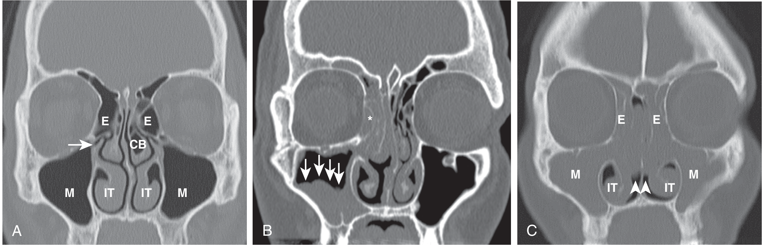
- "Obliterated bilaterally" = blocked on both sides by mucosal thickening.
- This is clinically important: it is both a result AND a cause of ongoing sinusitis - blockage prevents healing.
- Treatment often targets restoring OMC patency, sometimes surgically via Functional Endoscopic Sinus Surgery (FESS) if medical treatment fails.
- A septal spur is a sharp bony projection from the base of the septum, which can press against the adjacent turbinate and cause pain, headache, or obstruction.
- DNS is very common (present in up to 70-80% of people to varying degrees).
- The septum here is deviated toward the right, meaning the right nasal passage is narrower.
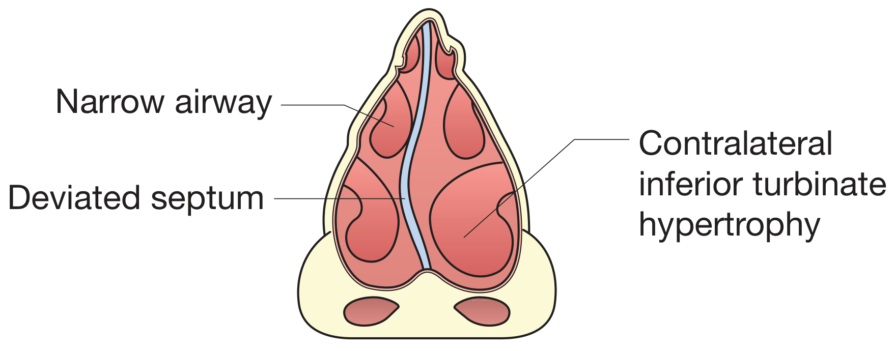
- Bilateral hypertrophy here is likely compensatory - the turbinate on the wider side (left, opposite to the DNS) enlarges to fill the extra space and maintain normal airway resistance, as explained in Cummings Otolaryngology.
- Causes include allergic rhinitis, chronic irritants, or the compensatory response to DNS.
- This is a major contributor to the patient's symptoms (nasal obstruction, mouth breathing, snoring).
- Nasal cavity is clear - no polyps, no foreign body.
- Bony outlines of sinuses are intact - no erosion, which rules out aggressive infection (fungal sinusitis) or tumor.
- Orbits and brain parenchyma appear normal - no spread of infection to the eye socket or brain, which is reassuring.
- Nasopharynx is unremarkable - the back of the throat behind the nose is normal.
Summary: What Does This All Mean?
| Finding | Plain English |
|---|---|
| Bilateral frontal, maxillary, ethmoidal sinusitis | Mild sinus infection/inflammation on both sides |
| OMC obliterated bilaterally | Drainage pathways blocked - sinuses can't clear properly |
| DNS right + septal spur | Crooked nose wall (bent right) with a bony spike |
| Bilateral inferior turbinate hypertrophy | Enlarged turbinates - narrowing both nostrils |
| Sphenoid normal; orbits/brain normal | No serious complications |
What Happens Next?
- Match symptoms to findings - Does the patient have nasal obstruction, facial pain/pressure, headache, post-nasal drip? These match the CT perfectly.
- Medical treatment first: Nasal saline irrigation, intranasal corticosteroid sprays (e.g. fluticasone), antihistamines if allergic, short course of antibiotics if bacterial infection suspected.
- Surgery if medical treatment fails:
- Septoplasty - to straighten the deviated septum
- Inferior turbinate reduction - to reduce turbinate size
- FESS (Functional Endoscopic Sinus Surgery) - to open blocked OMC and drain the sinuses These are often done together in a single procedure.
Important note: This explanation is for educational purposes. The patient should follow up with an ENT (Ear, Nose & Throat) specialist who can correlate these findings with the actual symptoms and decide on the best treatment approach.
- Cummings Otolaryngology Head and Neck Surgery, p. 570 - turbinate and septal obstruction
- Murray & Nadel's Textbook of Respiratory Medicine, p. 1612 - paranasal sinus anatomy and sinusitis CT