
Adenoids and JNA ENT MBBS 4TH YR
Reading File
Now let me search the medical library for both Adenoids and JNA (Juvenile Nasopharyngeal Angiofibroma) simultaneously.
Finding Sources
Finding Sources
Reading File
Reading File
Finding Sources
Finding Sources
Good. Now let me get comprehensive content on both topics from dedicated sections.
Finding Sources
Finding Sources
Reading File
Reading File
Now let me get the dedicated JNA section from Cummings and more adenoid content.
Reading File
Finding Sources
Excellent - I got detailed JNA content from Cummings. Let me get the rest of the JNA content and the adenoids dedicated section.
Reading File
Finding Sources
Reading File
Reading File
I now have comprehensive content on both topics. Let me also fetch the relevant images from the textbooks to include in the answer.
Loading Image
Loading Image
I now have all the content needed for a comprehensive ENT MBBS 4th year answer on both topics. Let me compile the complete notes.
ADENOIDS & JUVENILE NASOPHARYNGEAL ANGIOFIBROMA (JNA)
ENT - MBBS 4th Year
PART 1: ADENOIDS
Embryology
- Adenoids develop at 16 weeks as subepithelial infiltration of lymphocytes in the nasopharynx
- They form part of Waldeyer's ring (along with palatine tonsils, lingual tonsil, tubal tonsils)
(K.J. Lee's Essential Otolaryngology, p. 959)
Anatomy
- Located on the posterior wall of the nasopharynx (roof and posterior wall)
- Covered by ciliated pseudostratified columnar epithelium (respiratory type); luminal aspect has multiciliated and secretory cells
- Unlike palatine tonsils, adenoids have no crypts - they have surface folds/rugae
- They are not encapsulated and have no afferent lymphatics
Physiology / Role
- Part of the mucosal immune system; produce secretory IgA (sIgA)
- Act as first-line defense against inhaled antigens
- Adenoid size peaks at 4-10 years, then undergoes spontaneous involution at puberty
- In children with CRS (chronic rhinosinusitis):
- Adenoids serve as a bacterial reservoir - bacteria from middle meatal swabs and adenoidal core cultures are near-identical (S. pneumoniae, Group A strep, H. influenzae, S. aureus, coagulase-negative staphylococci)
- Positive predictive value of adenoid core culture for predicting middle meatal culture: 91.5%
- Biofilms cover 88-99% of adenoid surface in CRS children vs 0-6.5% in simple obstructive adenoid hypertrophy
- Lower IgA expression in CRS adenoids; higher TGF-β1, MMP-2, MMP-9
(Scott-Brown's ORL, Vol 2; Cummings Otolaryngology)
Adenoid Hypertrophy - Clinical Features
Age group: Most common between 4-10 years
Symptoms:
| System | Symptom |
|---|---|
| Nasal | Mouth breathing, nasal obstruction, hyponasal voice (rhinolalia clausa) |
| Otological | Recurrent otitis media, serous otitis media (glue ear), conductive hearing loss |
| Sleep | Snoring, obstructive sleep apnoea (OSA), restlessness during sleep, unusual sleep positions |
| Face | "Adenoid facies" - open mouth, high arched palate, elongated face, dull appearance |
| Systemic | Failure to thrive (in severe cases) |
Adenoid facies = characteristic look due to chronic mouth breathing: elongated face, open mouth, pinched nostrils, dental malocclusion.
Investigation:
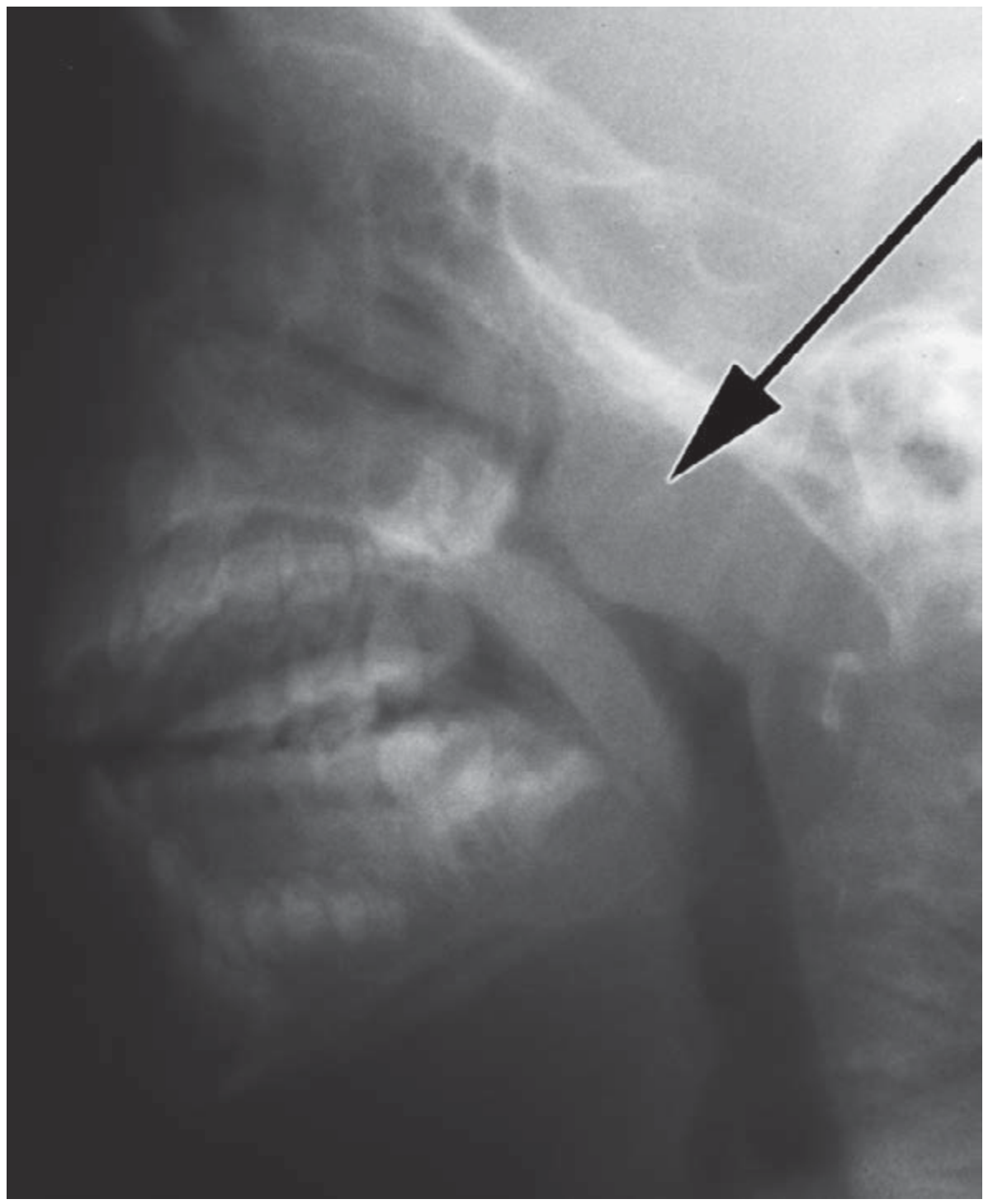
- Lateral soft tissue X-ray of nasopharynx - shows pad of adenoid tissue in postnasal space (arrow visible on soft tissue radiograph)
- Flexible nasendoscopy (gold standard)
- Note: adenoid size alone does not correlate with severity of CRS on CT
Adenoid and Otitis Media with Effusion (OME / Glue Ear)
- Adenoid hypertrophy causes Eustachian tube dysfunction → OME
- Adenoidectomy alone improves OME/recurrent AOM (due to both mechanical obstruction relief and elimination of bacterial reservoir)
- Added for children ≥4 years when distinct indications exist
Medical Treatment
- Intranasal corticosteroids may significantly improve nasal obstruction symptoms in moderate-to-severe adenoid hypertrophy and may reduce adenoid size (Cochrane evidence - limited but supportive)
Indications for Adenoidectomy
(Bailey & Love's Surgery, 28th Ed; KJ Lee's Otolaryngology)
- OSA associated with postnasal obstruction
- Recurrent acute otitis media or prolonged serous otitis media (>3 months duration)
- Recurrent rhinosinusitis
- Postnasal discharge / chronic adenoiditis
- Failure to thrive due to airway obstruction
Tonsillectomy indications (for comparison):
- 6-7 episodes of acute tonsillitis in 1 year
- 5 episodes/year for 2 years
- 3 episodes/year for 3 years (Paradise criteria)
- Peritonsillar abscess
Adenoidectomy - Operative Technique
- Patient supine, oropharynx exposed with Boyle-Davis mouth gag + Doughty's tongue plate
- Nasopharynx palpated; adenoid curette (St. Clair Thompson) used to scrape adenoid pad
- Haemostasis with post-nasal pack or suction cautery
- Important: Avoid sedative premedications and postoperative opiates in OSA patients (risk of respiratory depression)
PART 2: JUVENILE NASOPHARYNGEAL ANGIOFIBROMA (JNA)
Definition
A benign but locally invasive vascular tumour of the nasopharynx occurring almost exclusively in adolescent males. Accounts for <1% of all head and neck tumours.
(Cummings Otolaryngology Head & Neck Surgery)
Epidemiology
- Age: typically 10-25 years, predominantly males
- Extremely rare in females (some cases in maxillary sinus site)
- Androgen-dependent: androgen receptors (esp. estrogen receptors) present in tumour tissue - explains male predominance and puberty-related onset
Site of Origin
- Arises from the sphenopalatine foramen at the root of the pterygoid process on the lateral nasal wall
- This is the most important fact - the tumour originates here
Pathology
Macroscopy:
- Sessile, lobulated, rubbery, dark red to tan-gray
- Unencapsulated, sessile, wide-based attachment to nasopharynx
- No mucosal ulceration
Microscopy:
- Admixture of vascular tissue + fibrous stroma
- Vessel walls lack elastic fibres and have incomplete or absent smooth muscle → accounts for profuse bleeding
- Vascular cavities with endothelial layer but no muscle layer (unlike normal arteries)
- Similarities between nasal mucosal cells, genital erectile tissue, and vascular spaces
Growth Pattern
- Locally infiltrative, slow-growing
- Expands laterally via pterygopalatine fossa → infratemporal fossa
- Extends into nasopharynx and choanal space → anterior nasal cavity and sinuses
- Can expand to: maxillary/ethmoid sinuses, orbit, cheek, cavernous sinus
- Intracranial extension (middle/anterior cranial fossa) - uncommon but serious
Clinical Features
| Feature | Details |
|---|---|
| Age/sex | Adolescent males, 10-25 years |
| Epistaxis | Most common presentation - recurrent, severe, may lead to anaemia |
| Nasal obstruction | Usually unilateral |
| Other | Blood-stained sputum, serous otitis media, nasal discharge, facial swelling, proptosis, anosmia |
| Duration | Usually long history, often mild and innocuous initially |
Diagnosis
Endoscopy: Rubbery vascular mass protruding into anterior nasal space; bleeds excessively on contact - biopsy avoided due to risk of haemorrhage
CT with contrast:
- Enhancing soft tissue mass from nasopharynx/lateral nasal wall
- Widening of pterygopalatine fossa
- No bone erosion generally
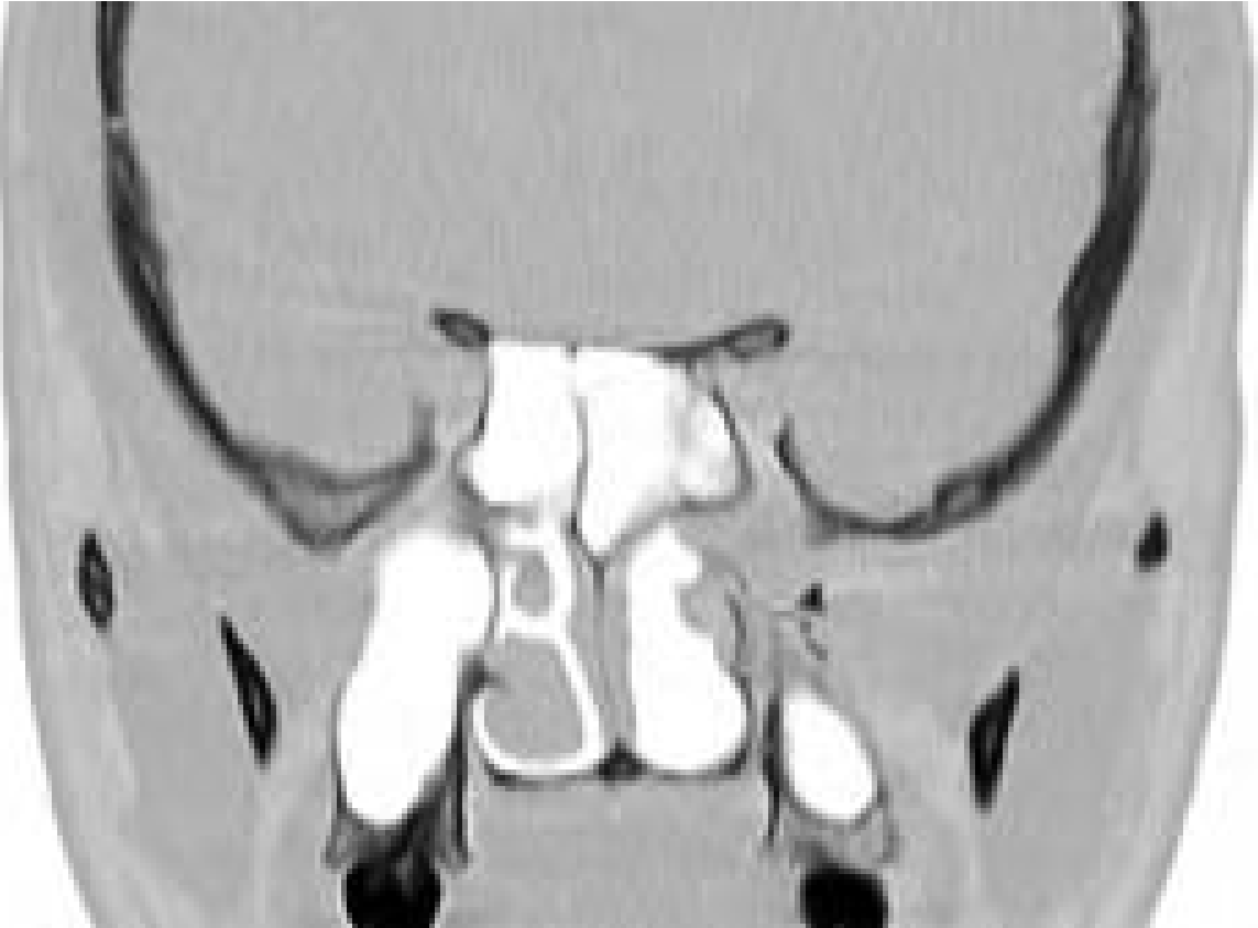
- Holman-Miller sign (classical): anterior bowing of posterior wall of maxillary sinus
MRI:
- Vascular tumour with flow voids within the mass
- Enhancement on gadolinium imaging
- Best for delineating intracranial extension and follow-up
MR Angiography / DSA (Digital Subtraction Angiography):
- Shows feeding vessels arising from branches of the external carotid artery (mainly internal maxillary artery)
- If intracranial spread: internal carotid artery also feeds the tumour
- DSA shows enlarged intratumoral vessels + intense inhomogeneous blush in venous phase
Histology: Final confirmation - required before definitive treatment
Chandler Staging (1984)
| Stage | Extent |
|---|---|
| Stage I | Confined to nasopharynx |
| Stage II | Extension into nasal cavity or sphenoid sinus |
| Stage III | Involvement of maxillary sinus, ethmoid sinus, infratemporal fossa, orbit, cheek, cavernous sinus |
| Stage IV | Intracranial extension |
Management
Primary treatment: Surgical excision
Pre-operative embolization:
- Well-established adjunct to induce tumour shrinkage, improve surgical access, reduce intra-operative blood loss
- Done transarterially with particles (PVA) or liquids (NBCA/Onyx)
- For tumours supplied by branches of external carotid - relatively straightforward
- Intracranial supply (internal carotid) increases risk of neurological deficit
- Direct tumour puncture technique with Onyx injection - improves devascularization
Surgical approaches:
- Traditional: Open approaches (transpalatal, lateral rhinotomy, midfacial degloving)
- Modern: Endoscopic surgical resection - now preferred for smaller tumours (Stage I-II) without spread; some consider preoperative embolization unnecessary for smaller endoscopic cases
- Large/intracranial tumours: Combined skull base approach (head and neck surgeon + neurosurgeon)
Radiotherapy:
- Used when surgery is contraindicated or for recurrence
- Primary radiation therapy has been reported
- Gamma knife used in some cases
Hormonal therapy (adjunct):
- Flutamide (androgen antagonist) - shown to reduce tumour size, may have role as pre-operative adjunct
Natural history:
- Spontaneous regression reported after cessation of androgen stimulation (post-puberty)
- Rare malignant transformation reported
Key Exam Points - JNA
| Feature | Value |
|---|---|
| Tumour type | Benign, locally invasive, vascular |
| Who | Adolescent males (10-25 years) |
| Origin site | Sphenopalatine foramen / lateral nasal wall |
| Classic sign on CT | Holman-Miller sign (anterior bowing of posterior maxillary wall) |
| Classic imaging finding | Flow voids on MRI |
| Feeding artery | Branches of external carotid (internal maxillary artery) |
| Biopsy in clinic | AVOID - risk of catastrophic haemorrhage |
| Pre-op adjunct | Embolization |
| Primary treatment | Surgical excision |
| Histology | Vascular spaces with absent smooth muscle + fibrous stroma |
| Staging | Chandler staging (I-IV) |
Comparison Table: Adenoids vs JNA
| Feature | Adenoids (Hypertrophy) | JNA |
|---|---|---|
| Type | Lymphoid hyperplasia | Benign vascular tumour |
| Age | Children, 4-10 years | Adolescent males, 10-25 years |
| Sex | Both | Almost exclusively male |
| Location | Roof/posterior nasopharynx | Sphenopalatine foramen |
| Main symptom | Nasal obstruction, OME | Epistaxis, nasal obstruction |
| Investigation | Lateral X-ray, nasendoscopy | CT, MRI, DSA |
| Classic sign | Adenoid facies | Holman-Miller sign |
| Treatment | Adenoidectomy | Surgery (±embolization) |
| Biopsy | Safe | DANGEROUS |
Sources: Cummings Otolaryngology Head and Neck Surgery; Bailey & Love's Short Practice of Surgery 28th Ed; Scott-Brown's Otorhinolaryngology Head & Neck Surgery; K.J. Lee's Essential Otolaryngology
This is a shared conversation. Sign in to Orris to start your own chat.