
Give the few identification points and give image histology anatomy
histology identification points mucous salivary gland hyaline cartilage cardiac muscle sensory ganglion thymus spleen aorta lymph node spotters
Histology Spotters - Complete Identification Guide
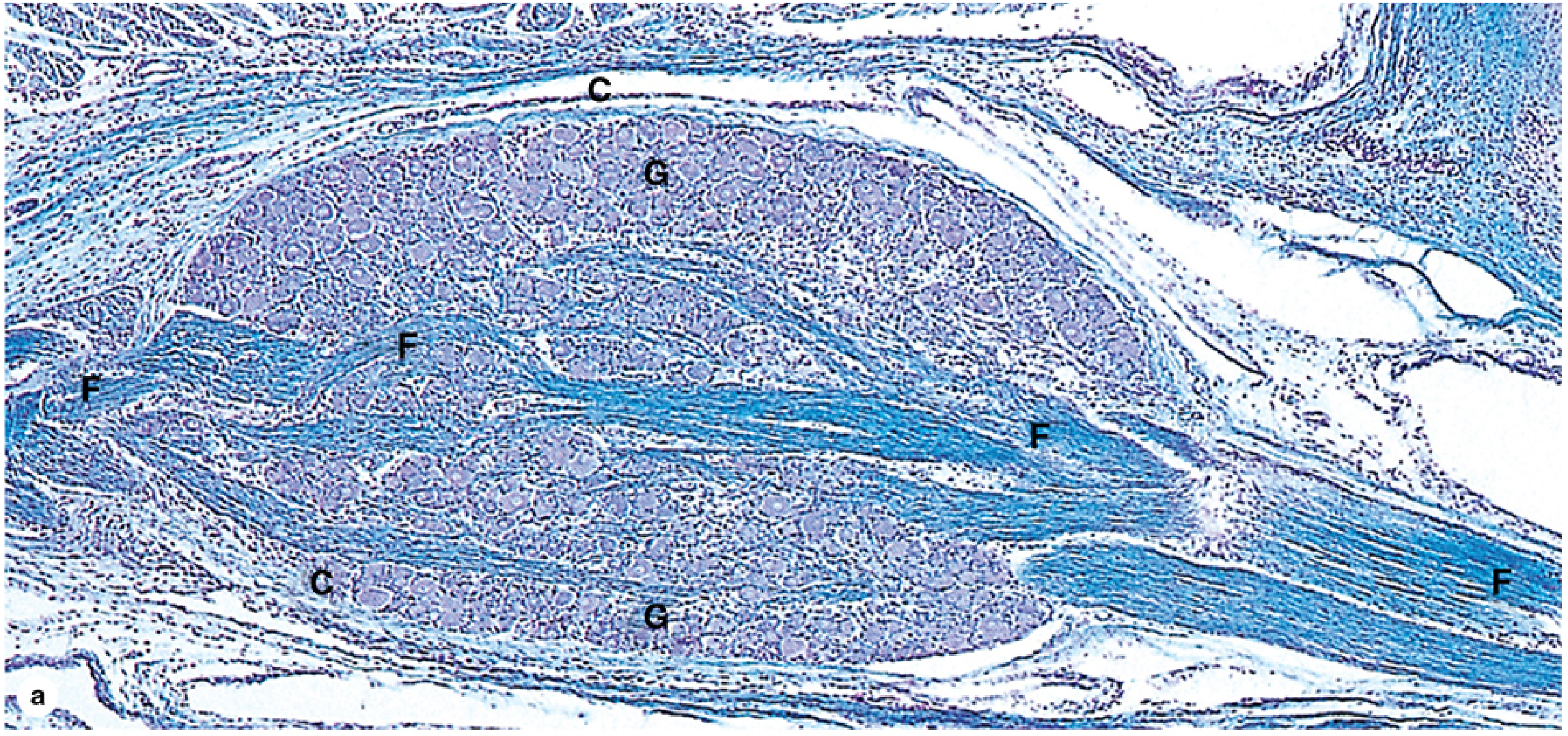
1. MUCOUS SALIVARY GLAND
- Mucous acini/tubules: Large, pale-staining cells with flattened basal nuclei pressed against the cell base (nucleus pushed to the periphery by mucin)
- Cytoplasm appears empty/foamy or lightly eosinophilic due to mucin washing out during processing
- No serous demilunes (or very few) - distinguishes it from submandibular gland
- Connective tissue septa divide the gland into lobules
- Ducts present: intercalated ducts, striated ducts, interlobular ducts
- PAS-positive staining (mucin is glycoprotein-rich)
- The sublingual gland is the classic "mucous salivary gland" specimen
2. HYALINE CARTILAGE ✓ (ticked on your list)
- Homogeneous, glassy/amorphous matrix (no visible fibers under H&E - collagen type II fibrils are invisible)
- Chondrocytes in lacunae (spaces within the matrix)
- Chondrocytes may appear in isogenous groups (2-4 cells sharing a lacuna = cell nests)
- Perichondrium present (outer fibrous + inner chondrogenic layer) - EXCEPT on articular surfaces
- Basophilic matrix (GAG-rich, sulfated proteoglycans)
- Territorial matrix (darker, immediately around lacunae) vs. interterritorial matrix (lighter, between cell nests)
- No blood vessels, lymphatics, or nerves within the cartilage itself
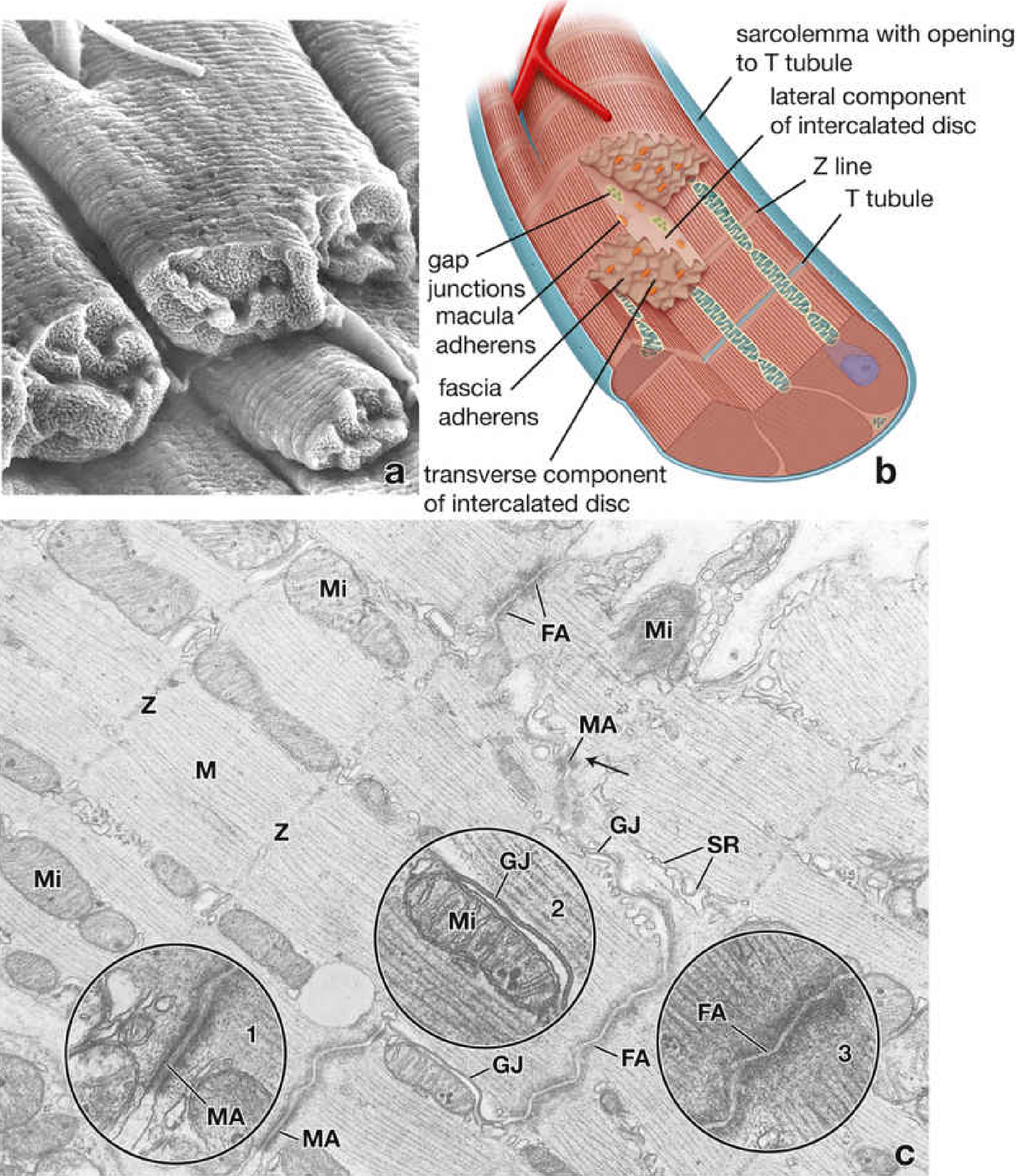
3. CARDIAC MUSCLE
- Intercalated discs - the single most diagnostic feature; appear as darkly-staining transverse lines crossing the fibers in a step-like/staircase pattern (unique to cardiac muscle)
- Cells are branched (fork and rejoin neighboring cells)
- Single central nucleus (or occasionally binucleate), oval, centrally placed
- Cross-striations (A, I, Z bands) visible - like skeletal muscle
- Abundant mitochondria in the perinuclear sarcoplasm (reflect high energy demand)
- Endomysium rich in capillaries between fibers
- NO neuromuscular junctions (involuntary)
4. SENSORY GANGLION (Dorsal Root / Spinal Ganglion)
- Large, rounded/spherical neuron cell bodies (pseudounipolar neurons) arranged in groups or scattered throughout
- Each neuron is surrounded by a single layer of satellite cells (small, flattened cells forming a distinct capsule/halo around each neuron)
- Neuron cell body has a large, pale, centrally-placed nucleus with a prominent nucleolus
- Nissl bodies (clumped RER) visible in cytoplasm
- Myelinated nerve fibers (axon bundles) running between ganglionic neurons
- Connective tissue capsule surrounds the whole ganglion
- No synaptic contacts on cell bodies (unlike autonomic ganglia)
- Variable neuron sizes (large and small cells present)
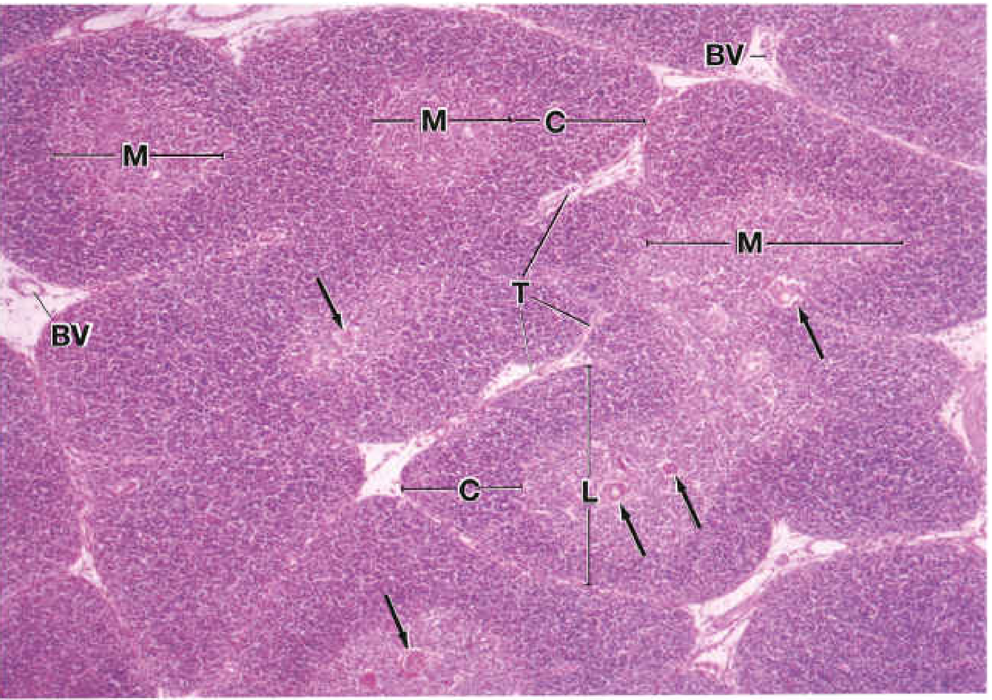
5. THYMUS
- Bilobed structure with a connective tissue capsule and septa dividing it into lobules
- Each lobule has two distinct zones:
- Cortex - DARK (densely packed small lymphocytes / thymocytes)
- Medulla - PALE (fewer, more mature lymphocytes, epithelial cells)
- Hassall's corpuscles (thymic corpuscles) in the medulla - concentric whorls of flattened keratinized epithelial cells - PATHOGNOMONIC
- Blood-thymus barrier in the cortex (no antigen access)
- Epithelial reticular cells form the supporting framework
- No germinal centers (no B cells, no follicles)
- In young individuals: large and prominent; involutes with age (replaced by fat)
6. SKIN L.S (Longitudinal Section - Thick/Glabrous Skin e.g. Palm/Sole)
- Epidermis with 5 layers (thick skin): Stratum basale, spinosum, granulosum, lucidum (clear layer - unique to thick skin), corneum
- Thick stratum corneum (deeply eosinophilic, anucleate squames)
- Stratum lucidum - clear/translucent band between granulosum and corneum
- Dermal papillae project up into epidermis (irregular interface)
- Dermis: Papillary (loose CT) + Reticular (dense irregular CT)
- Eccrine sweat glands (coiled secretory portion in deep dermis/hypodermis; dark cuboidal cells)
- Sweat duct opening at surface as a spiral pore
- No hair follicles, no sebaceous glands (in thick skin)
- Meissner's corpuscles in dermal papillae; Pacinian corpuscles (concentric laminae/"onion bulb") in deep dermis/hypodermis
7. SKIN T.S (Transverse Section - Thin/Hairy Skin)
- Thinner epidermis - only 4 layers (no stratum lucidum)
- Hair follicles in cross-section (appear as rounded structures in dermis) - KEY identifier vs. thick skin
- Sebaceous glands attached to hair follicles (pale, lipid-laden cells)
- Arrector pili muscle (smooth muscle bundle attached to follicle)
- Both eccrine and apocrine sweat glands may be present
- Dermis: papillary + reticular
- Nerve fibers and hair follicle nerve endings
8. UMBILICAL CORD
- Wharton's jelly - abundant pale-staining myxomatous/mucoid connective tissue (stellate fibroblasts in a loose, gelatinous matrix rich in hyaluronic acid) - KEY identifier
- Two umbilical arteries (thick muscular walls, no internal elastic lamina) - round, with narrow lumen and thick wall
- One umbilical vein (larger diameter, thinner wall, wider lumen)
- No nerves, no lymphatics
- Covered by amniotic epithelium (simple squamous/cuboidal)
- The 2 arteries carry deoxygenated blood TO placenta; 1 vein carries oxygenated blood FROM placenta
9. PLACENTA
- Chorionic villi - the dominant histological feature; finger-like projections floating in intervillous spaces
- Cytotrophoblast cells (inner layer, distinct cell borders, pale staining)
- Syncytiotrophoblast (outer layer, no cell borders, multiple nuclei - syncytium; darkly staining)
- Hofbauer cells (fetal macrophages) in villous stroma
- Fetal blood vessels within the villous stroma
- Intervillous space contains maternal blood (lacunae)
- Fibrinoid deposits (pink amorphous material) on villous surfaces
- Decidua basalis may be present at base
10. SPLEEN
- Capsule (with trabeculae extending inward)
- White pulp - lymphoid tissue arranged as Malpighian corpuscles (spherical aggregates of lymphocytes around a central arteriole = PALS - periarteriolar lymphoid sheath); may have germinal centers
- Central arteriole eccentrically placed within the white pulp (not truly "central")
- Red pulp - the large, abundant pale/pink area filled with blood sinusoids and splenic cords (cords of Billroth) - erythrocytes, macrophages, plasma cells
- Marginal zone between white and red pulp
- No lymphatic sinuses (unlike lymph node)
- Trabeculae contain trabecular arteries and veins
11. AORTA (Elastic Artery)
- Tunica intima: endothelium + thin subendothelial CT
- Tunica media: THICK, with numerous concentric elastic laminae (wavy, darkly-staining pink lines on H&E; jet black with Verhoeff's / orcein stain)
- Multiple elastic fenestrated membranes (40-60 layers) in the media - KEY feature distinguishing it from muscular arteries
- Smooth muscle cells between elastic lamellae
- Tunica adventitia: relatively thin, loose CT with vasa vasorum (blood vessels supplying the vessel wall) and nervi vasorum
- No distinct internal or external elastic lamina (as seen in muscular arteries) - instead, multiple elastic sheets
- Very thick media relative to total wall
12. LYMPH NODE ✓ (ticked on your list)
- Capsule of dense CT with subcapsular sinus just beneath it (lymph flows into this sinus from afferent lymphatics)
- Cortex (outer): contains primary follicles (uniform lymphocytes) and secondary follicles (with pale germinal centers = active B cell response)
- Paracortex (deep cortex / inner cortex): T lymphocyte zone, high endothelial venules (HEV) present
- Medulla: medullary cords (plasma cells, macrophages) separated by medullary sinuses
- Afferent lymphatics enter at multiple points on the capsule; ONE efferent lymphatic exits at the hilum
- Germinal centers = active B-cell proliferation, differentiation to plasma cells
- Hilum: blood vessels + efferent lymphatic
Quick Comparison Table
| Specimen | Single Best Identifier |
|---|---|
| Mucous Salivary Gland | Pale mucous acini + flat basal nuclei |
| Hyaline Cartilage | Glassy matrix + lacunae + chondrocytes |
| Cardiac Muscle | Intercalated discs (step-like dark lines) |
| Sensory Ganglion | Large neurons + satellite cell capsules |
| Thymus | Hassall's corpuscles in medulla |
| Skin L.S (Thick) | 5 layers including stratum lucidum |
| Skin T.S (Thin) | Hair follicles + sebaceous glands |
| Umbilical Cord | Wharton's jelly + 2 arteries + 1 vein |
| Placenta | Syncytiotrophoblast + chorionic villi |
| Spleen | White pulp + central arteriole in PALS |
| Aorta | Multiple concentric elastic laminae in media |
| Lymph Node | Capsule + follicles + subcapsular sinus + medullary sinuses |
Histology diagram and points
Histology Spotters - Diagrams & Identification Points
1. MUCOUS SALIVARY GLAND
- Predominantly pale/empty-looking tubules - mucin washes out in processing
- Flattened basal nuclei - pushed to cell base by apical mucin granules
- Cells arranged as tubuloacinar units (not round acini)
- Connective tissue septa dividing gland into lobules
- Intralobular and interlobular ducts visible between tubules
- No (or very few) serous demilunes - distinguishes purely mucous from mixed glands
- PAS-positive staining (mucin = glycoprotein)
2. HYALINE CARTILAGE
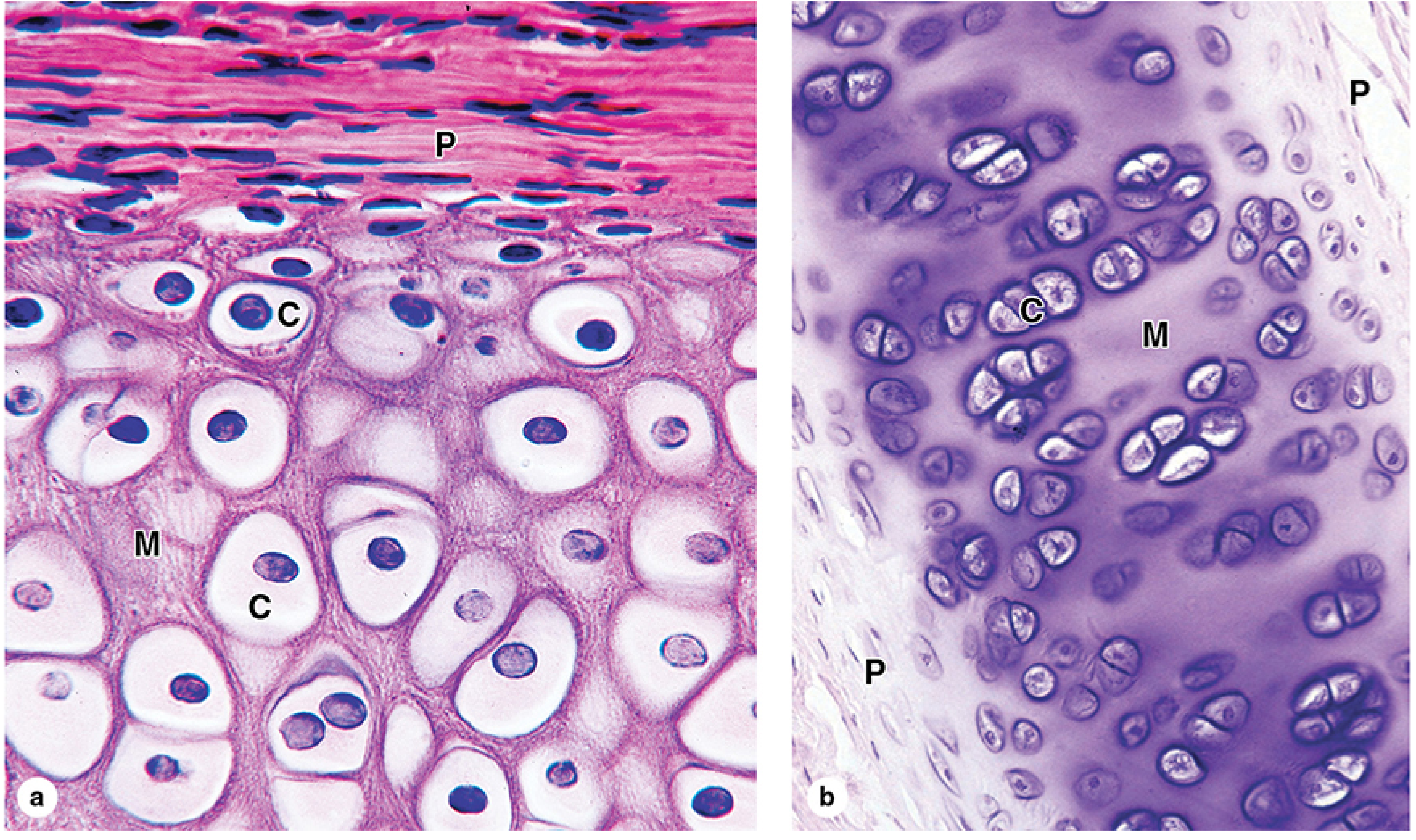
- Homogeneous, glassy amorphous matrix - no visible fibers (type II collagen invisible on H&E)
- Chondrocytes in lacunae (spaces within matrix)
- Isogenous groups - 2-4 chondrocytes sharing a lacuna (from mitotic division)
- Perichondrium - outer fibrous layer + inner chondrogenic layer (ABSENT on articular surfaces)
- Territorial matrix (dark, around each lacuna) vs interterritorial matrix (pale, between groups)
- Basophilic matrix due to sulfated GAGs (proteoglycans)
- No blood vessels, no nerves within cartilage itself
3. CARDIAC MUSCLE
- Intercalated discs - darkly-staining transverse lines in a step/staircase pattern - PATHOGNOMONIC
- Branched cells - fibers fork and reconnect with adjacent cells
- Single central oval nucleus (occasionally binucleate)
- Cross-striations (A, I, Z bands) visible - like skeletal muscle
- Abundant perinuclear mitochondria (pale halo around nucleus)
- Rich endomysium with capillaries between fibers
- No neuromuscular junctions; no satellite cells
4. SENSORY GANGLION (Dorsal Root Ganglion)
- Connective tissue capsule - well-defined, thick
- Large, rounded pseudounipolar neurons - largest cells in the section
- Each neuron surrounded by a ring of small satellite cells (flat, elongated nuclei forming a halo)
- Large pale nucleus with prominent nucleolus ("owl eye" appearance)
- Nissl bodies (blue granules of rough ER) in cytoplasm
- Myelinated nerve fiber bundles running between neuron clusters
- Variable cell size (large and small neurons present)
- No synapses on cell bodies (unlike autonomic ganglia)
5. THYMUS
- Capsule and septa dividing gland into incomplete lobules
- Dark cortex - densely packed small lymphocytes (thymocytes)
- Pale medulla - fewer, more mature lymphocytes + epithelial reticular cells
- Hassall's corpuscles in medulla - concentric whorls of keratinized epithelial cells (PATHOGNOMONIC)
- Center of large Hassall's corpuscles may show amorphous keratinization
- No germinal centers (no B cells, no follicles)
- Involutes with age (replaced by adipose tissue)
6. SKIN L.S (Thick Skin - Palm/Sole)
- 5 epidermal layers (from deep to surface): Basale - Spinosum - Granulosum - Lucidum - Corneum
- Stratum lucidum - clear translucent band unique to thick skin (between granulosum and corneum)
- Thick stratum corneum - anucleate eosinophilic squames
- No hair follicles, no sebaceous glands
- Dermal papillae - tall, closely packed finger-like projections into epidermis
- Eccrine sweat glands - coiled, two cell types (dark secretory + pale myoepithelial)
- Spiral sweat duct - duct opens as a spiral pore on the surface ridge
- Meissner's corpuscles in dermal papillae; Pacinian corpuscles in deep dermis
7. SKIN T.S (Thin Skin - Hairy Skin)
- 4 epidermal layers only - NO stratum lucidum
- Thinner stratum corneum than thick skin
- Hair follicles in dermis - KEY distinguishing feature from thick skin
- Sebaceous glands - pale lipid-laden cells attached to hair follicle (pilosebaceous unit)
- Arrector pili muscle - smooth muscle bundle running from follicle to dermis
- Eccrine + apocrine sweat glands both present
- Papillary + reticular dermis + hypodermis with fat
- Melanin pigment may be seen in stratum basale cells
8. UMBILICAL CORD
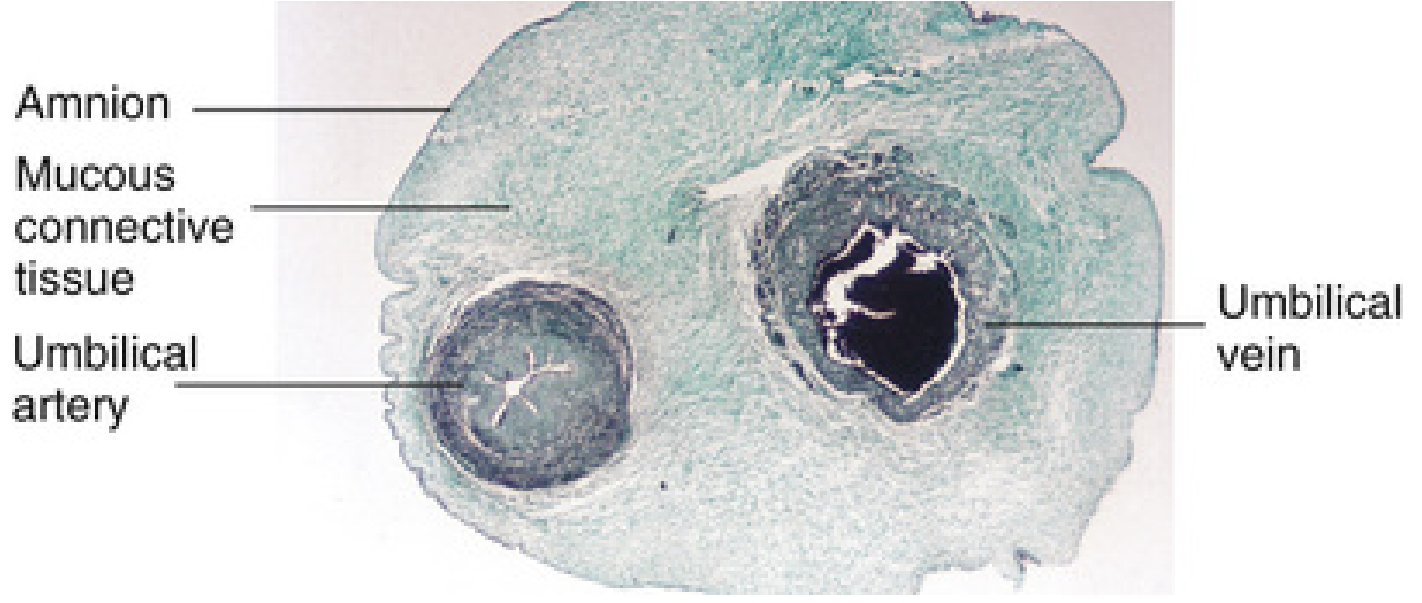
- Wharton's jelly - abundant pale gelatinous mucoid stroma (stellate fibroblasts in hyaluronic acid matrix) - PATHOGNOMONIC
- 2 umbilical arteries - thick muscular walls, small round/star-shaped lumen
- 1 umbilical vein - larger lumen, thinner wall (easy to distinguish)
- Outer covering = amniotic epithelium (simple cuboidal/squamous)
- No nerves, no lymphatics within the cord
- Vessels are longer than cord length - causes spiraling/coiling
- Arteries carry deoxygenated blood to placenta; vein carries oxygenated blood from placenta
9. PLACENTA
- Chorionic villi - dominant feature; finger-like projections in an intervillous space
- Syncytiotrophoblast - outer layer, NO cell borders, multiple nuclei evenly spaced (or in syncytial knots), dark eosinophilic - present throughout gestation
- Cytotrophoblast - inner layer, individual cells with distinct borders (prominent in early placenta; sparse at term)
- Syncytial knots - clusters of nuclei on villous surface (increases with maturation)
- Fetal blood vessels within villous stroma
- Hofbauer cells (fetal macrophages) in villous stroma
- Intervillous space contains maternal blood
- Fibrinoid deposits on villous surfaces (pink amorphous material)
10. SPLEEN
- Capsule + trabeculae extending into the organ
- White pulp - spherical dark aggregates of lymphocytes = Malpighian corpuscles
- Central arteriole eccentrically placed within each white pulp nodule (PALS = periarteriolar lymphoid sheath of T-cells)
- Germinal centers may be present in white pulp (B cell zones)
- Red pulp (majority of organ) - pale/pink, contains venous sinusoids and splenic cords (cords of Billroth) with erythrocytes + macrophages
- Marginal zone between white and red pulp
- No subcapsular sinus (unlike lymph node)
- Trabeculae contain trabecular arteries and veins
11. AORTA
- Tunica intima - endothelium + thin subendothelial layer
- Tunica media - VERY THICK, dominated by ~50 concentric elastic laminae alternating with smooth muscle layers (wavy pink lines on H&E)
- Elastic laminae stain jet black with Verhoeff's stain (orcein/resorcin-fuchsin)
- No distinct internal/external elastic lamina - replaced by multiple sheets (unlike muscular arteries)
- Tunica adventitia - thin, loose CT with vasa vasorum (vessels within the wall)
- Media-to-lumen ratio is high
- Smooth muscle cells lie between elastic lamellae
- Wall thickness >2 mm at aortic root
12. LYMPH NODE
- Capsule of dense CT + subcapsular sinus immediately beneath (afferent lymph drains here first)
- Outer cortex - lymphoid follicles: primary (uniform dark lymphocytes) or secondary (with pale germinal centers = active B cells)
- Germinal centers - pale spherical areas with large proliferating B cells, tingible body macrophages
- Paracortex (deep cortex) - T lymphocyte zone, contains high endothelial venules (HEV)
- Medullary cords - contain plasma cells and macrophages
- Medullary sinuses - separate medullary cords; connect to efferent lymphatic at hilum
- Afferent lymphatics enter through capsule (multiple); one efferent lymphatic at hilum
- Hilum - indentation where blood vessels + efferent lymphatic exit
At-a-Glance: One-Word Diagnostic Feature
| # | Specimen | Clincher Feature |
|---|---|---|
| 1 | Mucous Salivary Gland | Pale tubules + flat basal nuclei |
| 2 | Hyaline Cartilage | Glassy matrix + lacunae |
| 3 | Cardiac Muscle | Intercalated discs |
| 4 | Sensory Ganglion | Large neurons + satellite cell halos |
| 5 | Thymus | Hassall's corpuscles |
| 6 | Skin L.S (Thick) | Stratum lucidum + no hair follicles |
| 7 | Skin T.S (Thin) | Hair follicles + sebaceous glands |
| 8 | Umbilical Cord | Wharton's jelly + 2A + 1V |
| 9 | Placenta | Syncytiotrophoblast on villi |
| 10 | Spleen | Central arteriole in PALS |
| 11 | Aorta | Multiple elastic laminae in thick media |
| 12 | Lymph Node | Subcapsular sinus + follicles + medullary sinuses |
Only histology diagram and points
Histology diagram of Mucous Salivary Gland (sublingual gland), H&E stain, labeled: pale mucous acini/tubules with empty cytoplasm, flattened basal nuclei pushed to cell periphery, intralobular duct, connective tissue septum dividing lobules, myoepithelial cells. Clean medical illustration style with clear labels and arrows.
Histology diagram of Hyaline Cartilage, H&E stain, labeled: perichondrium (outer fibrous layer), chondroblasts at periphery, chondrocytes in lacunae, isogenous groups (2-4 cells per lacuna), homogeneous glassy amorphous matrix, territorial matrix (dark around cells), interterritorial matrix (pale). Clean medical illustration with clear labels and arrows.
Histology diagram of Cardiac Muscle, H&E stain, labeled: intercalated discs (dark step-like transverse lines), branched muscle fibers, central oval nucleus, cross striations (A band, I band, Z line), abundant mitochondria perinuclear, endomysium with capillaries. Clean medical illustration with clear labels and arrows.
Histology diagram of Sensory Ganglion (dorsal root ganglion), H&E stain, labeled: dense connective tissue capsule, large pseudounipolar neuron cell body, satellite cells forming halo around each neuron, large pale nucleus with prominent nucleolus, Nissl bodies in cytoplasm, myelinated nerve fiber bundles. Clean medical illustration with clear labels and arrows.
Histology diagram of Thymus gland, H&E stain, labeled: capsule, trabecula, lobule, dark cortex with densely packed thymocytes, pale medulla, Hassall's corpuscles (concentric whorls of keratinized epithelial cells), epithelioreticular cells, blood vessel. Clean medical illustration with clear labels and arrows.
Histology diagram of Thick Skin longitudinal section (palm/sole), H&E stain, labeled: 5 epidermal layers from bottom to top: stratum basale, stratum spinosum, stratum granulosum, stratum lucidum (clear pale band), stratum corneum (thick anucleate), dermal papillae, papillary dermis, reticular dermis, eccrine sweat gland coiled in dermis, sweat duct spiraling to surface. Clean medical illustration with clear labels and arrows.
Histology diagram of Thin Skin transverse section (hairy skin), H&E stain, labeled: 4 epidermal layers (basale, spinosum, granulosum, corneum - no lucidum), hair follicle cross section in dermis, sebaceous gland attached to follicle, arrector pili smooth muscle, eccrine sweat gland, papillary and reticular dermis, hypodermis with adipose tissue. Clean medical illustration with clear labels and arrows.
Histology diagram of Umbilical Cord cross section, labeled: outer amniotic epithelium, Wharton's jelly (pale gelatinous mucoid connective tissue with stellate fibroblasts), two umbilical arteries (thick muscular walls, small lumen, star-shaped), one umbilical vein (thin wall, large lumen). Clean medical illustration with clear labels and arrows.
Histology diagram of Placenta, H&E stain, labeled: chorionic villus (cross section), syncytiotrophoblast (outer layer, no cell borders, multiple nuclei), cytotrophoblast cells (inner layer, distinct cell borders), fetal blood vessels in villous stroma, Hofbauer cells (macrophages), intervillous space with maternal blood, syncytial knots, fibrinoid deposits. Clean medical illustration with clear labels and arrows.
Histology diagram of Spleen, H&E stain, labeled: capsule, trabecula, white pulp (Malpighian corpuscle - dark lymphoid nodule), central arteriole eccentrically placed, PALS periarteriolar lymphoid sheath, germinal center, marginal zone, red pulp (splenic sinusoids and cords of Billroth), macrophages. Clean medical illustration with clear labels and arrows.
Histology diagram of Aorta (elastic artery) cross section, labeled: tunica intima with endothelium, tunica media very thick with multiple concentric wavy elastic laminae alternating with smooth muscle layers (about 50 laminae), tunica adventitia thin with vasa vasorum. Verhoeff elastic stain appearance showing black wavy lines. Clean medical illustration with clear labels and arrows.
Histology diagram of Lymph Node, H&E stain, labeled: dense CT capsule, subcapsular sinus (just beneath capsule), afferent lymphatic vessel, outer cortex with primary follicle, secondary follicle with germinal center (pale) and mantle zone, paracortex (deep cortex, T cell zone) with high endothelial venule, medullary cords (plasma cells), medullary sinuses, hilum with efferent lymphatic and blood vessels. Clean medical illustration with clear labels and arrows.