
Sequencial events of a cardiac cycle including concurrent pressure volume change and generation of heart sound
Reading File
Now let me search the medical library for cardiac cycle content in parallel:
Finding Sources
Finding Sources
Searching Images
cardiac cycle pressure volume changes heart sounds Wiggers diagram
Excellent. Now let me read the key sections from Guyton & Hall and Costanzo Physiology - the best physiology sources - in parallel:
Reading File
Reading File
Reading File
Excellent content retrieved. Let me now get more detailed content on the individual phases and the pressure-volume loop:
Reading File
Reading File
Finding Sources
I found a cardiac cycle diagram. Let me fetch that image and read the pressure-volume loop section:
Loading Image
Reading File
Reading File
Let me also fetch the pressure-volume loop diagram:
Loading Image
Reading File
I now have all the information needed for a comprehensive, well-sourced answer. Let me compose the full response.
The Cardiac Cycle: Sequential Events, Pressure-Volume Changes, and Heart Sounds
The cardiac cycle spans from the beginning of one heartbeat to the beginning of the next. At a heart rate of 72 beats/min, the total duration is approximately 0.83 seconds (systole ~0.3 s, diastole ~0.5 s). The cycle is initiated by spontaneous depolarization of the sinoatrial (SA) node, which triggers atrial then ventricular contraction after a 0.1 s AV nodal delay.
The diagram below (from Costanzo Physiology) is the definitive Wiggers-style reference - study each phase (A through G) against pressure, volume, venous pulse, and heart sounds simultaneously:
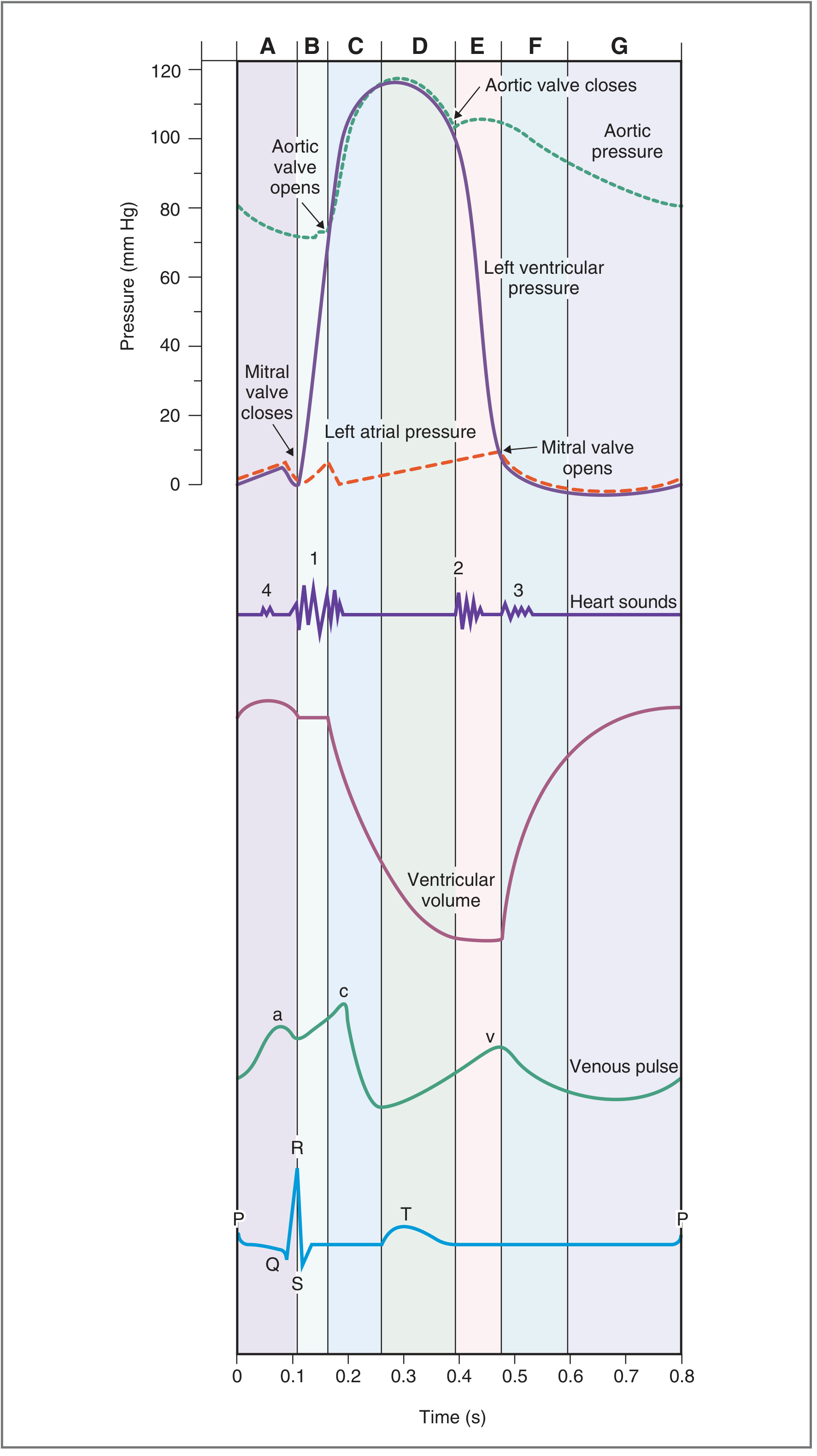
The Seven Sequential Phases
Phase A - Atrial Systole
| Parameter | Event |
|---|---|
| ECG | P wave (atrial depolarization) followed by PR interval |
| Valves | Mitral (AV) valve already open; aortic valve closed |
| Pressure | Left atrial pressure rises; small "blip" in LV pressure as blood is actively added |
| Volume | LV volume increases (final ~20-30% of ventricular filling) |
| Heart Sound | S4 (fourth heart sound) - not audible in normal adults; heard when ventricular compliance is decreased (e.g., LV hypertrophy) |
The atria act as primer pumps: atrial contraction produces the "a wave" on the venous (JVP) pulse. The ventricle was already filling passively before this phase - atrial systole contributes roughly the last 20% of end-diastolic volume (EDV). The EDV at end of this phase is approximately 120 mL.
Phase B - Isovolumetric Ventricular Contraction (IVC)
| Parameter | Event |
|---|---|
| ECG | QRS complex (ventricular depolarization) |
| Valves | Mitral valve closes (when LV pressure exceeds LA pressure); aortic valve still closed |
| Pressure | LV pressure rises steeply from ~10 mmHg toward ~80 mmHg; all other pressures relatively unchanged |
| Volume | Constant at ~120 mL (EDV) - both AV and semilunar valves are closed |
| Heart Sound | S1 ("lub") - caused by vibration of the taut mitral (and tricuspid) valve leaflets at closure, plus reverberation of blood and ventricular walls |
This is the most energy-expensive phase per unit time. The ventricle builds up pressure without shortening. S1 is split because the mitral valve closes slightly before the tricuspid valve. The "c wave" appears on the venous pulse, caused by bulging of the tricuspid valve into the right atrium.
Phase C - Rapid Ventricular Ejection
| Parameter | Event |
|---|---|
| ECG | ST segment (ventricular depolarization complete, repolarization not yet started) |
| Valves | Aortic valve opens (when LV pressure exceeds aortic diastolic pressure ~80 mmHg) |
| Pressure | LV pressure rises to its peak (~120 mmHg); aortic pressure rises to its maximum (~120 mmHg) |
| Volume | LV volume drops rapidly; most of the stroke volume (~70%) is ejected in this phase |
| Heart Sound | None |
Blood is driven from the LV into the aorta by a LV-to-aorta pressure gradient. Left atrial pressure begins to rise as blood returns from the lungs (filling for the next cycle). The venous "x descent" (tricuspid annulus pulled down) occurs here.
Phase D - Reduced Ventricular Ejection
| Parameter | Event |
|---|---|
| ECG | T wave (ventricular repolarization begins) |
| Valves | Aortic valve still open; mitral still closed |
| Pressure | LV pressure starts to fall (ventricle beginning to relax); aortic pressure also starts to fall as runoff into arteries exceeds inflow from ventricle |
| Volume | LV volume continues to decrease, but at a slower rate, reaching the end-systolic volume (ESV) ~50 mL |
| Heart Sound | None |
By the end of phase D, stroke volume = EDV - ESV = 120 - 50 = 70 mL. Ejection fraction = 70/120 = ~58%.
Phase E - Isovolumetric Ventricular Relaxation (IVR)
| Parameter | Event |
|---|---|
| ECG | After end of T wave (full repolarization) |
| Valves | Aortic valve closes (when LV pressure falls below aortic pressure); mitral valve still closed |
| Pressure | LV pressure falls steeply from ~100 mmHg to near zero; aortic pressure shows the dicrotic notch (incisura) at the point of aortic valve closure |
| Volume | Constant at ~50 mL (ESV) - all valves closed |
| Heart Sound | S2 ("dub") - caused by sudden closure and elastic recoil of the semilunar valves (aortic then pulmonary), producing blood reverberation in the aortic/pulmonary roots |
S2 is shorter (0.11 s) and higher pitched than S1 (0.14 s) because the semilunar valves are tauter than AV valves. Physiological splitting of S2 on inspiration: inspiration delays pulmonic valve closure (increased RV filling via Frank-Starling mechanism prolongs RV ejection), widening the A2-P2 interval. The venous "v wave" (atrial filling against closed tricuspid valve) peaks here.
Phase F - Rapid Ventricular Filling
| Parameter | Event |
|---|---|
| ECG | Isoelectric baseline |
| Valves | Mitral valve opens (when LV pressure falls below LA pressure) |
| Pressure | LV pressure low but constant (compliant relaxed ventricle); aortic pressure continues to fall as blood runs off to periphery |
| Volume | LV volume increases rapidly from ~50 mL to ~100 mL |
| Heart Sound | S3 (third heart sound) - caused by oscillation of blood reverberating between ventricular walls as blood rushes in from the atrium |
S3 is normal in children and young adults but in adults over ~40 it indicates volume overload (e.g., systolic heart failure, mitral/tricuspid regurgitation). The venous "y descent" occurs as the tricuspid valve opens. The atria had been filling throughout systole (phases B-E), so they are now full and empty rapidly into the ventricle.
Phase G - Reduced Ventricular Filling (Diastasis)
| Parameter | Event |
|---|---|
| ECG | Isoelectric baseline; cycle ends with next P wave |
| Valves | Mitral valve still open |
| Pressure | LV and LA pressures equalize at low values |
| Volume | LV volume increases gradually from ~100 to ~120 mL |
| Heart Sound | None (silence until next P wave triggers atrial systole) |
This is the quiet period where pressure and volume equilibrate across the open mitral valve. At faster heart rates, this phase is the first to be shortened or abolished.
Summary Table: All Phases at a Glance
| Phase | ECG Event | Valves | LV Pressure | LV Volume | Heart Sound |
|---|---|---|---|---|---|
| A - Atrial Systole | P wave | MV open, AoV closed | Slight rise | ↑ to EDV (120 mL) | S4 (pathological) |
| B - IVC | QRS | MV closes | ↑↑ (10→80 mmHg) | Constant (120 mL) | S1 |
| C - Rapid Ejection | ST segment | AoV opens | ↑ to max (~120 mmHg) | ↓↓ rapidly | None |
| D - Reduced Ejection | T wave | AoV open | ↓ slightly | ↓ to ESV (50 mL) | None |
| E - IVR | Post-T | AoV closes | ↓↓ (100→7 mmHg) | Constant (50 mL) | S2 + dicrotic notch |
| F - Rapid Filling | Isoelectric | MV opens | Low, constant | ↑↑ rapidly | S3 (normal in young) |
| G - Diastasis | Pre-P | MV open | Equalized, low | ↑ slowly to ~120 mL | None |
Pressure-Volume Loop
The P-V loop plots LV pressure (y-axis) against LV volume (x-axis), traversing counterclockwise. It captures all four phases geometrically:
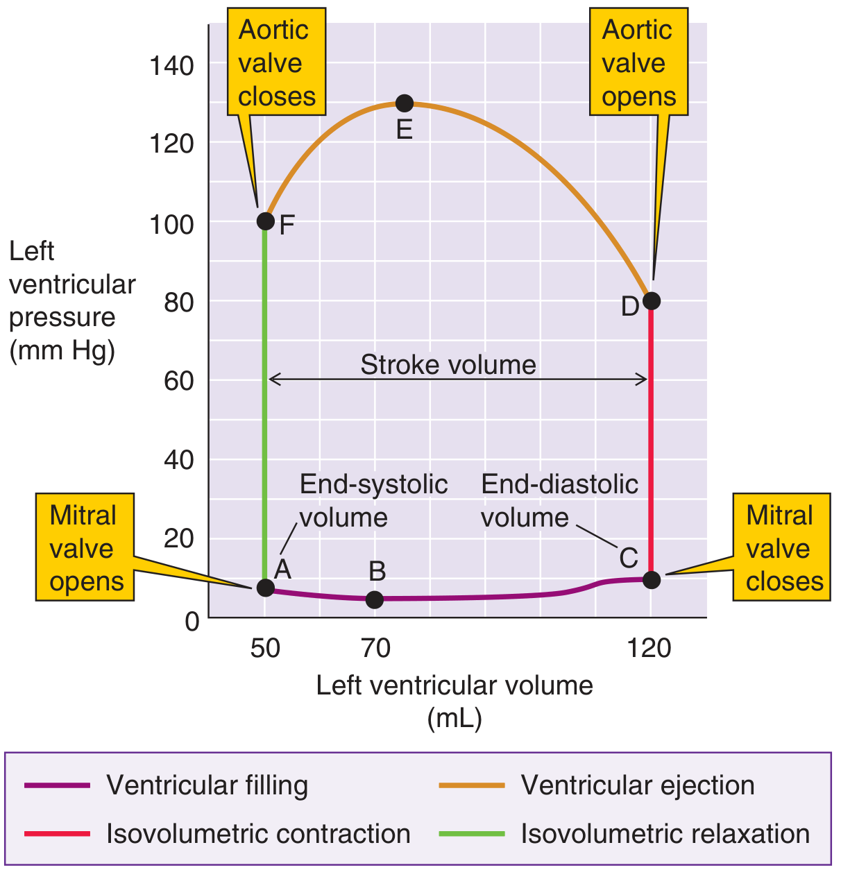
| Segment | Phase | Description |
|---|---|---|
| A → B | Early rapid filling | Mitral valve opens; volume rises 50→70 mL, pressure slightly falls (ventricle still relaxing) |
| B → C | Late filling (diastasis + atrial systole) | Volume rises 70→120 mL (EDV) with modest pressure rise (high compliance) |
| C → D | Isovolumetric contraction | Vertical line at 120 mL; pressure rises 10→80 mmHg (mitral closed, aortic closed) |
| D → E | Ventricular ejection (rapid + reduced) | Aortic valve open; volume falls 120→50 mL; pressure peaks at ~130 mmHg then falls |
| F → A | Isovolumetric relaxation | Vertical line at 50 mL; pressure falls 100→7 mmHg (both valves closed) |
The area enclosed within the loop = net stroke work done by the LV per beat. The width of the loop at any level = stroke volume (70 mL at baseline).
Heart Sounds: Mechanism Summary
| Sound | Timing | Cause | Clinical Note |
|---|---|---|---|
| S1 ("lub") | Start of systole (IVC) | Mitral (mainly) + tricuspid valve closure; vibration of taut leaflets, chordae, blood, and ventricular wall | Duration ~0.14 s; split = M1 before T1 |
| S2 ("dub") | End of systole (IVR) | Aortic + pulmonary valve closure; elastic recoil of semilunar leaflets; blood reverberates in aortic root | Duration ~0.11 s; higher pitch than S1; physiological splitting on inspiration (A2-P2) |
| S3 | Early diastole (rapid filling) | Blood oscillating between ventricular walls during rapid inflow; low frequency | Normal in children/<40 yrs; in adults = volume overload/systolic HF |
| S4 | Late diastole (atrial systole) | Atrium contracting against stiff/hypertrophied ventricle; very low frequency | Always pathological in adults; = decreased ventricular compliance |
Key Numbers to Remember (at 72 beats/min)
- Total cycle duration: 0.83 s
- Systole (B-E): ~0.3 s; Diastole (F-A): ~0.5 s
- EDV: ~120 mL | ESV: ~50 mL | Stroke volume: ~70 mL | EF: ~58%
- Peak LV systolic pressure: ~120 mmHg | Aortic diastolic pressure: ~80 mmHg
- Duration S1: ~0.14 s | Duration S2: ~0.11 s
Sources: Costanzo Physiology 7th Edition, pp. 160-162; Guyton and Hall Textbook of Medical Physiology, Chapters 9 & 23; Medical Physiology (Boron & Boulpaep), p. 761-762
Basal ganglion function connection and parkinson
The basal ganglia are a group of subcortical nuclei (striatum, globus pallidus, subthalamic nucleus, substantia nigra) that regulate voluntary movement via direct and indirect pathways. In the direct pathway, striatal neurons inhibit GPi/SNr, releasing the thalamus to activate the cortex (movement facilitation). In the indirect pathway, striatal neurons inhibit GPe, which disinhibits the STN, which then excites GPi/SNr, ultimately suppressing thalamic output (movement inhibition). Dopamine from the substantia nigra pars compacta (SNc) facilitates the direct pathway (D1 receptors) and inhibits the indirect pathway (D2 receptors), net effect = promoting movement. In Parkinson's disease, SNc neurons degenerate, dopamine is lost, the indirect pathway becomes overactive, and the thalamus is excessively suppressed, causing the hallmark hypokinetic features.
Searching sources...
Finding Sources
Finding Sources
Searching Images
basal ganglia direct indirect pathway dopamine Parkinson disease circuit diagram
Reading File
Reading File
Finding Sources
Finding Sources
Reading File
Reading File
Reading File
Finding Sources
Finding Sources
Reading File
Finding Sources
Finding Sources
Reading File
Reading File
Reading File
Excellent - I have a Harrison's basal ganglia circuit diagram. Let me fetch the key images and also read the Neuroanatomy through Clinical Cases section for the circuit description:
Loading Image
Loading Image
Reading File
I now have all the material needed for a comprehensive, well-illustrated answer. Let me compose the full response.
Basal Ganglia: Function, Connections, and Parkinson's Disease
1. Components and Anatomy
The basal ganglia are a collection of subcortical gray matter nuclei deep within the cerebral hemispheres. The main components are:
| Structure | Division | Role |
|---|---|---|
| Caudate nucleus | Input (striatum) | Receives cortical input; cognitive/limbic loops |
| Putamen | Input (striatum) | Receives cortical input; motor loop |
| Caudate + Putamen = Striatum (neostriatum) | Major input nucleus | All BG inputs arrive here |
| Globus Pallidus externa (GPe) | Intrinsic/relay | Intermediate relay in indirect pathway |
| Globus Pallidus interna (GPi) | Output | Major inhibitory output to thalamus |
| Subthalamic Nucleus (STN) | Intrinsic | Excitatory relay in indirect pathway |
| Substantia Nigra pars compacta (SNc) | Modulatory input | Dopaminergic; projects to striatum |
| Substantia Nigra pars reticulata (SNr) | Output | Acts in parallel with GPi; output to thalamus |
The striatum receives virtually all inputs. The GPi and SNr are the two major output nuclei. Their output is tonically inhibitory (GABAergic) to the thalamus - this is the fundamental principle on which the entire circuitry rests.
2. Functions of the Basal Ganglia
The basal ganglia do not directly activate muscles or project to the spinal cord. Their role is modulatory:
- Motor control - facilitation and inhibition of voluntary movements; initiation, execution, and termination of learned motor tasks
- Action selection - selecting one motor program while suppressing competing ones ("go/stop" gating)
- Motor learning and habit formation - procedural memory (how to ride a bike)
- Cognitive functions - working memory, attention, planning (via prefrontal loops)
- Limbic/affective functions - motivation, reward processing (via nucleus accumbens/ventral striatum)
- Oculomotor control - via the superior colliculus (SNr projections)
3. The Cortico-Basal Ganglia-Thalamo-Cortical Loop
All information flows in reentrant closed loops: Cortex → Striatum → Output nuclei → Thalamus → Cortex (back to the same cortical area that initiated the signal). There are at least five parallel loops:
| Loop | Cortical Origin | Striatal Region | Function |
|---|---|---|---|
| Motor | Motor/premotor cortex | Putamen | Voluntary limb movement |
| Oculomotor | Frontal eye fields | Caudate (body) | Saccadic eye movements |
| Prefrontal | Dorsolateral PFC | Caudate (head) | Spatial working memory |
| Orbitofrontal | Orbital/medial PFC | Caudate (ventromedial) | Motivation, social behavior |
| Limbic | Anterior cingulate, hippocampus | Nucleus accumbens/ventral striatum | Emotion, reward |
4. The Direct and Indirect Pathways
This is the central circuit model. The diagram below (from Stahl's Essential Psychopharmacology) shows the complete "go" (direct) and "stop" (indirect) pathways with their neurotransmitters:
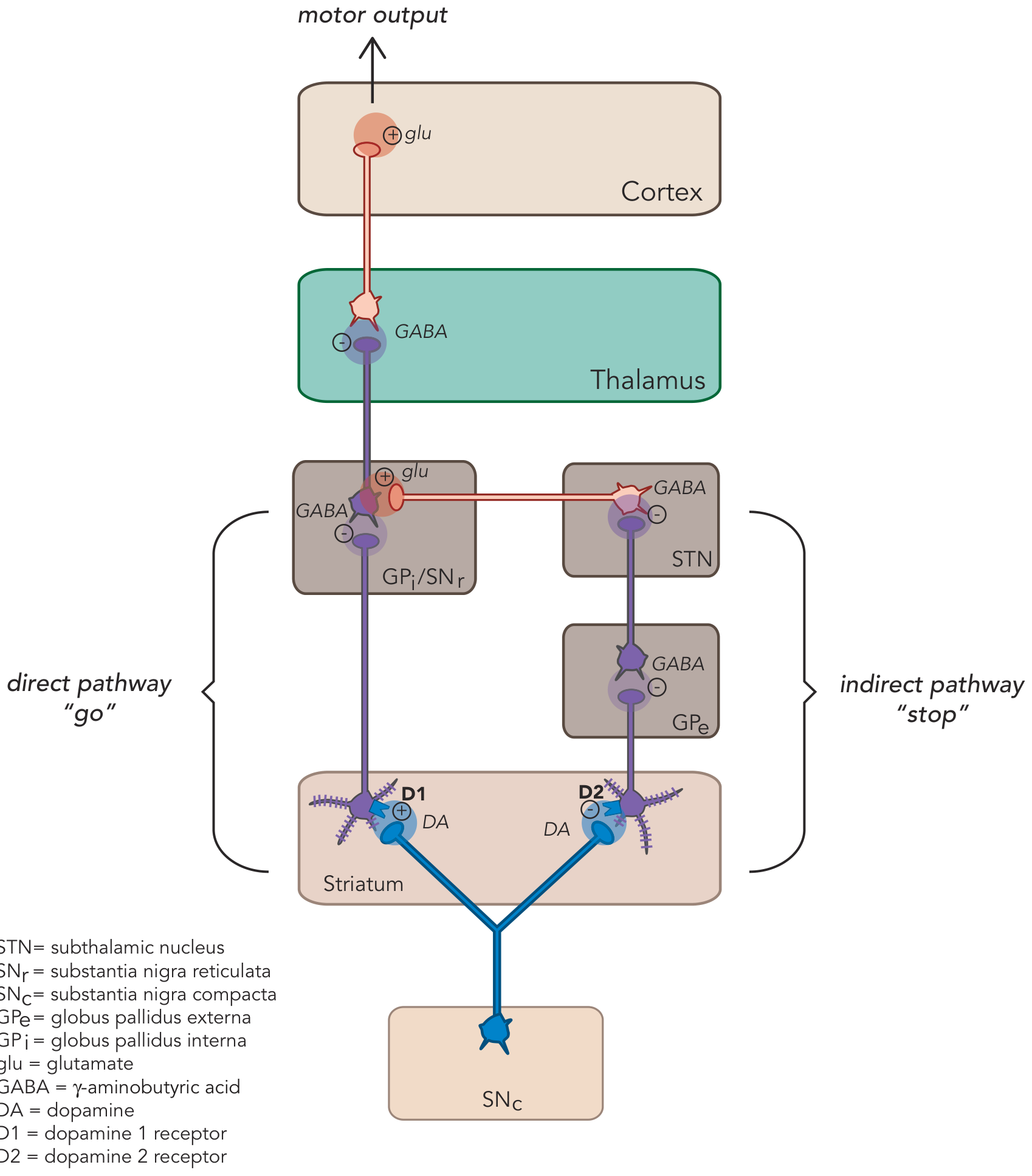
Direct Pathway ("Go" - Movement Facilitating)
Cortex →(+Glu)→ Striatum (D1 neurons) →(-GABA)→ GPi/SNr →(-GABA)→ Thalamus →(+Glu)→ Cortex
Step-by-step logic:
- Cortex excites striatal D1 neurons (+)
- Striatal D1 neurons inhibit GPi/SNr (-)
- GPi/SNr are now less active, so they release their inhibition of the thalamus (double negative = positive)
- Thalamus is now free to excite the motor cortex (+)
- Net effect: Movement is facilitated ("GO")
Neurotransmitters in direct pathway: striatal neurons contain GABA + Substance P
Indirect Pathway ("Stop" - Movement Inhibiting)
Cortex →(+Glu)→ Striatum (D2 neurons) →(-GABA)→ GPe →(-GABA)→ STN →(+Glu)→ GPi/SNr →(-GABA)→ Thalamus →(+Glu)→ Cortex
Step-by-step logic:
- Cortex excites striatal D2 neurons (+)
- Striatal D2 neurons inhibit GPe (-)
- GPe, now less active, releases its inhibition of STN (double negative = +)
- STN is disinhibited and actively excites GPi/SNr with glutamate (+)
- GPi/SNr are overactive and strongly inhibit the thalamus (-)
- Thalamus is suppressed, so less excitation reaches the cortex
- Net effect: Movement is suppressed ("STOP")
Neurotransmitters in indirect pathway: striatal neurons contain GABA + Enkephalin
Memory Trick
"Indirect Inhibits" - the indirect pathway inhibits movement; the direct pathway facilitates it. Their outputs are balanced and opposite.
5. Dopamine Modulation: The Master Regulator
Dopamine from SNc projects to the striatum (the nigrostriatal pathway) and acts on two populations of receptors with opposite effects:
| Receptor | Pathway | Effect of DA | Net Effect |
|---|---|---|---|
| D1 | Direct | Excites D1 neurons → activates direct pathway | ↑ Movement (Go) |
| D2 | Indirect | Inhibits D2 neurons → dampens indirect pathway | ↓ Stop signal (also = Go) |
Both D1 activation and D2 inhibition push in the same direction - toward movement facilitation. This is why dopamine = "pro-movement." Loss of dopamine from either mechanism tips the balance toward the stop signal.
6. Parkinson's Disease: The Circuit Fails
Pathology
In Parkinson's disease, dopaminergic neurons of the SNc degenerate progressively. By the time motor symptoms appear, approximately 60-80% of SNc neurons are already lost (and 80% of striatal dopamine is depleted). The degenerating neurons accumulate Lewy bodies (alpha-synuclein aggregates).
The diagram below (from Harrison's Principles of Internal Medicine, 22nd edition) contrasts the circuit in the normal state (A), PD (B), and levodopa-induced dyskinesia (C):
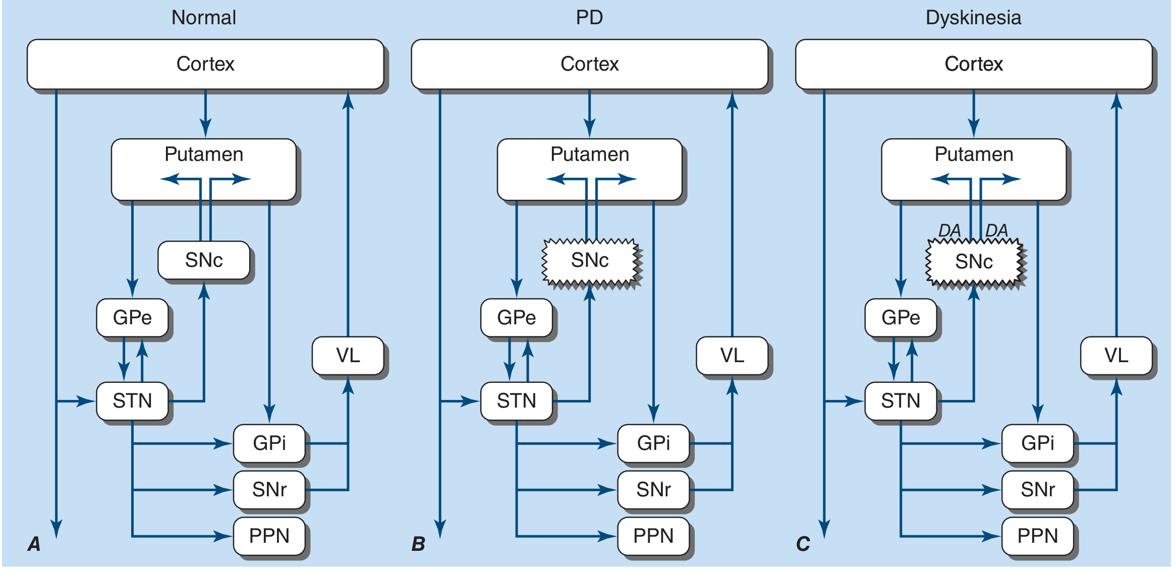
What Goes Wrong in the Circuit
Without dopamine:
| Pathway | Change | Consequence |
|---|---|---|
| Direct | Loss of D1 stimulation → striatum less active → GPi/SNr more active → thalamus more suppressed | Less "GO" signal |
| Indirect | Loss of D2 inhibition → striatum more active → GPe more inhibited → STN disinhibited → GPi/SNr more active (from two sources now) → thalamus even more suppressed | "STOP" signal dominates |
Bottom line: GPi/SNr become overactive, excessively inhibiting the thalamus, which then fails to excite the motor cortex adequately → hypokinesia/bradykinesia.
Additionally, the dopamine-ACh balance in the striatum is disrupted: dopamine normally inhibits ACh interneurons. Loss of dopamine leads to relative cholinergic excess in the striatum, contributing to tremor and rigidity. This is why anticholinergic drugs provide partial symptomatic relief.
Clinical Features (TRAP)
| Feature | Mechanism |
|---|---|
| Tremor (resting, 4-6 Hz, "pill-rolling") | Oscillations in basal ganglia-thalamo-cortical loop without adequate dopamine dampening |
| Rigidity (cogwheel/leadpipe) | Increased tonic contraction; loss of BG-mediated reciprocal inhibition |
| Akinesia/Bradykinesia | Failure of direct pathway "GO" signal; inability to initiate/scale movement |
| Postural instability | Loss of postural righting reflexes; cholinergic pedunculopontine nucleus (PPN) degeneration |
Other features from non-dopaminergic neurodegeneration (per Braak staging):
- Micrographia (small handwriting)
- Hypophonia (soft voice), dysphagia
- Anosmia (early - olfactory bulb affected first)
- REM sleep behavior disorder (RBD - often precedes motor symptoms)
- Autonomic dysfunction: orthostatic hypotension, constipation
- Cognitive impairment / PD dementia (late stages)
7. Braak Staging of PD Pathology
Lewy body pathology spreads in a predictable pattern (ascending):
| Stage | Regions Affected | Clinical Correlate |
|---|---|---|
| 1-2 | Olfactory bulb, dorsal motor nucleus of vagus | Anosmia, constipation, RBD (prodromal) |
| 3-4 | SNc, basal forebrain, amygdala | Motor symptoms emerge (Stage 3) |
| 5-6 | Neocortex (temporal, prefrontal) | Cognitive impairment, dementia |
8. Treatment Strategies (Targeting the Circuit)
| Treatment | Mechanism | Target |
|---|---|---|
| Levodopa (L-DOPA) | Dopamine precursor; crosses BBB, converted to DA | Restores D1/D2 signaling |
| Dopamine agonists (pramipexole, ropinirole) | Directly stimulate D2 (and D3) receptors | Bypass depleted SNc |
| MAO-B inhibitors (selegiline, rasagiline) | Reduce DA breakdown in striatum | Prolong synaptic DA |
| COMT inhibitors (entacapone) | Reduce peripheral L-DOPA breakdown | Extend L-DOPA effect |
| Anticholinergics (benztropine) | Reduce relative cholinergic excess | Address ACh-DA imbalance |
| Deep Brain Stimulation (DBS) of STN or GPi | High-frequency stimulation effectively inhibits overactive STN/GPi | Restores thalamic output |
| Levodopa-induced dyskinesia | Chronic D2 stimulation → hypersensitivity → GPi/STN underactive → thalamus over-excited | Diagram C above |
Summary: The Core Logic
NORMAL:     SNc-DA     → D1 activates direct ("GO") + D2 inhibits indirect ("STOP")
                       → Balanced thalamic output → normal movement

PARKINSON:  SNc-DA ↓↓  → Direct pathway WEAK + Indirect pathway OVERACTIVE
                       → GPi/SNr OVERACTIVE → Thalamus SUPPRESSED
                       → Motor cortex UNDER-ACTIVATED → Bradykinesia / Rigidity / Tremor
Sources: Costanzo Physiology 7th Edition; Neuroanatomy through Clinical Cases 3rd Edition; Harrison's Principles of Internal Medicine 22nd Edition; Stahl's Essential Psychopharmacology; Adams and Victor's Principles of Neurology 12th Edition; Bradley and Daroff's Neurology in Clinical Practice
This is a shared conversation. Sign in to Orris to start your own chat.