
35. What is Tracheostomy? What are the types, indications and complications? (10 Marks) (SMIMER)***** *Tracheostomy. (6 Marks) (NAMO) *Indications and types of Tracheostomy. (6 Marks) (GMC) *Tracheostomy - indications. (6 Marks) (KMCRI) *Complications of tracheostomy. (6 Marks) (VALMED)
Reading File
Now let me search the medical library for tracheostomy content:
Finding Sources
Finding Sources
Excellent results. Let me now read the most comprehensive sources - Sabiston (surgical textbook) and Scott-Brown's (ENT) plus Roberts & Hedges (complications):
Reading File
Reading File
Reading File
Now let me read the Sabiston section on complications and Scott-Brown's Vol 2 for the complete tracheostomy section:
Reading File
Reading File
Reading File
Now I have excellent content. Let me read the complications section from Sabiston more thoroughly and fetch the anatomy diagram image:
Reading File
Reading File
I now have comprehensive content from authoritative textbooks. Let me also fetch the anatomy image that was referenced:
Loading Image
I now have everything needed to write a comprehensive 10-mark exam answer. Here it is:
Tracheostomy
Definition
Tracheostomy is a surgical procedure in which an opening (stoma) is created through the anterior wall of the neck into the trachea (between the 2nd and 3rd, or 3rd and 4th tracheal rings), and a tube is inserted to maintain a patent airway. It is the most common surgical procedure performed in critically ill patients requiring prolonged mechanical ventilation.
- Roberts and Hedges' Clinical Procedures in Emergency Medicine, p. 203
- Sabiston Textbook of Surgery, p. 960
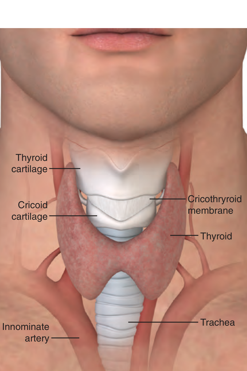
Types of Tracheostomy
1. Based on Urgency
| Type | Description |
|---|---|
| Emergency (Acute) Tracheostomy | Done urgently to secure a compromised airway (e.g., acute laryngeal obstruction, failed intubation) |
| Elective (Planned) Tracheostomy | Performed in a controlled setting for anticipated prolonged airway needs |
2. Based on Technique
A. Open (Surgical) Tracheostomy
- Traditional method performed in the operating room (or bedside)
- A horizontal skin incision is made, strap muscles are retracted, thyroid isthmus is divided if necessary, and a window is cut in the tracheal wall between the 2nd-3rd or 3rd-4th rings
- The anterior tracheal wall is sutured to the skin to maintain the tract
B. Percutaneous Dilatational Tracheostomy (PDT)
- Preferred in ICU patients; performed at the bedside under bronchoscopic guidance
- A Seldinger-based technique: needle insertion, guidewire passage, then serial or graduated dilation before tube placement
- First described by Ciaglia et al. in 1985
- Advantages over open: decreased wound infection, cost-effective, avoids OR transport
- Periprocedural mortality < 0.2%; major complication rate ~0.15%
- Meta-analyses show equivalence to open technique with fewer wound infections
3. Based on Duration/Intent
| Type | Feature |
|---|---|
| Temporary | For reversible conditions; tube removed after recovery (decannulation) |
| Permanent | After total laryngectomy; stoma is the definitive airway |
4. Based on Age Group
- Adult tracheostomy - standard technique
- Paediatric tracheostomy - tubes rarely have inflatable cuffs; higher risk of complications (granulation tissue, suprastomal collapse, subglottic stenosis)
Indications for Tracheostomy
Indications fall into three broad categories:
1. Upper Airway Obstruction
- Foreign body obstruction not amenable to retrieval
- Severe angioedema (anaphylaxis, hereditary)
- Upper airway tumors (larynx, hypopharynx)
- Significant maxillofacial trauma / bilateral mandible fractures
- Acute laryngeal infections (severe croup, epiglottitis) - when intubation fails
- Burns or inhalation injury causing supraglottic swelling
- Bilateral vocal cord palsy
2. Prolonged Mechanical Ventilation
- When a patient is predicted to require ventilatory support for >2 weeks
- Facilitates weaning from ventilator (reduced dead space, less resistance, less sedation)
- Easier nursing care and oral hygiene
- Allows speech with specialized valves (Passy-Muir)
3. Neurologic Conditions Preventing Safe Extubation
- Brain injury (acute TBI or progressive neurological disease)
- Spinal cord injury (especially cervical; patients with halo fixation)
- Severe agitation or delirium
- Prolonged altered mental status
- Poor cough reflex / inability to protect airway from aspiration
4. Other Specific Indications
- Head and neck surgery (laryngectomy, radical neck dissection)
- Elective protection of airway after major oral/pharyngeal surgery
- Sleep apnea (rare, refractory cases)
- Bronchial toilet - clearing secretions in patients who cannot cough effectively
(Source: Sabiston Textbook of Surgery, Table 47.1, p. 960)
Contraindications
Most contraindications are relative and often temporary:
| Contraindication |
|---|
| Recent anterior neck surgery (<7 days) |
| High ventilator settings (FiO₂ >50%, PEEP >10 cmH₂O) |
| Hemodynamic instability |
| Elevated intracranial pressure |
| Significant bleeding risk / coagulopathy |
| Local infection or malignancy at proposed site |
| Predicted early mortality |
Complications of Tracheostomy
Complications are classified as immediate, early (days to weeks), and late (>3 weeks).
16-20% of patients experience early complications; 6-8% experience late complications. Emergency tracheostomy carries a higher complication rate than elective.
A. Immediate (Intra-operative) Complications
| Complication | Notes |
|---|---|
| Hemorrhage | From anterior jugular veins, thyroid isthmus; commonest immediate complication |
| False passage / misplacement | Tube placed outside trachea - life-threatening |
| Apnea / cardiac arrest | Loss of hypoxic drive in COPD patients; vagal reflex |
| Hypoxia | Prolonged procedure time |
| Injury to adjacent structures | Recurrent laryngeal nerve, esophagus |
| Subcutaneous emphysema | Air tracking into neck tissues |
| Pneumothorax / pneumomediastinum | Especially in children or obese patients |
B. Early Complications (Days to Weeks)
| Complication | Notes |
|---|---|
| Hemorrhage (postoperative) | Secondary to vessel erosion |
| Tube dislodgement / displacement | Especially before tract matures (first 4-5 days); most dangerous early complication |
| Tube obstruction | Mucus plugging; prevented by regular humidification and suctioning |
| Subcutaneous emphysema | |
| Wound / soft tissue infection | |
| Pneumothorax, pneumomediastinum | |
| Aspiration | Cuff leak or swallowing dysfunction |
C. Late Complications (>3 Weeks)
| Complication | Notes |
|---|---|
| Tracheal stenosis | Most important late complication; incidence ~6% after PDT; subclinical stenosis in 40% |
| Tracheomalacia | Weakening and collapse of tracheal cartilage |
| Tracheo-innominate artery fistula | Rare but life-threatening; massive hemorrhage; occurs when cuff erodes into innominate artery (usually at day 7-14) |
| Tracheo-esophageal fistula | From posterior wall erosion; presents with aspiration |
| Granulation tissue formation | Can cause obstruction |
| Infection | Pneumonia, aspiration, stomal infection |
| Equipment failure | Cuff leak, tube fracture |
| Dysphagia and swallowing difficulty | |
| Difficult decannulation | Due to stoma scarring or patient dependency |
| Persistent tracheo-cutaneous fistula | Stoma fails to close after decannulation |
Summary Table
| Aspect | Key Points |
|---|---|
| Definition | Surgical opening into trachea for airway access |
| Site | Between 2nd-3rd or 3rd-4th tracheal rings |
| Types | Emergency/Elective; Open/Percutaneous; Temporary/Permanent |
| Main Indications | Airway obstruction, prolonged ventilation, neurological |
| Most dangerous immediate complication | Hemorrhage / false passage |
| Most important early complication | Tube displacement (before day 5) |
| Most important late complication | Tracheal stenosis / tracheo-innominate fistula |
Sources:
- Sabiston Textbook of Surgery, Chapter 47 (TRACHEOSTOMY), pp. 960-961
- Roberts and Hedges' Clinical Procedures in Emergency Medicine, Chapter 7, pp. 202-204
- Scott-Brown's Otorhinolaryngology Head & Neck Surgery Vol 1, p. 405
- Scott-Brown's Otorhinolaryngology Head & Neck Surgery Vol 2, pp. 395-396
This is a shared conversation. Sign in to Orris to start your own chat.