

Give a diag acc to first year mbbs books
structure of eyeball labeled diagram anatomy cross section
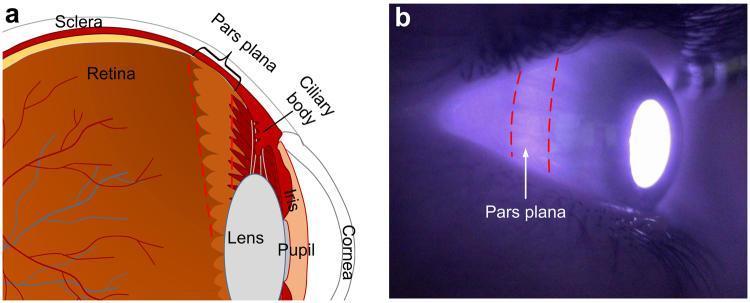
This composite educational graphic details the anatomy and visualization of the pars plana for ophthalmic imaging. (a) An anatomical diagram shows a cross-section of the human eye. The pars plana is identified as the smooth, posterior portion of the ciliary body, located between the retinal ora serrata and the ciliary processes. Key labeled structures include the sclera, retina with vasculature, lens, iris, pupil, and cornea. (b) A clinical photograph displays the eyeball under near-infrared (850 nm) transillumination. The image demonstrates the high transmittance of the pars plana, which appears as a distinctively bright, circumferential band approximately 3–4 mm posterior to the limbus, demarcated by red dashed lines. This educational material illustrates the clinical significance of the pars plana as a strategic anatomical window for trans-scleral illumination in non-mydriatic fundus photography due to its relative lack of muscle, blood vessels, and heavy pigmentation compared to surrounding ocular tissues.
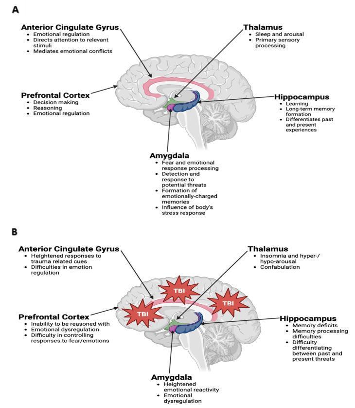
Educational comparison diagram illustrating the functional anatomy of the limbic system in health and post-traumatic brain injury (TBI). Part A (Normal State) displays a sagittal cross-section of the human brain, identifying five key structures: the anterior cingulate gyrus (emotional regulation, attention), prefrontal cortex (decision-making, reasoning), amygdala (fear processing, threat detection), thalamus (sleep, arousal), and hippocampus (learning, long-term memory). Each structure is color-coded and spatially mapped within the medial cerebrum. Part B (Post-TBI State) utilizes the same anatomical template but includes red 'TBI' impact icons localized to the frontal and parietal regions. This section details pathological functional changes, such as emotional dysregulation in the prefrontal cortex and amygdala, heightened trauma responses in the anterior cingulate gyrus, sleep disturbances (insomnia) in the thalamus, and memory deficits or threat differentiation difficulties in the hippocampus. The diagram serves as a clinical education tool to explain the pathophysiology of neuropsychiatric sequelae, such as PTSD, following physical or psychological trauma.
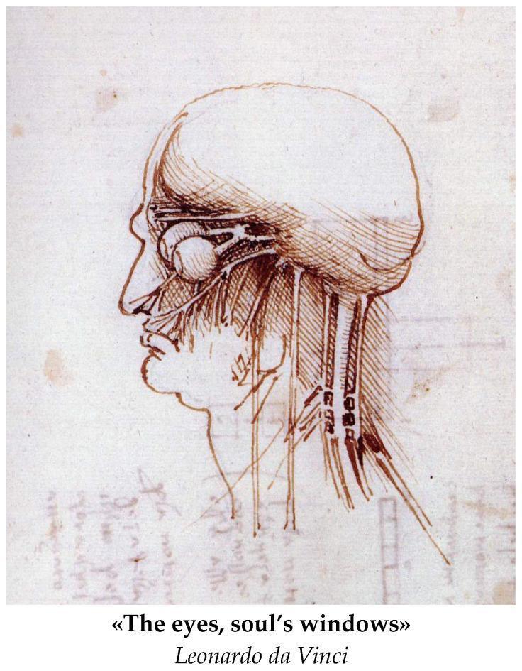
Anatomical Diagram: A historical medical illustration by Leonardo da Vinci, depicting a right lateral profile of the human head and neck. The drawing serves as an early study of gross anatomy and neuroanatomy. It features a detailed representation of the orbit, showing the eyeball as a spherical structure with attached extraocular muscles or neural pathways extending posteriorly toward the brain. The cranium is outlined, with fine hatching and cross-hatching used to provide topographical depth to the skull and facial features. The nasal cavity and oral region are visible in profile. Extending inferiorly from the base of the skull through the neck are several vertical, parallel structures representing major vessels (carotid arteries/jugular veins) and possibly nerves or muscular bundles. The illustration highlights the early Renaissance focus on understanding the relationship between surface anatomy and underlying physiological structures, particularly the visual system's connection to the central nervous system.
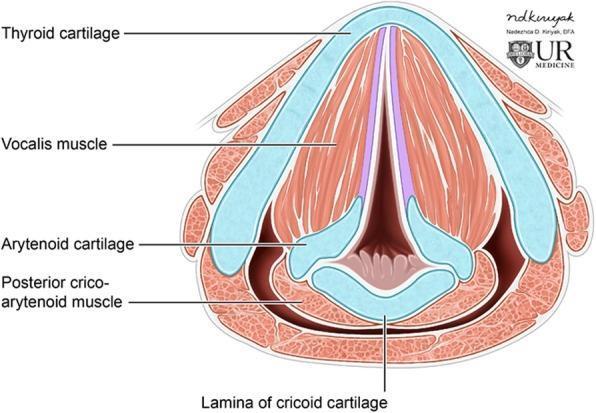
Anatomical diagram of the larynx in axial cross-section at the level of the glottis, illustrating the spatial relationship between cartilaginous structures and intrinsic musculature. The thyroid cartilage forms a prominent, V-shaped anterior boundary. Located posteriorly and inferiorly is the lamina of the cricoid cartilage, which supports the paired arytenoid cartilages. The true vocal folds extend from the anterior midline of the thyroid cartilage to the vocal processes of the arytenoid cartilages, creating the glottic opening. Key intrinsic muscles are labeled, including the vocalis muscle, which runs parallel within the vocal folds, and the posterior cricoarytenoid muscle, situated at the posterior aspect of the cricoid lamina. This illustration is optimized for educational use in otolaryngology and radiology, specifically for correlating axial CT imaging anatomy with laryngeal structure and function, such as vocal fold apposition and glottic patency.
eyeball cross section layers coats fibrous vascular nervous tunic anatomy
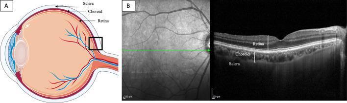
This composite educational graphic illustrates human ocular anatomy and diagnostic imaging. Section A presents a sagittal schematic diagram of the eye, highlighting the three posterior layers: the retina (inner), the choroid (middle vascular layer), and the sclera (outer fibrous layer). A black box indicates the posterior pole region being analyzed. Section B features a Spectral Domain Optical Coherence Tomography (SD-OCT) scan centered on the fovea. The left side of panel B shows a grayscale infrared fundus image with a green horizontal line indicating the scan acquisition path. The right side displays the corresponding cross-sectional OCT B-scan, which provides high-resolution visualization of the retinal architecture, the underlying choroidal layer with its characteristic vascular luminal and stromal spaces, and the interface with the sclera. Labels on the B-scan confirm the stratified arrangement of the retina, choroid, and sclera, making this a foundational resource for understanding posterior segment imaging and choroidal thickness assessment.
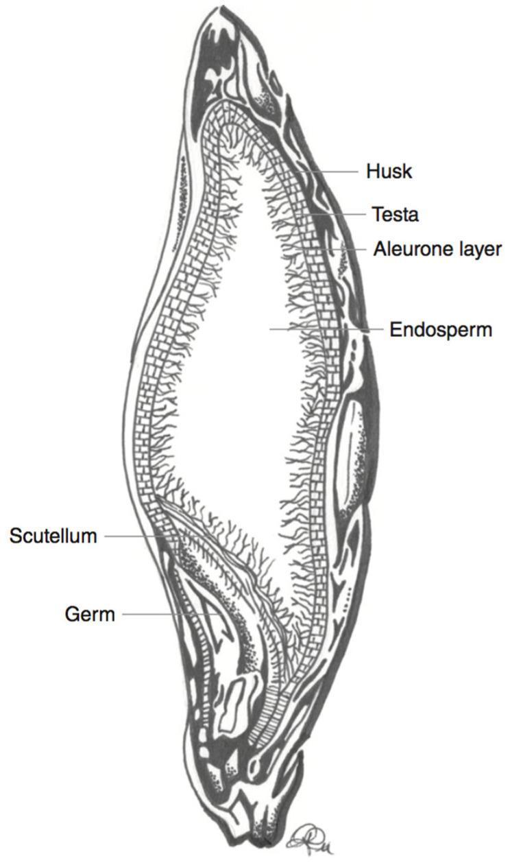
This medical-botanical schematic illustration depicts a longitudinal cross-section of a barley kernel (Hordeum vulgare), serving as a model for cereal grain anatomy relevant to human nutrition and dietetics. The diagram identifies key structural components involved in nutrient distribution, specifically the localization of anthocyanins and fiber. The anatomical layers are labeled from the periphery to the interior: the outermost fibrous Husk, the thin protective Testa, and the Aleurone layer composed of structured, brick-like cells. The largest central region is the Endosperm, characterized by a grainy, fibrous texture. At the base of the kernel, the embryo-related structures, the Scutellum and the Germ, are clearly demarcated. This diagram is educationally significant for understanding the localization of bioactive compounds like phenolic pigments, which are primarily concentrated in the outer layers (pericarp and aleurone) and are often lost during commercial milling processes.
Low-power brightfield histology section stained with hematoxylin and eosin reveals multiple well-circumscribed, smooth-surfaced nodules composed of dense eosinophilic hyalinized fibroconnective tissue embedded within the tunica of a vascular wall. The nodules are sparsely cellular, containing few fibroblasts within a homogeneous, acellular-appearing extracellular matrix rich in collagen. Surrounding tunica shows fibrous thickening with accentuated eosinophilia and limited intervening stroma. No necrosis, calcification, hemorrhage, or acute inflammatory infiltrate is evident. The overall pattern is characteristic of hyalinization and fibrous remodeling of a vascular tunic, more consistent with chronic degenerative change or organized scar tissue than with neoplastic processes. The nodules’ smooth, rounded margins and lack of cellular atypia argue against malignant vascular tumors. Diagnostic significance rests on recognizing hyalinized nodules within tunica as a remodeling or scar-related phenomenon; correlation with clinical vascular risk factors and gross anatomy is advised. Differential considerations include hyaline arteriolosclerosis, fibrous intimal hyperplasia, fibrous nodules in tunica, and organized scar tissue of prior injury. Clinically, this finding could reflect long-standing hypertension or diabetes with vascular remodeling. Educational and reference material usage includes histology teaching, QA pathology slides, and comparative studies of vascular wall pathology. This image serves as a concise reference for teaching vascular histomorphology.
labeled diagram eyeball anterior posterior segment lens vitreous humor optic nerve
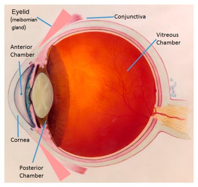
This medical anatomical diagram provides a cross-sectional view of the human eye, illustrating key ocular structures and chambers. In the anterior segment, labeled components include the eyelid with the meibomian gland, the cornea, and the fluid-filled anterior and posterior chambers. The iris and crystalline lens are visible, demarcating the boundary between the anterior and posterior sections. The posterior segment is dominated by the vitreous chamber, depicted with a reddish-orange hue representing the fundus. Within this chamber, the retinal vasculature is shown branching across the inner surface, with the optic nerve head visible at the posterior pole. Additional labels identify the conjunctiva and the layers of the ocular wall. This illustration is designed for basic to intermediate medical education, emphasizing the spatial relationships between ocular compartments and providing clinical context for conditions such as meibomian gland dysfunction, dacryocystitis, and retinal health.
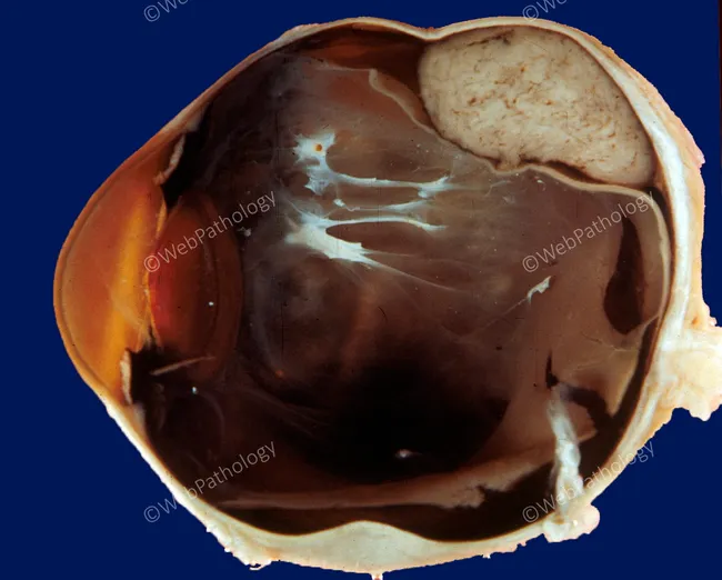
Comprehensive Description: Gross pathology photograph of an enucleated eyeball, oriented in sagittal (anterior–posterior) section. The globe reveals a large, dark brown posterior chamber with exposed sclera and choroid; the retina shows extensive detachment with folded, ribbon-like fibrous membranes extending from the periphery toward the posterior pole. Subretinal white-to-creamy ridges represent fibrous scarring or subretinal exudates. The vitreous cavity is relatively clear in this view, with minimal inflammatory debris; the lens is partially visible on the left, while the optic nerve stump is at the posterior aspect. The anterior segment is not fully captured in this slice. Overall, findings are compatible with advanced rhegmatogenous retinal detachment with secondary fibrovascular proliferation and traction. No acute suppurative endophthalmitis is evident in this section, though chronic inflammatory changes cannot be excluded. The image illustrates disruption of retinal architecture, detachment geometry, and posterior segment pathology that can produce severe, permanent vision loss. Clinically, this supports history of long-standing retinal detachment or intraocular process necessitating enucleation for blind painful eye, trauma, or suspicion of intraocular tumor. Educational relevance includes gross-pathology to histology correlation and informs ophthalmology education, retinal pathology, and surgical decision-making. This image also serves as a teaching aid for histopathology correlation in academic and clinical settings.
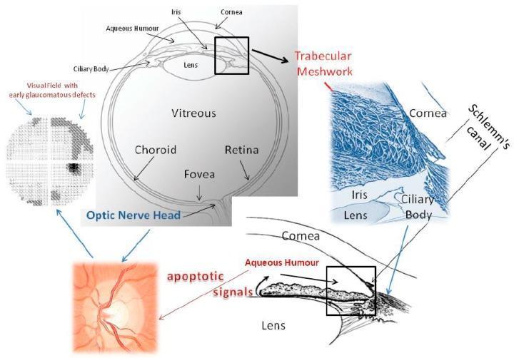
This composite educational diagram illustrates the pathophysiology of glaucoma, focusing on the aqueous humor outflow pathway and its impact on the posterior segment. A central cross-sectional anatomical diagram identifies the cornea, iris, lens, ciliary body, vitreous, retina, fovea, and optic nerve head. An inset provides a high-magnification view of the iridocorneal angle, detailing the trabecular meshwork and Schlemm’s canal. A 3D blue-tinted microscopic representation highlights the fibrous structure of the trabecular meshwork. The diagram demonstrates the functional relationship between malfunctioning trabecular meshwork and the generation of 'apoptotic signals' that travel to the optic nerve head. The clinical consequences are represented by a fundus photograph of an optic nerve head with neuroretinal rim thinning and a corresponding visual field plot showing early glaucomatous arcuate defects. Arrows indicate the flow of aqueous humor and the progression of damage from the anterior chamber to the retinal ganglion cells, emphasizing the mechanio-biological link between increased intraocular pressure and irreversible visual loss.
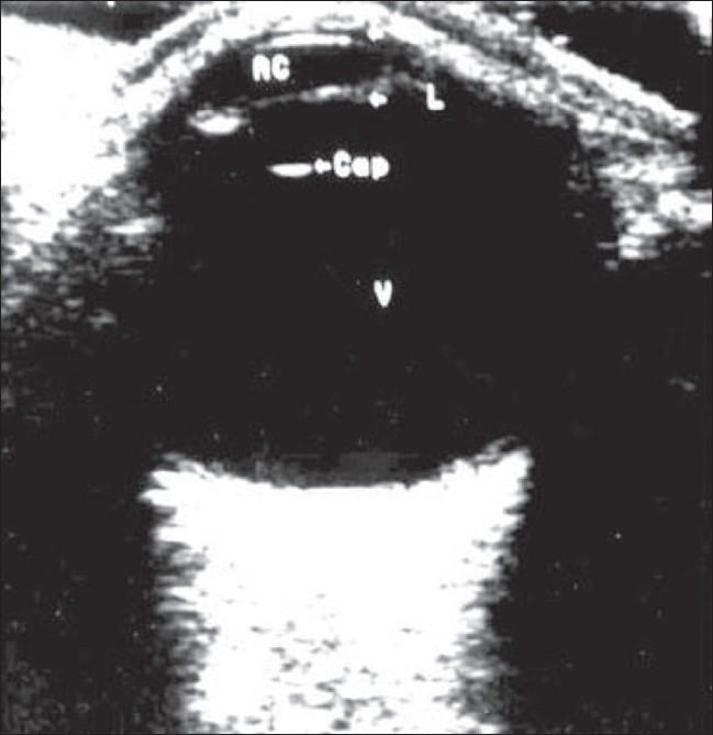
**Imaging Modality:** Ophthalmic B-scan ultrasonography. **Anatomical Region:** Globe (eyeball), axial cross-section. **Observations:** The image demonstrates the normal anatomical structures of the eye. The anterior segment displays the anterior chamber (labeled AC) and the crystalline lens (L). A distinct, curvilinear hyperechoic signal represents the posterior lens capsule (labeled ‘cap’). The posterior segment is dominated by a large, homogeneous, anechoic (black) space representing a clear vitreous chamber (V), devoid of opacities, membranes, or hemorrhages. The posterior wall shows a smooth, continuous hyperechoic curvilinear interface corresponding to the retina-choroid-sclera complex. **Characteristic Visual Features:** - **Anechoic Vitreous:** Indicates the absence of vitreous syneresis, debris, or posterior vitreous detachment (PVD). - **Lens Landmarks:** Clear visualization of the lens boundaries and the acoustic interface of the posterior capsule. - **Intact Posterior Contour:** Maintains a normal spherical configuration without evidence of staphyloma, retinal detachment, or choroidal thickening. **Clinical Significance:** This image serves as a baseline reference for normal ocular ultrasonography, used to rule out intraocular pathology such as vitreous hemorrhage, retinal detachment, or intraocular tumors.
Structure of the Eyeball - Labeled Diagram
Labeled Diagram
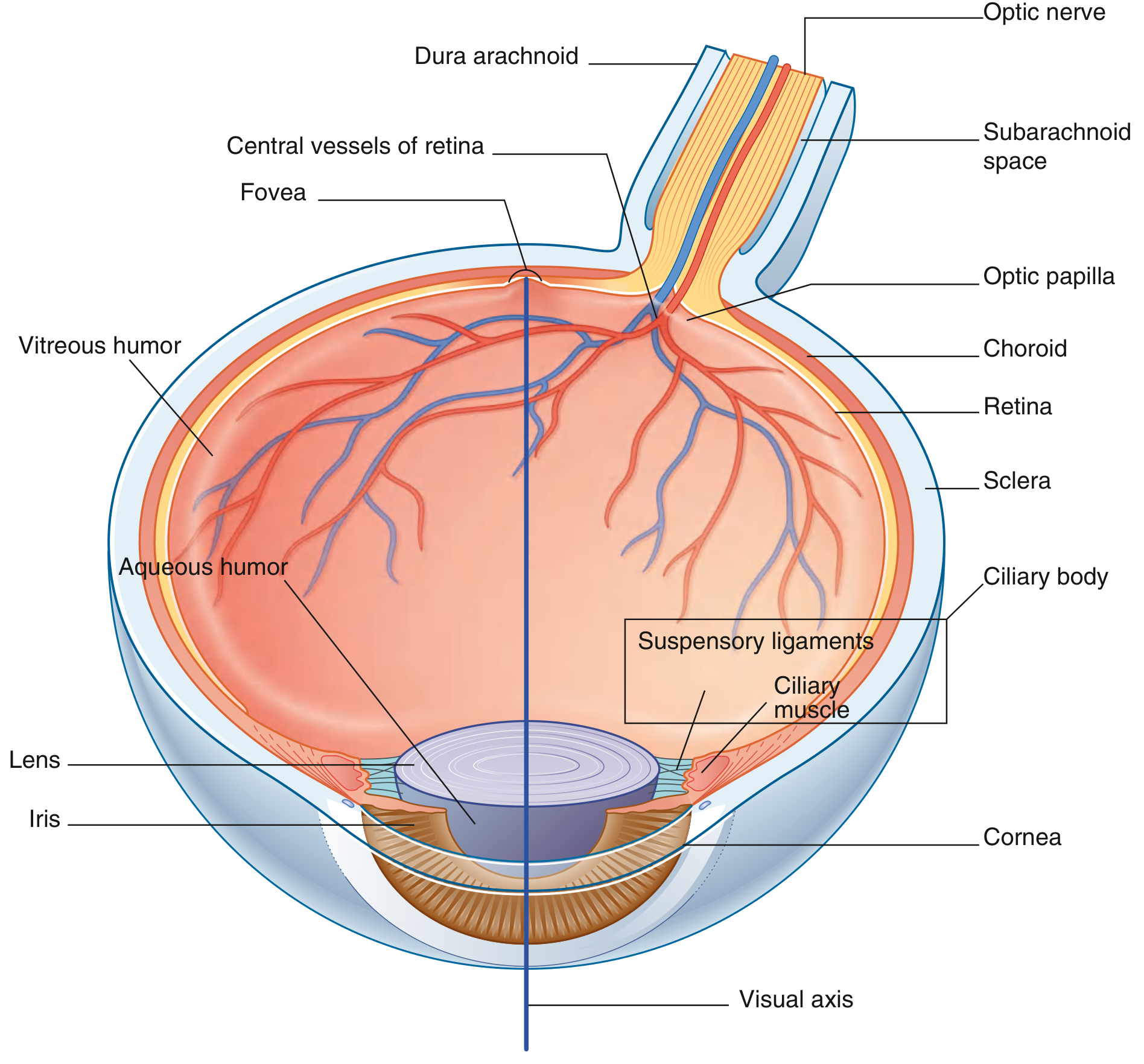
Overview
THREE COATS (TUNICS) OF THE EYEBALL
1. Fibrous Coat (Outer Layer)
| Structure | Details |
|---|---|
| Sclera (posterior 5/6) | Dense white fibrous tissue; maintains shape; gives attachment to extraocular muscles |
| Cornea (anterior 1/6) | Transparent, avascular; refracts light entering the eye; most powerful refracting medium |
- The junction between sclera and cornea is the corneoscleral junction (limbus)
2. Vascular Coat / Uveal Tract (Middle Layer)
| Structure | Details |
|---|---|
| Choroid | Highly vascular; nourishes outer retinal layers; contains melanin (absorbs stray light) |
| Ciliary body | Contains ciliary muscle + ciliary processes; produces aqueous humor; controls lens shape (accommodation) via suspensory ligaments (zonule of Zinn) |
| Iris | Coloured diaphragm with central aperture = pupil; contains sphincter pupillae (miosis - CN III) and dilator pupillae (mydriasis - sympathetic) |
3. Nervous Coat (Inner Layer)
| Structure | Details |
|---|---|
| Retina | Innermost layer; contains photoreceptors (rods and cones); converts light to nerve impulses |
| Optic disc (optic papilla) | Point where optic nerve exits; no photoreceptors = blind spot |
| Macula lutea | Area of maximum visual acuity; centre = fovea centralis |
| Pigment epithelium | Absorbs and refracts light; interdigitates with photoreceptors |
REFRACTIVE MEDIA (Contents of the Eyeball)
| Medium | Location | Function |
|---|---|---|
| Cornea | Anterior | Most powerful refraction (~43 D) |
| Aqueous humor | Anterior and posterior chambers | Nourishes avascular cornea and lens; maintains intraocular pressure |
| Lens | Behind iris, in front of vitreous | Biconvex; focuses light (accommodation) |
| Vitreous humor | Posterior to lens | Gel-like; maintains shape; supports retina |
CHAMBERS OF THE EYE
- Anterior chamber - between cornea and iris; filled with aqueous humor
- Posterior chamber - between iris and lens; filled with aqueous humor
- Vitreous chamber - posterior to lens; filled with vitreous humor (largest compartment)
OPTIC NERVE
- Exits the eyeball posteromedially
- Surrounded by dura mater, arachnoid, and pia mater (meningeal sheaths)
- Central retinal artery and vein run within it
- The subarachnoid space around it is continuous with the cranial subarachnoid space
PHOTORECEPTORS (Retinal Cells) - AN 41.3
| Rods | Cones | |
|---|---|---|
| Number | ~120 million | ~6 million |
| Location | Peripheral retina | Concentrated at fovea |
| Function | Dim light / night vision | Color vision / high acuity |
| Pigment | Rhodopsin (visual purple) | Iodopsin (3 types: red, green, blue) |
Quick Memory Aid (3 Coats)
Fibrous (Sclera + Cornea) → Vascular (Choroid + Ciliary body + Iris) → Nervous (Retina)
Another diag easy
simple labeled eye anatomy diagram cornea iris lens retina sclera choroid
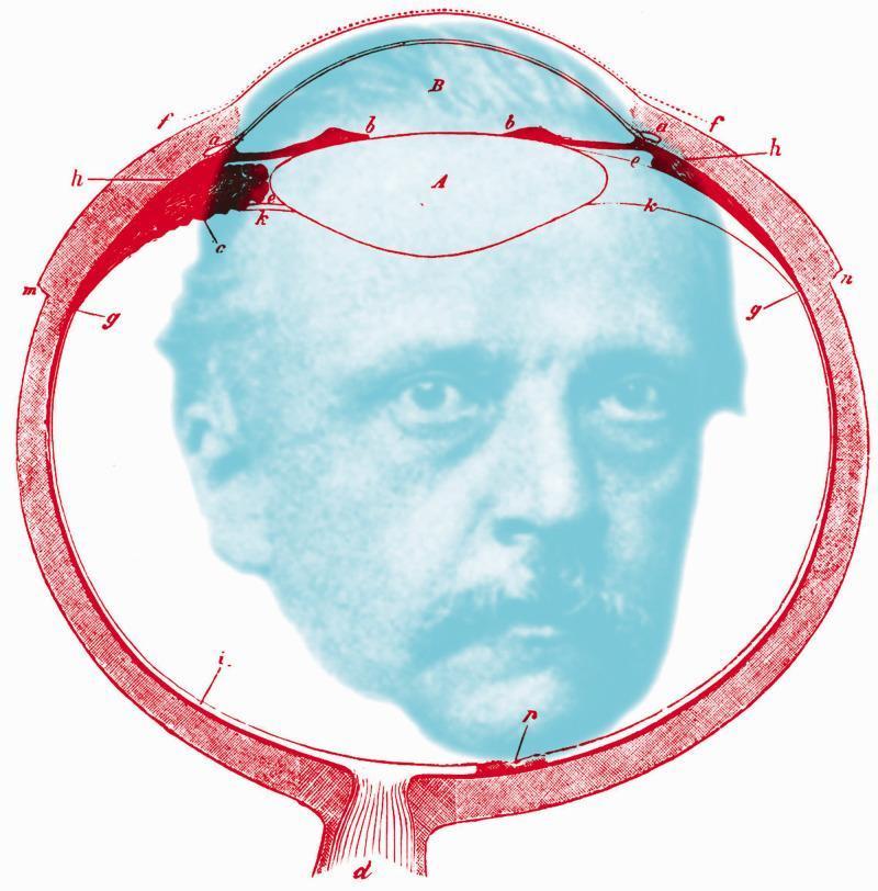
This educational illustration is an anatomical diagram of the human eye in sagittal cross-section, overlaid with a portrait of Hermann von Helmholtz. The diagram, rendered in red linework, depicts essential ophthalmic structures including the cornea (labeled B), the crystalline lens (labeled A), and the ciliary body (b). Various alphabetical annotations (c, d, e, f, g, h, i, k, m, n) indicate specific histological and anatomical regions, such as the sclera, retina, choroid layers, and the optic nerve exit (d). The lens is shown in its anatomical position behind the anterior chamber and iris. The image serves as a conceptual tribute to physiological optics, bridging the anatomy of vision with the historical figure responsible for major advancements in the field, such as the invention of the ophthalmoscope and the primary treatise on physiological optics. The spatial arrangement emphasizes the internal ocular environment, with the retinal lining and posterior segment framing the central portrait.
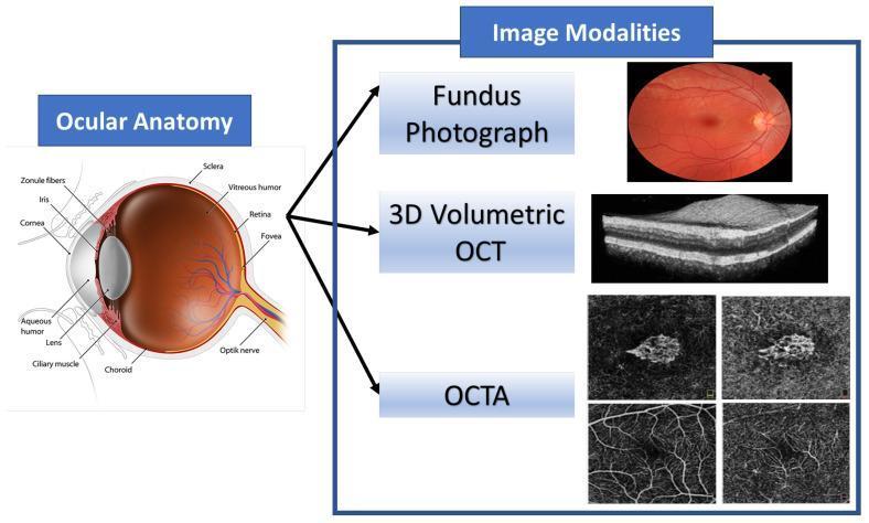
This educational graphic combines an anatomical diagram of the human eye with corresponding ophthalmic imaging modalities. The 'Ocular Anatomy' section features a detailed sagittal cross-section identifying key structures: the cornea, aqueous humor, iris, pupil, biconvex lens, zonule fibers, and ciliary muscle in the anterior segment; and the vitreous humor, retina, fovea, choroid, sclera, and optic nerve in the posterior segment. The 'Image Modalities' section illustrates diagnostic tools used to visualize these structures. A color Fundus Photograph provides a surface view of the retina, optic disc, and vasculature. A 3D Volumetric Optical Coherence Tomography (OCT) scan displays a high-resolution, grayscale cross-sectional view of retinal layers, highlighting tissue stratification. Finally, Optical Coherence Tomography Angiography (OCTA) panels show non-invasive, depth-resolved images of the retinal and choroidal microvasculature. This composite serves as a reference for correlating anatomical landmarks with clinical diagnostic imaging used in the management of retinal diseases such as Age-related Macular Degeneration (AMD).
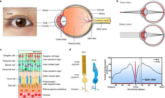
Educational medical illustration of ocular anatomy and the visual process. (a) A clinical photograph of a human eye paired with a sagittal schematic of the eyeball, labeling the cornea, sclera, iris/pupil, lens, ciliary body, vitreous body, choroid, retina, optic disc, and optic nerve. (b) A comparison of visual accommodation, showing the lens thickening for close vision and thinning for distant vision to focus light precisely on the retina. (c) A cross-sectional anatomical diagram of the retina, detailing the cellular layers from the choroid and retinal pigment epithelium through the photoreceptor layer (rods and cones), horizontal, bipolar, amacrine, and ganglion cell layers. (d) A specialized illustration of rod and cone cell morphology (left) and a comparative line graph showing photoreceptor density across the retinal eccentricity. The graph highlights the sharp peak of cone density at the fovea and the high density of rods in the peripheral (nasal and temporal) retina, with both photoreceptor types absent at the optic disc (blind spot).
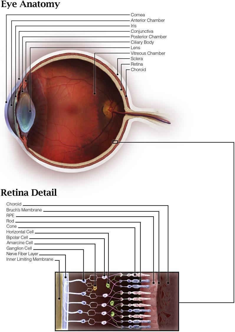
This medical illustration presents a detailed anatomical overview of the human eye, featuring a primary cross-sectional diagram and a magnified 'Retina Detail' inset. The main diagram identifies key anterior structures including the cornea, anterior chamber, iris, and lens, alongside the posterior vitreous chamber. The globe's wall is depicted in three distinct layers: the outer sclera, the vascular choroid, and the inner light-sensitive retina. The optic nerve is visible at the posterior pole. The lower 'Retina Detail' inset provides a high-resolution view of retinal microanatomy, illustrating the stratified organization from the outer choroid and Bruch's membrane to the inner limiting membrane. This section highlights the sequence of retinal pigment epithelium (RPE), photoreceptors (rods and cones), and the complex neural network involving horizontal, bipolar, amacrine, and ganglion cells. The nerve fiber layer is shown as the innermost cellular component, conveying signals toward the optic nerve. This illustration serves as an educational resource for understanding ocular histology and the blood-retinal barrier.
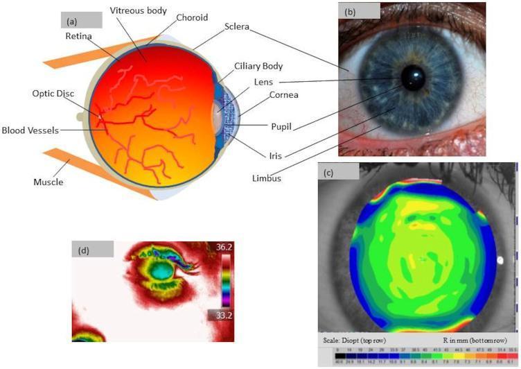
This composite educational image illustrates the anatomical and physiological features of a healthy human eye. Panel (a) is a cross-sectional anatomical diagram labeling key structures: retina, optic disc, blood vessels, vitreous body, choroid, sclera, ciliary body, lens, cornea, pupil, iris, and limbus. Panel (b) shows a high-resolution clinical photograph of the anterior segment, highlighting the iris pattern, pupil, and corneal clarity. Panel (c) is a corneal topography map displaying the anterior curvature in Diopters (refractive power) and millimeters (radius of curvature), with color-coded gradients indicating surface regularities. Panel (d) presents an infrared thermographic image showing surface temperature distribution. It highlights a physiological gradient where the central cornea is cooler (approximately 33.2°C) compared to the vascularized scleral and periocular regions (reaching 36.2°C). This multi-modal representation serves to teach ocular anatomy, corneal geometry, and thermal physiology for medical and optometric education.
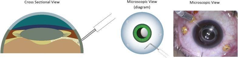
This composite educational graphic illustrates the surgical technique of pneumatic dissection of the anterior hyaloid membrane (AHM) during ophthalmic surgery. It consists of three panels: a cross-sectional diagram, a top-down microscopic diagram, and a real-time intraoperative surgical photograph. The cross-sectional view details the ocular anatomy, including the cornea, anterior chamber, iris, crystalline lens, and the posterior segment layers (sclera and choroid/retina). A 30-gauge needle is shown entering through the pars plana, with the tip positioned posterior to the lens. The top-down diagram illustrates the needle orientation relative to the iris and limbus. The clinical photograph demonstrates the actual surgical field, showing an eye with multiple sclerotomy cannulas (trocars) in place and a needle-mounted syringe performing the air injection near the pupillary margin. The primary educational focus is on the safe entry and positioning of the needle tip behind the lens within the vitreous cavity to facilitate AHM dissection while avoiding iatrogenic damage to the lens or retina.
Diagram 1 - Full Eyeball with Retinal Layers + Photoreceptors
- Panel a - External eye + cross-section with all key labels (sclera, choroid, retina, lens, cornea, pupil, ciliary body, vitreous body, optic disc, optic nerve)
- Panel b - Accommodation - lens shape for close vs. distant vision
- Panel c - All retinal cell layers in order (must know for AN 41.3)
- Panel d - Rod vs. cone distribution graph (rods = periphery, cones = fovea)
Diagram 2 - Clean Cross-Section with All Structures Labeled
For your record/notes, draw the cross-section and label these in order (outer to inner):
Give
What do you want me to give you?
A clean, detailed anatomical diagram of a human eyeball in horizontal cross-section for first year MBBS anatomy students. Show a sagittal cut through the middle of the eyeball. Clearly label all the following structures with neat leader lines: Cornea, Sclera, Iris, Pupil, Lens, Ciliary body, Ciliary muscle, Suspensory ligaments (zonule of Zinn), Anterior chamber (aqueous humor), Posterior chamber, Vitreous humor, Choroid, Retina, Pigment epithelium, Fovea centralis, Macula lutea, Optic disc (blind spot), Optic nerve, Central artery and vein of retina, Dura mater sheath, Visual axis. Use a white background, medical textbook illustration style, color-coded layers (blue outer fibrous coat, orange/red middle vascular coat, yellow inner nervous coat), clean sans-serif font labels. Title at top: "Structure of the Eyeball - Horizontal Section"
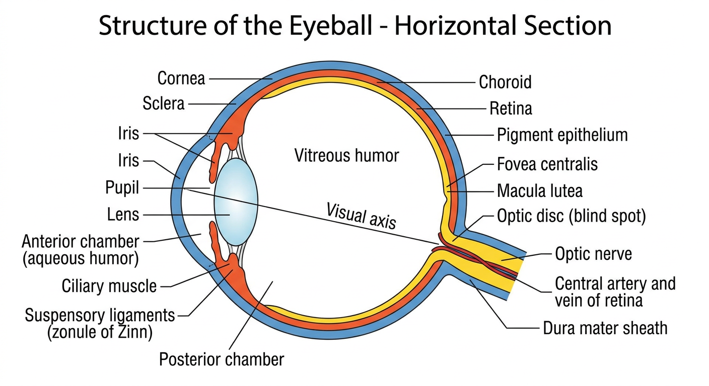
- Outer coat - Sclera + Cornea
- Middle coat - Choroid + Ciliary body + Iris
- Inner coat - Retina with fovea, optic disc
- Contents - Lens, aqueous humor, vitreous humor
- Posterior - Optic nerve with its meningeal sheaths