
penile block explain
penile nerve block anatomy technique
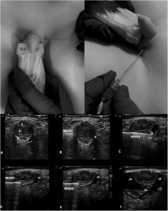
Educational composite image illustrating the ultrasound-guided dorsal penile nerve block (US-DPNB) technique. The upper panels show clinical photographs of the procedural setup, featuring a high-frequency linear ultrasound probe (13-6 MHz) in a sterile sleeve and the insertion of a block needle into the suprapubic/penile base region. The lower section consists of a six-panel ultrasound sequence (A-F) in transverse cross-section. Image A identifies the anatomy: corpus spongiosum (1), corpora cavernosa (2), and Buck’s fascia (3). Images B and C demonstrate the hyperechoic needle advancing toward the target (X) beneath Buck’s fascia. Images D through F show the real-time administration of local anesthetic (LA), visualized as an expanding anechoic/hypoechoic area surrounding the dorsal nerves and vascular structures. This material serves as a clinical guide for regional anesthesia in urological procedures, emphasizing needle trajectory and pharmacological spread relative to penile internal anatomy.
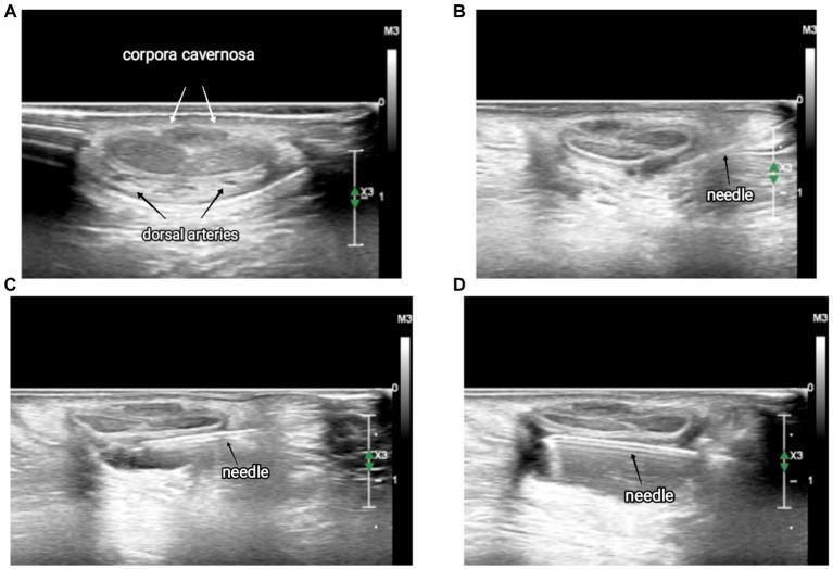
This set of four transverse-view ultrasound images (labeled A-D) illustrates the anatomy and procedural technique for an ultrasound-guided dorsal penile nerve block. Panel A displays the baseline sonoanatomy, identifying the paired corpora cavernosa as central, oval-shaped structures with a medium-gray echotexture, and the smaller, hypoechoic dorsal arteries located inferiorly. Panels B, C, and D demonstrate the dynamic advancement of a hyperechoic (bright) echoic needle using an in-plane approach. The sequence shows the needle entering from the lateral aspect (B), advancing through the fascia to the midline (C), and finally extending beneath the corpora cavernosa toward the contralateral nerve (D). The clinical focus is on visualizing the fascia and vascular structures to ensure precise anesthetic deposition and avoid intravascular injection. This clinical imaging is essential for pediatric anesthesia education regarding safe, regional block techniques for penile procedures.
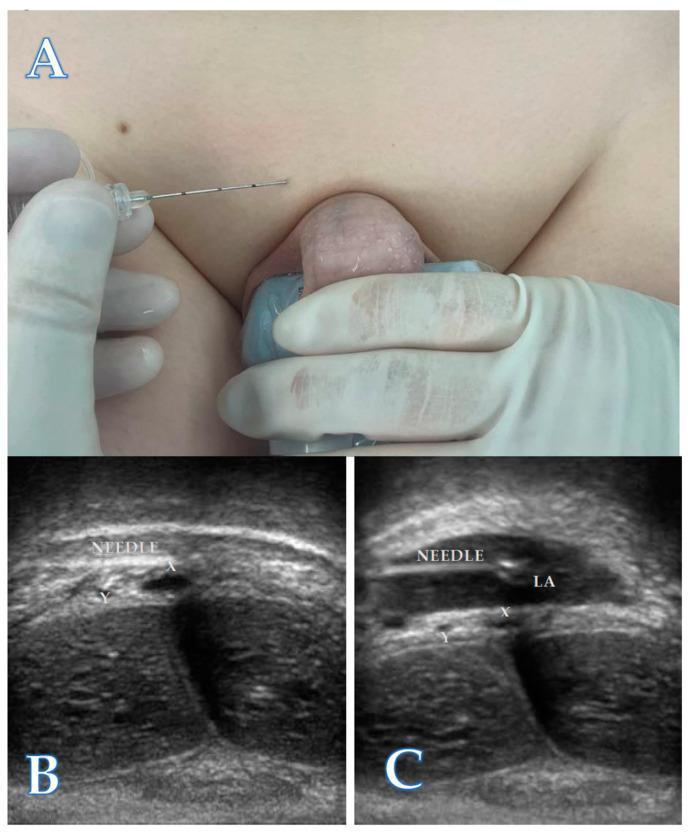
This composite educational image illustrates the technique for an ultrasound-guided dorsal penile nerve block (DPNB). Panel A is a clinical photograph demonstrating the patient positioning and probe placement. A linear ultrasound transducer is positioned in a coronal plane at the base of the penis, while a 22-gauge needle is being advanced using an in-plane, lateral-to-medial approach. Panels B and C are corresponding ultrasound scans. Panel B shows the pre-injection anatomy of the neurovascular bundle, identifying the deep dorsal penile vein (X) and dorsal penile arteries (Y) located deep to Buck's fascia. The needle tip is visible approaching these structures. Panel C demonstrates the post-injection state, where a well-defined hypoechoic area (LA) indicates the successful distribution of local anesthetic within the neurovascular sheath, surrounding the vein (X) and arteries (Y). This visualization confirms correct drug placement for anesthesia during procedures like circumcision. The content is designed for intermediate medical trainees learning regional anesthesia and pediatric urology techniques.
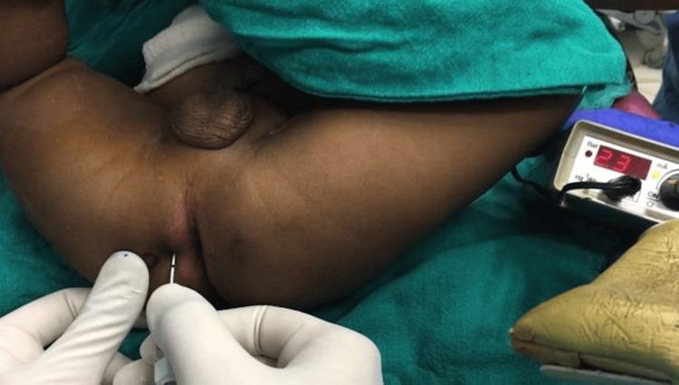
This clinical procedural photograph demonstrates the technique for a pediatric pudendal nerve block (PNB) using nerve stimulator guidance. The patient is positioned in the lithotomy position, with the perineal region exposed under sterile conditions. A clinician, wearing sterile gloves, is seen performing the block by inserting a specialized nerve stimulator needle (typically 22-24 gauge) into the perineum. The needle is positioned at approximately the 3 o'clock location relative to the anus, midway between the anus and the ischial tuberosity. To the right of the procedural field, a nerve stimulation device is visible, displaying a digital output of '2.3 mA', which is used to localize the nerve by monitoring for motor responses (e.g., penile movement). The image highlights key anatomical landmarks and the precise needle trajectory required for regional anesthesia in the pudendal distribution, commonly used for postoperative pain management in penile surgeries such as hypospadias repair. This content is intended for intermediate to advanced medical training in pediatric anesthesia and pain management.
Penile Block
Nerve Supply (Why It Works)
- A minor contribution also comes from the perineal nerve (ventral surface), which is why a ring block or additional ventral injection may be added for complete coverage.
Anatomical Layers (Critical for Technique)
- Skin
- Scarpa's fascia (superficial to the subpubic space)
- Subpubic space - the target injection space
- Buck's fascia (the deeper fascial envelope around the corpora)
- Dorsal nerve, artery, and vein under Buck's fascia
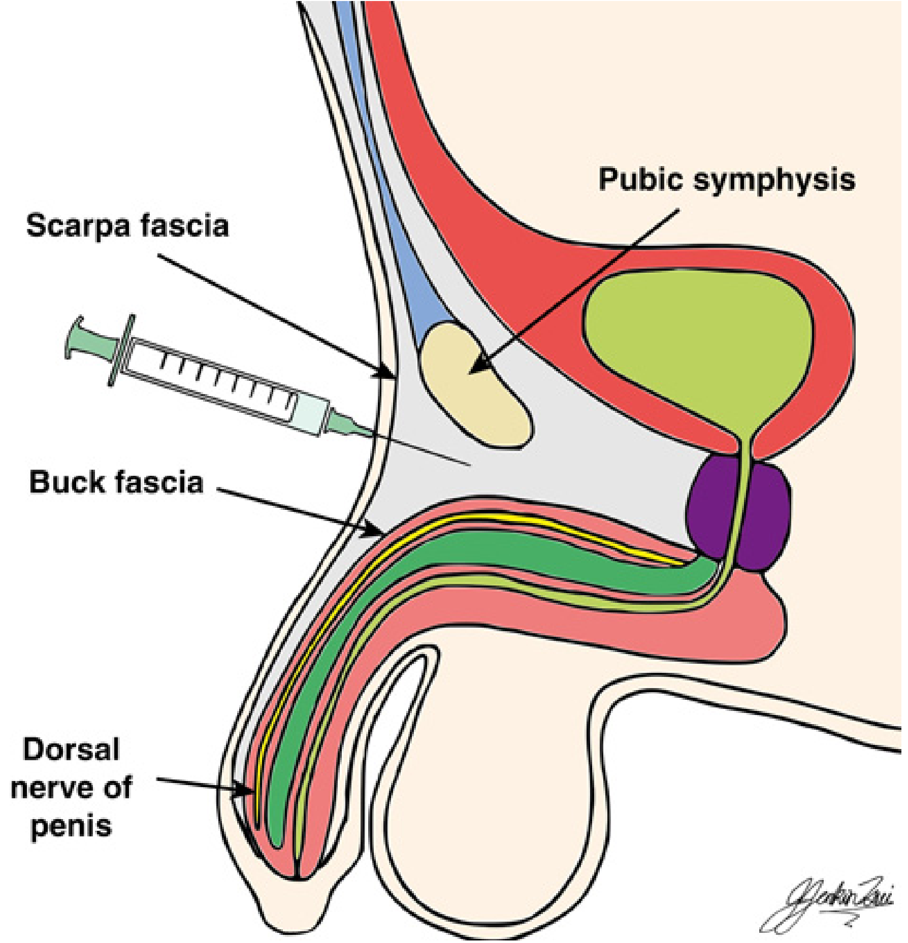
The safest injection plane is between skin and Buck's fascia (in the subpubic space, just piercing Scarpa's fascia). Injecting below Buck's fascia risks arterial injury and intracavernous injection, which mimics an IV bolus of local anesthetic.
Techniques
1. Subpubic (Dorsal Penile Nerve Block) - Most Common
- Patient supine. Prepare skin.
- Identify two injection points just below and medial to the pubic spine (approximately 10 o'clock and 2 o'clock relative to the base of the penis).
- Apply gentle traction on the penis to tension Scarpa's fascia (helps feel the fascial click).
- Insert a 25-27 gauge needle perpendicular to the skin at each point, directed caudally at ~15° from the midline.
- Advance until the pubic bone is contacted, then "walk" the needle off the inferior edge of the pubis.
- A subtle fascial click is felt as the needle passes through Scarpa's fascia into the subpubic space.
- Aspirate to confirm no blood.
- Inject local anesthetic at each site.
- Adults: 5 mL of 0.5% bupivacaine on each side (move needle depth by 1 cm during injection for spread)
- Children: 0.1 mL/kg per side (max 5 mL per side), 0.25% bupivacaine
- Infants: 0.5-1 mL per side
2. Ring Block (Circumferential Infiltration)
- Raise two skin wheals at the dorsolateral base of the penis.
- Inject 5 mL of local anesthetic superficially and deep along the lower border of the pubic ramus on each side.
- An additional ~5 mL (adults) is infiltrated subcutaneously around the ventral/undersurface of the shaft to complete the ring.
- Total dose: 20-25 mL of 0.75% lidocaine or 0.25% bupivacaine in adults.
3. Ultrasound-Guided Penile Block
- Advantage: Reduces failure rate, decreases first-hour postoperative pain, and delays time to first analgesic requirement vs. the fascial-click technique.
- Disadvantage: Adds ~10 minutes to procedure duration.
Local Anesthetic Choice
| Agent | Concentration | Onset | Duration |
|---|---|---|---|
| Bupivacaine | 0.25% (child) / 0.5% (adult) | Slow | Long (12-24 h analgesia) |
| Lidocaine | 1-2% | Fast | Moderate |
| Mixed (bupivacaine + lidocaine) | Equal volumes | Fast onset + long duration | Best for circumcision |
NEVER use epinephrine (adrenaline) in a penile block. The penis is an end-arterial structure with no collateral circulation. Epinephrine causes vasospasm and can result in ischemia/necrosis of the penis.
Indications
- Circumcision (most common - adults and children)
- Phimosis repair
- Paraphimosis reduction
- Hypospadias repair (penile block preferred over caudal - lower fistula rate; 0% vs 19.2% in one study)
- Foreskin stuck in zipper
- Glans/shaft surgery
- Neonatal circumcision (more effective than EMLA cream or sucrose)
Contraindications
- Epinephrine-containing solutions (absolute contraindication)
- Active skin infection at the puncture site
- Allergy to local anesthetics
- Coagulopathy (relative)
Complications
| Complication | Cause |
|---|---|
| Hematoma | Injury to dorsal penile artery (especially with midline puncture) |
| Local anesthetic systemic toxicity (LAST) | Intracavernous injection (equivalent to IV injection) |
| Block failure | Incomplete coverage of ventral surface (perineal nerve not blocked) |
| Penile ischemia/necrosis | Use of epinephrine-containing solution |
| Infection | Poor aseptic technique |
Key Points to Remember
- Nerve enters only along the dorsum - a ring block is often unnecessary but improves completeness.
- Inject in the subpubic space deep to Scarpa's fascia, but NOT below Buck's fascia (risk: arterial injury + cavernous injection).
- No epinephrine - ever.
- Bilateral injection is required (two dorsal nerves).
- Penile block is preferred over caudal block for hypospadias repair due to lower postoperative fistula rates.
- US guidance improves efficacy over landmark technique.