
So orris i need to clear ls 4 by tomorrow 15 morning 9am now its 14th 4am i will sleep at 11am and wake up at 7pm i am 5th sem aboard medical student it has Virology Parasitology and immunology and Bacteriology i completed Virology now rest calculate and say
Your Timeline (July 13-15)
Sleep/Wake Schedule
| Period | Time |
|---|---|
| Sleep | July 14, 11:00 AM |
| Wake up | July 14, 7:00 PM |
| Sleep again (assumed ~2-3 hrs) | July 15, ~2-3 AM |
| Wake up for exam | July 15, ~6-7 AM |
Available Study Hours
Total usable study time: ~19-20 hours
Subjects Remaining
- Parasitology
- Immunology
- Bacteriology
Study Allocation
Session 1 - Tonight (11:53 PM → 11:00 AM) = ~11 hours
| Subject | Time | Hours |
|---|---|---|
| Parasitology | 12:00 AM - 4:30 AM | 4.5 hrs |
| Short break | 4:30 AM - 4:45 AM | 15 min |
| Immunology | 4:45 AM - 8:30 AM | 3.75 hrs |
| Short break | 8:30 AM - 8:45 AM | 15 min |
| Bacteriology (Part 1 - High yield) | 8:45 AM - 10:45 AM | 2 hrs |
| Wind down/review notes | 10:45 AM - 11:00 AM | 15 min |
| SLEEP | 11:00 AM | - |
Session 2 - Evening (7:00 PM → 2:00 AM) = ~7 hours
| Subject | Time | Hours |
|---|---|---|
| Bacteriology (Part 2) | 7:00 PM - 11:00 PM | 4 hrs |
| Full revision sweep | 11:00 PM - 1:30 AM | 2.5 hrs |
| Wind down | 1:30 AM - 2:00 AM | 30 min |
| SLEEP | 2:00 AM | - |
Exam Morning (6:30 AM → 8:45 AM) = ~1.5 hrs
- Quick notes scan only - no new topics
- Focus on mnemonics, classifications, and organisms you're shaky on
Priority Topics (High-Yield for LS4)
Key Tips
- Don't re-read - revise. Use short notes, mnemonics, previous year questions
- Parasitology and Immunology are more memory-based - do them first when your brain is fresh
- Bacteriology is vast - focus on high-yield organisms only, skip rare ones
- Set alarms - sleep deprivation kills recall more than skipping one topic
No i have 18 hrs only
Corrected Timeline
18 Hours Split Across 3 Subjects
| Subject | Hours | Reason |
|---|---|---|
| Parasitology | 5 hrs | Moderate load - lifecycles, species |
| Immunology | 5 hrs | Conceptual + memory heavy |
| Bacteriology | 7 hrs | Heaviest subject, most organisms |
| Revision buffer | 1 hr | Final sweep before exam |
| Total | 18 hrs | ✅ |
Session 1 - Tonight (Now → 11:00 AM)
| Block | Subject | Duration |
|---|---|---|
| 12:00 AM - 5:00 AM | Parasitology | 5 hrs |
| 5:00 AM - 5:15 AM | Break | 15 min |
| 5:15 AM - 10:15 AM | Immunology | 5 hrs |
| 10:15 AM - 11:00 AM | Bacteriology start | 45 min |
| 11:00 AM | SLEEP | - |
Session 2 - After Waking (7:00 PM → ~3:00 AM)
| Block | Subject | Duration |
|---|---|---|
| 7:00 PM - 1:00 AM | Bacteriology (remaining 6.25 hrs) | 6 hrs |
| 1:00 AM - 2:00 AM | Full Revision | 1 hr |
| 2:00 AM - 3:00 AM | Wind down / light notes | 1 hr |
| ~3:00 AM | SLEEP | - |
Exam Morning (6:30 - 8:45 AM)
- Mnemonics only
- Classifications, organism comparisons
- No new topics
High-Yield Focus Per Subject
- Malaria - Plasmodium species, lifecycle, cerebral malaria
- Leishmania types (cutaneous/visceral)
- Entamoeba histolytica vs dispar
- Taenia solium vs saginata, Echinococcus
- Toxoplasma, Cryptosporidium, Giardia
- Hypersensitivity I-IV with examples
- Complement (classical vs alternate vs lectin)
- T cell vs B cell differentiation, CD markers
- MHC I vs II
- Primary immunodeficiencies (DiGeorge, SCID, Bruton's)
- Vaccines - live vs killed
- Gram +ve: Staph, Strep, Clostridium, Bacillus, Listeria
- Gram -ve: Neisseria, E.coli, Salmonella, Shigella, Vibrio, H.pylori
- Mycobacterium TB - full detail
- Spirochetes: Treponema, Leptospira, Borrelia
- Toxins: exotoxin vs endotoxin table
- Culture media (high yield for exams)
I have started Bacteriology my goal is to complete by morning 9am now its 4 am
Bacteriology Sprint: 4:00 AM → 9:00 AM = 5 Hours
5-Hour Bacteriology Block Plan
| Time | Topic | Duration |
|---|---|---|
| 4:00 - 4:45 AM | Gram +ve Cocci - Staph + Strep | 45 min |
| 4:45 - 5:15 AM | Gram +ve Bacilli - Clostridium, Bacillus, Listeria, Corynebacterium | 30 min |
| 5:15 - 5:20 AM | Quick break | 5 min |
| 5:20 - 6:05 AM | Gram -ve - Neisseria, Enterobacteriaceae (E.coli, Salmonella, Shigella, Klebsiella) | 45 min |
| 6:05 - 6:35 AM | Gram -ve continued - Vibrio, H.pylori, Pseudomonas, Haemophilus, Bordetella | 30 min |
| 6:35 - 6:40 AM | Quick break | 5 min |
| 6:40 - 7:20 AM | Mycobacterium - TB full detail + Leprosy | 40 min |
| 7:20 - 7:50 AM | Spirochetes + Miscellaneous - Treponema, Leptospira, Borrelia, Rickettsia, Chlamydia, Mycoplasma | 30 min |
| 7:50 - 8:30 AM | High-yield tables - Toxins, Culture media, Stains, Virulence factors | 40 min |
| 8:30 - 9:00 AM | Rapid fire revision - Weak points only | 30 min |
What to Focus ON vs SKIP
✅ DO cover (exam favorites)
- Staph aureus - toxins (TSST, exfoliatin, coagulase), MRSA
- Strep - classification (alpha/beta/gamma), Lancefield groups, Rheumatic fever
- Clostridium - tetani, perfringens, botulinum, difficile (mechanisms!)
- Salmonella vs Shigella differences (key table)
- Mycobacterium TB - cord factor, Ghon complex, PPD, culture (LJ medium)
- Neisseria - gonorrhoeae vs meningitidis differences
- Vibrio cholerae - rice water stool, El Tor biotype
- Treponema pallidum - VDRL vs TPHA
- Rickettsia - Weil-Felix reaction
- Chlamydia - types (A-C trachoma, D-K genital, L1-L3 LGV)
- Exotoxin vs Endotoxin comparison table - always in exams
❌ SKIP or skim only
- Rare organisms (Brucella, Francisella, Pasteurella)
- Detailed biochemical tests
- Long historical context
- Antibiotic mechanisms (unless specifically in your syllabus)
Must-Know Quick Tables
| Gram +ve | Gram -ve |
|---|---|
| Staph, Strep, Bacillus, Clostridium, Listeria, Coryne | Neisseria, E.coli, Salmonella, Shigella, Vibrio, H.pylori, Pseudomonas |
| Neither: Mycobacterium (AFB), Mycoplasma (no wall), Chlamydia, Rickettsia (intracellular), Treponema (dark field) |
| Organism | Medium |
|---|---|
| TB | LJ medium |
| Cholera | TCBS, Alkaline peptone water |
| Diphtheria | Tellurite, Loeffler's |
| Staph | Mannitol salt agar |
| Strep | Blood agar |
| Neisseria | Chocolate agar, Thayer-Martin |
| Bordetella | Bordet-Gengou |
| Salmonella/Shigella | MacConkey, XLD, DCA |
This is the topics
cd /home/daytona/workspace/attachments/c6ec6246-d110-440e-90a7-f81027173941/ && python3 -c " from pypdf import PdfReader reader = PdfReader('Life Sciences IV ENG.docx.pdf') print(f'Pages: {len(reader.pages)}') text = '' for page in reader.pages: text += page.extract_text() + '\n' print(text) "
Your LS4 Syllabus - What's Actually in the Exam
✅ VIROLOGY - DONE
Remaining Topics Breakdown
🦠 BACTERIOLOGY (Clinical Microbiology - Weeks 2-9)
| Week | Topic |
|---|---|
| W2 | Staphylococcal infections, Pneumococcal infections |
| W3 | Streptococcal & Enterococcal infections |
| W4 | Mycobacterium Tuberculosis (Acid-fast bacteria) |
| W5 | Meningococcal infections, Haemophilus, Moraxella catarrhalis |
| W6 | Mycoplasma, Chlamydia, Legionella, Bordetella pertussis |
| W7 | Gram-negative infections (E.coli, Salmonella, Shigella, Vibrio, Klebsiella - Ch 161,163,165,166,167) |
| W8 | Treponema pallidum (Syphilis) |
| W9 | Lyme Disease, Leptospirosis |
🦟 PARASITOLOGY (Weeks 13-14)
| Week | Topic |
|---|---|
| W13 | Intestinal Flagellates, Hemoflagellates, Amebas, Blood Sporozoans (Malaria, Leishmania, Giardia, Entamoeba, Toxoplasma) |
| W14 | Helminths - Cestodes (Taenia, Echinococcus), Trematodes (Schistosoma), Nematodes (Ascaris, Hookworm, Filaria) |
🛡️ IMMUNOLOGY (Weeks 1-15)
| Week | Topic |
|---|---|
| W1 | Immune Receptors & Signal Transduction |
| W2 | Lymphocyte Development & Antigen Receptor Gene Rearrangement |
| W3 | Activation of T Lymphocytes |
| W4 | Differentiation & Functions of CD4+ Effector T Cells |
| W5 | Differentiation & Functions of CD8+ Effector T Cells |
| W6 | B Cell Activation & Antibody Production |
| W7 | Effector Mechanisms of Humoral Immunity |
| W8 | Specialized Immunity at Epithelial Barriers & Immune Privileged Tissues |
| W9 | Immunologic Tolerance & Autoimmunity |
| W10 | Immunity to Microbes |
| W11 | Transplantation Immunology |
| W12 | Immunity to Tumors |
| W13 | Hypersensitivity Disorders |
| W14 | Allergy |
| W15 | Congenital & Acquired Immunodeficiencies |
Revised 18-Hour Plan (You're on Bacteriology now at 4 AM)
Session 1 - Tonight (4:00 AM → 11:00 AM) = 7 hrs
| Time | Topic | Focus |
|---|---|---|
| 4:00 - 5:00 AM | Staph + Pneumococcus | TSST, coagulase, MRSA, S.pneumo virulence |
| 5:00 - 5:45 AM | Strep + Enterococcus | Lancefield groups, Rheumatic fever, Group B Strep |
| 5:45 - 6:30 AM | TB (Mycobacterium) | Cord factor, Ghon complex, LJ medium, PPD |
| 6:30 - 7:00 AM | Neisseria + Haemophilus + Moraxella | Chocolate agar, Thayer-Martin, capsule types |
| 7:00 - 7:40 AM | Mycoplasma + Chlamydia + Legionella + Bordetella | No cell wall (Mycoplasma), Chlamydia types, Legionnaire's, whooping cough |
| 7:40 - 8:20 AM | Gram -ve Enterics - E.coli, Salmonella, Shigella, Vibrio, Klebsiella | Toxins, serotypes, rice-water stool |
| 8:20 - 9:00 AM | Spirochetes - Syphilis (VDRL/TPHA), Lyme (Borrelia), Leptospira | Stages of syphilis, Weil's disease |
| 9:00 AM → Wind down, light nap | ✅ Bacteriology done |
Session 2 - Evening (7:00 PM → ~3:00 AM) = 8 hrs
| Time | Topic | Focus |
|---|---|---|
| 7:00 - 8:30 PM | Parasitology Part 1 | Malaria lifecycle + Plasmodium species, Leishmania (VL/CL/MCL), Giardia, Entamoeba, Toxoplasma |
| 8:30 - 9:30 PM | Parasitology Part 2 | Cestodes (Taenia solium vs saginata, Echinococcus), Trematodes (Schistosoma), Nematodes (Ascaris, Hookworm, Filaria, Pinworm) |
| 9:30 - 9:40 PM | Break | |
| 9:40 - 11:10 PM | Immunology Part 1 | T cell activation, CD4 vs CD8, B cell activation, Antibody classes |
| 11:10 PM - 12:30 AM | Immunology Part 2 | Hypersensitivity I-IV + Allergy (IgE mast cell), Transplantation, Tumor immunity |
| 12:30 - 1:30 AM | Immunology Part 3 | Tolerance + Autoimmunity, Immunodeficiencies (DiGeorge, SCID, Bruton's, AIDS) |
| 1:30 - 2:30 AM | Full Rapid Revision | All weak points, mnemonics, comparison tables |
| 2:30 - 3:00 AM | Wind down | |
| ~3:00 AM SLEEP |
Exam Morning July 15 (6:30 AM - 8:45 AM)
- Scan your key comparison tables only
- No new topics
Key Point
Okay start teaching i completed Staph now start from strep
🦠 STREPTOCOCCUS & ENTEROCOCCUS
Exam-Focused Teaching | ~45 minutes
STEP 1: THE BIG PICTURE - Classification
Two Classification Systems - Know BOTH:
| Type | Hemolysis | Example |
|---|---|---|
| β-hemolytic | Complete (clear zone) | S. pyogenes (GAS), S. agalactiae (GBS) |
| α-hemolytic | Partial (green zone) | S. pneumoniae, Viridans strep |
| γ-hemolytic | None | Enterococcus |
| Group | Species | Disease |
|---|---|---|
| A | S. pyogenes | Pharyngitis, impetigo, rheumatic fever |
| B | S. agalactiae | Neonatal meningitis, maternal sepsis |
| C, G | S. dysgalactiae | Pharyngitis, skin infections |
| D | Enterococcus faecalis/faecium | UTI, endocarditis, nosocomial |
| No group | Viridans (S. mutans, S. mitis) | Dental caries, SBE |
| No group | S. pneumoniae | Pneumonia, meningitis, otitis |
STEP 2: GROUP A STREP (S. pyogenes) - The Most Important
Virulence Factors (HIGH YIELD)
| Factor | Function |
|---|---|
| M protein (>150 types) | Anti-phagocytic; type-specific immunity; molecular mimicry → rheumatic fever |
| Hyaluronic acid capsule | Binds CD44 on epithelial cells; anti-phagocytic |
| Streptolysin O (SLO) | Oxygen-labile; lyses RBCs, WBCs; antigen → ASO titre (used to diagnose post-strep disease) |
| Streptolysin S (SLS) | Oxygen-stable; causes β-hemolysis on blood agar; NOT antigenic |
| Streptokinase | Dissolves clots (fibrinolysin) |
| Hyaluronidase | "Spreading factor" - breaks down connective tissue |
| DNase (streptodornase) | Degrades DNA in pus |
| Pyrogenic exotoxins (SPE A, B, C) | Superantigens → scarlet fever rash, toxic shock |
| C5a peptidase | Destroys complement chemoattractants → blocks neutrophil recruitment |
Mnemonic for GAS toxins: "Some Sinister Killers Hide, Dodging Poison" Streptolysin, Streptokinase, Hyaluronidase, DNase, Pyrogenic exotoxins
Clinical Diseases of GAS
A) PHARYNGITIS ("Strep Throat")
- Sudden onset fever, sore throat, NO cough (cough = viral)
- Tonsillar exudate, tender anterior cervical lymph nodes
- Centor Score (1 point each): Fever + No cough + Tender cervical nodes + Tonsillar exudate
- 0-1 points → no test, no antibiotics
- 2-3 points → rapid antigen test
- 4 points → treat empirically
- Treatment: Penicillin V (first line) or amoxicillin
B) SCARLET FEVER
- Pharyngitis + diffuse sandpaper-like rash (blanches on pressure)
- Strawberry tongue (white initially, then red)
- Pastia's lines - rash accentuated in skin folds (axilla, groin)
- Caused by pyrogenic exotoxin (SPE) - superantigen
- Circumoral pallor (pale around mouth)
C) SKIN INFECTIONS
| Disease | Features |
|---|---|
| Impetigo | Superficial; golden-crusted lesions; face/limbs |
| Erysipelas | Upper dermis + lymphatics; fiery red, RAISED borders, face/legs; ONLY GAS |
| Cellulitis | Subcutaneous; pink, diffuse, NON-raised borders |
| Necrotizing fasciitis | "Flesh-eating disease"; deep fascia; rapid spread; surgical emergency |
D) TOXIC SHOCK SYNDROME (Streptococcal TSS)
- Hypotension + multi-organ failure
- Often from necrotizing fasciitis/wound infection
- Caused by SPE superantigens (vs Staph TSS caused by TSST-1)
Post-Streptococcal Sequelae (VERY HIGH YIELD)
🔥 RHEUMATIC FEVER
- Occurs 2-4 weeks after GAS pharyngitis (NOT skin infection)
- Mechanism: molecular mimicry - M protein antibodies cross-react with cardiac tissue
- Jones Criteria - need 2 Major OR 1 Major + 2 Minor:
| MAJOR (JONES) | MINOR |
|---|---|
| Joints - migratory polyarthritis | Fever |
| ❤️ Carditis (pancarditis - all 3 layers) | Elevated ESR/CRP |
| Overactive (Chorea - Sydenham's) | Prolonged PR interval on ECG |
| Nodules (subcutaneous) | Arthralgia (only if joints not used as major) |
| Erythema marginatum (rash) | Previous rheumatic fever |
| Supporting evidence: elevated ASO titre |
Mnemonic: JONES = Joints, cOrditis, chNoreA (Chorea), Nodules, Erythema marginatum, Supporting (ASO)
- Treatment: Penicillin + aspirin (arthritis) + steroids (carditis)
- Prophylaxis: Monthly Benzathine Penicillin G injections for years (prevents recurrence)
🔥 POST-STREPTOCOCCAL GLOMERULONEPHRITIS (PSGN)
- Occurs 1-3 weeks after pharyngitis OR 3-6 weeks after skin infection
- Immune complex deposition in glomeruli → nephritic syndrome
- Hematuria, proteinuria, hypertension, oliguria
- Low complement (C3↓), elevated ASO titre
- Spontaneously resolves - antibiotics do NOT prevent it (unlike RF)
STEP 3: GROUP B STREP (S. agalactiae)
- Normal flora of vagina (~25% women colonized)
- CAMP test positive - key lab ID
- Main victims: Neonates + pregnant women + elderly + immunocompromised
| Disease | Timing | Notes |
|---|---|---|
| Early-onset neonatal | Within 7 days of birth | Pneumonia, sepsis, meningitis - acquired during delivery |
| Late-onset neonatal | 1 week - 3 months | Meningitis - acquired from environment/breast milk |
| Maternal | Peripartum | Puerperal sepsis, chorioamnionitis |
- Prevention: Screen all pregnant women at 35-37 weeks; give IV Penicillin during labor if positive
STEP 4: S. PNEUMONIAE (Pneumococcus) - α-hemolytic, No Lancefield group
- Lancet-shaped diplococci (pairs, not chains)
- Quellung reaction - capsule swells with specific antisera (used for ID)
- Optochin sensitive + bile soluble (distinguishes from viridans)
- Polysaccharide capsule = main virulence factor; anti-phagocytic
| Disease | Notes |
|---|---|
| Lobar pneumonia | Most common cause in adults; "rusty sputum" |
| Meningitis | #1 cause in adults; high mortality |
| Otitis media | #1 cause in children |
| Sinusitis | Common |
| Septicemia | Especially in asplenic patients |
- Treatment: Penicillin (if sensitive), Ceftriaxone
- Vaccines: PCV13 (conjugate, children) + PPSV23 (polysaccharide, adults/elderly)
STEP 5: VIRIDANS STREPTOCOCCI - α-hemolytic
- Part of normal oral flora
- Optochin resistant + bile insoluble (vs S. pneumoniae)
- S. mutans → dental caries
- S. mitis, S. sanguis → Subacute Bacterial Endocarditis (SBE) after dental procedures
- Enter bloodstream during dental work → colonize damaged heart valves
STEP 6: ENTEROCOCCUS - γ-hemolytic (Group D)
Key Properties:
- Grow in 6.5% NaCl (salt-tolerant - distinguishes from Strep)
- Grow at 10-45°C and at high pH
- Hydrolyze esculin in bile (esculin test +ve)
- PYR test positive
- Normal GI flora
Clinical Diseases:
| Disease | Notes |
|---|---|
| UTI | Most common; especially catheter-associated, nosocomial |
| Endocarditis | Often in elderly men with GI/GU procedures |
| Bacteremia | Hospital-acquired |
| Intra-abdominal infections | Post-surgical |
THE KEY ISSUE - Antibiotic Resistance:
| Type | Resistance | Treatment |
|---|---|---|
| Normal | Intrinsic resistance to low-dose penicillin | Penicillin/Ampicillin + Aminoglycoside (synergy) |
| VRE (Vancomycin-Resistant Enterococcus) | vanA or vanB gene; hospital nightmare | Linezolid or Daptomycin |
E. faecium is more likely to be VRE than E. faecalis. VRE = major nosocomial problem in ICUs.
QUICK COMPARISON TABLE (High Yield)
| Feature | S. pyogenes (GAS) | S. agalactiae (GBS) | S. pneumoniae | Enterococcus |
|---|---|---|---|---|
| Group | A | B | None | D |
| Hemolysis | β | β | α | γ (none) |
| Key test | ASO titre | CAMP test +ve | Optochin sensitive, Quellung | Grows in 6.5% NaCl |
| Disease | Pharyngitis, RF, PSGN | Neonatal meningitis | Lobar pneumonia | UTI, Endocarditis |
| Toxin | SPE, Streptolysin O | - | Pneumolysin | - |
| Treatment | Penicillin | Penicillin | Penicillin/Ceftriaxone | Ampicillin ± Aminoglycoside; Linezolid (VRE) |
⚡ LAST-MINUTE MNEMONICS
- ASO titre rises in GAS pharyngitis but NOT in skin infection (DNase-B rises in skin)
- RF after pharyngitis (not skin) - "throat to heart"
- PSGN after skin OR throat
- Penicillin does NOT prevent PSGN but DOES prevent RF
- Erysipelas = ONLY GAS (raised borders, upper dermis)
- Necrotizing fasciitis = GAS Type 1 (flesh-eating, surgical emergency)
- VRE = E. faecium > faecalis
Okay start teaching me immunology topics
🛡️ IMMUNOLOGY - COMPLETE EXAM GUIDE
All 15 Weeks | Exam-Ready | Based on Abbas & Lichtman
PART 1: THE FOUNDATIONS (Weeks 1-2)
W1 - Immune Receptors & Signal Transduction
The Two Arms of Immunity
| Innate Immunity | Adaptive Immunity |
|---|---|
| Fast (minutes-hours) | Slow (days-weeks) |
| Non-specific | Antigen-specific |
| No memory | Has memory |
| PRRs (Pattern Recognition Receptors) | TCR, BCR |
| Neutrophils, Macrophages, NK cells, Complement | T cells, B cells |
Key Receptors to Know:
| Receptor | Location | Recognizes | Example |
|---|---|---|---|
| TLRs (Toll-Like Receptors) | Cell surface/endosome | PAMPs (bacterial LPS, viral dsRNA) | TLR4 → LPS; TLR3 → viral dsRNA |
| NOD-like receptors | Cytoplasm | Intracellular bacteria | NOD2 → Crohn's disease when mutated |
| RIG-I | Cytoplasm | Viral RNA | Antiviral response |
- Recognizes peptide + MHC (cannot recognize free antigen alone)
- Linked to CD3 complex for signal transduction
- Signal cascade: TCR → CD3 → ZAP-70 → PLC-γ → IP3 + DAG → Ca²⁺ + PKC → NFAT + NFκB → gene transcription → IL-2 production
- Surface IgM/IgD
- Recognizes free antigen (no MHC needed)
- Linked to Igα/Igβ (CD79a/b) for signaling
Key signal molecule: IL-2 = the master T cell growth factor. Produced by activated T cells → drives proliferation (autocrine).
W2 - Lymphocyte Development & Antigen Receptor Gene Rearrangement
T Cell Development (in Thymus)
Bone Marrow stem cell → Thymus
↓
Double Negative (DN): CD4⁻ CD8⁻ → TCR gene rearrangement (VDJ)
↓
Double Positive (DP): CD4⁺ CD8⁺
↓
POSITIVE SELECTION (survive if TCR recognizes self-MHC)
↓
NEGATIVE SELECTION (die if TCR binds self-MHC too strongly → prevents autoimmunity)
↓
Single Positive: CD4⁺ OR CD8⁺ → exit to periphery
B Cell Development (in Bone Marrow)
Stem cell → Pro-B cell → Pre-B cell → Immature B cell → Mature B cell (IgM+IgD)
                              ↓
                    VDJ recombination of heavy chain
                    VJ recombination of light chain
MHC (Major Histocompatibility Complex) - EXTREMELY HIGH YIELD
| Feature | MHC Class I | MHC Class II |
|---|---|---|
| Found on | ALL nucleated cells | APCs only (DCs, macrophages, B cells) |
| Presents to | CD8⁺ T cells | CD4⁺ T cells |
| Peptide source | Cytosolic (endogenous) - viruses, tumors | Endosomal (exogenous) - bacteria, phagocytosed |
| Molecules | HLA-A, HLA-B, HLA-C | HLA-DR, HLA-DP, HLA-DQ |
| Co-receptor | CD8 binds α3 domain | CD4 binds β2 domain |
| Peptide length | 8-9 amino acids | 10-20 amino acids |
Mnemonic: "8 × CD8 = Class I" (8 aa peptide, CD8+ T cell) "4 × CD4 = Class II" (CD4+ T cell, exogenous antigen)
PART 2: T CELL IMMUNITY (Weeks 3-5)
W3 - Activation of T Lymphocytes
The Two-Signal Rule (CRITICAL)
| Signal | What it is | Example |
|---|---|---|
| Signal 1 | TCR + CD3 recognizes peptide-MHC | Antigen-specific |
| Signal 2 | Co-stimulation | CD28 (on T cell) + B7/CD80/CD86 (on APC) |
Sequence of T Cell Activation:
- DC captures antigen in tissues → migrates to lymph node
- Naive T cell in lymph node recognizes peptide-MHC on DC (Signal 1)
- CD28-B7 interaction (Signal 2)
- T cell produces IL-2 → autocrine proliferation
- Clonal expansion → thousands of daughter cells
- Differentiation into effector T cells + memory T cells
Key Co-stimulatory & Inhibitory Molecules:
| Molecule | On | Binds | Effect |
|---|---|---|---|
| CD28 | T cell | B7 (CD80/86) on APC | ✅ Activation |
| CTLA-4 | Activated T cell | B7 (CD80/86) | ❌ Inhibition (checkpoint) |
| PD-1 | T cell | PD-L1 on cells | ❌ Inhibition (checkpoint) |
CTLA-4 and PD-1 are immune checkpoints - cancer immunotherapy (ipilimumab, pembrolizumab) blocks these to unleash T cells against tumors.
W4 - CD4⁺ Effector T Cells (T Helper Cells)
| Subset | Induced by | Produces | Function |
|---|---|---|---|
| Th1 | IL-12, IFN-γ | IFN-γ, TNF | Activates macrophages → kills intracellular bacteria (TB, Leishmania) |
| Th2 | IL-4 | IL-4, IL-5, IL-13 | B cell help → IgE production, eosinophil activation → helminth immunity + ALLERGY |
| Th17 | IL-6, TGF-β | IL-17, IL-22 | Neutrophil recruitment → extracellular bacteria, fungi |
| Treg | TGF-β | TGF-β, IL-10 | Suppress immune responses → FOXP3 transcription factor |
| Tfh | IL-21 | IL-21 | Help B cells in germinal centers → antibody production |
Mnemonic for Th1 vs Th2:
- Th1 = "Think intracellular" → IFN-γ → macrophage activation
- Th2 = "Two allergies" → IL-4/5 → IgE/eosinophils → worms + allergy
W5 - CD8⁺ Effector T Cells (Cytotoxic T Lymphocytes - CTLs)
- Recognize peptide + MHC Class I (on ALL nucleated cells)
- Kill virally infected cells and tumor cells
Killing Mechanisms:
| Mechanism | How |
|---|---|
| Perforin | Punches pores in target cell membrane |
| Granzymes | Enter through perforin pores → activate caspases → apoptosis |
| Fas-FasL | CTL expresses FasL → binds Fas on target → apoptosis |
| TNF | Also contributes to target cell death |
Key: CTLs kill by apoptosis, not necrosis. They do NOT use phagocytosis.
PART 3: B CELL & ANTIBODY IMMUNITY (Weeks 6-7)
W6 - B Cell Activation & Antibody Production
Two Types of B Cell Responses:
- Antigen: Protein antigens
- Requires CD4+ T helper cell (Tfh)
- CD40L (T cell) + CD40 (B cell) = essential interaction
- Results in: Class switching, affinity maturation, memory B cells, plasma cells
- Antigen: Polysaccharides, lipopolysaccharides (no protein)
- No T cell help needed
- Only IgM produced (no class switching)
- No memory cells (reason why polysaccharide vaccines are weak in infants)
Germinal Center Reaction (key events):
- Activated B cell enters germinal center in lymph node
- Somatic hypermutation → mutate Ig variable regions → higher affinity antibodies
- Affinity maturation → B cells with best antigen-binding selected (others die by apoptosis)
- Class switch recombination → IgM → IgG, IgA, or IgE
- Output: Plasma cells (antibody factories) + Memory B cells
Antibody Classes - HIGH YIELD:
| Isotype | Key Features |
|---|---|
| IgM | First antibody produced; pentamer; best complement activator; does NOT cross placenta |
| IgG | Most abundant in blood; crosses placenta (maternal immunity to fetus); opsonization; neutralization |
| IgA | Secretory form (dimer) in mucosa (saliva, tears, breast milk, GI tract); first line in mucosal immunity |
| IgE | Lowest serum concentration; binds mast cells/basophils; mediates Type I hypersensitivity + parasite immunity |
| IgD | On naive B cell surface; function not fully known |
W7 - Effector Mechanisms of Humoral Immunity
| Mechanism | How | What for |
|---|---|---|
| Neutralization | Antibody blocks pathogen from binding host cell | Viruses, toxins |
| Opsonization | IgG coats bacteria → Fc receptors on phagocytes grab them | Bacteria |
| Complement activation | IgM/IgG activates classical pathway → MAC → lysis | Bacteria |
| ADCC (Antibody-Dependent Cell Cytotoxicity) | IgG coats target → NK cells bind Fc → kill target | Virus-infected cells, tumors |
| Mast cell degranulation | IgE on mast cells cross-linked by antigen → histamine release | Type I hypersensitivity |
Complement Pathways:
| Pathway | Triggered by | First component |
|---|---|---|
| Classical | IgM or IgG-antigen complex | C1q |
| Lectin (MBL) | Mannose on bacterial surface | MBL |
| Alternative | Spontaneous C3 hydrolysis; bacterial surfaces | C3 |
- C3b = opsonin (coats bacteria for phagocytosis)
- C3a, C5a = anaphylatoxins (mast cell degranulation, inflammation)
- C5a = most potent chemotactic factor
- MAC (C5b-9) = membrane attack complex → lysis
W8 - Specialized Immunity at Epithelial Barriers
- IgA is the key antibody at mucosal surfaces
- Secretory IgA = dimer linked by J chain + secretory component (protects from proteases)
- Sites: gut (GALT), respiratory tract (BALT), genitourinary tract
- Eye (anterior chamber)
- Brain (blood-brain barrier; CNS)
- Testis
- Placenta/fetus
- Mechanisms: TGF-β, IL-10, FasL expression, regulatory T cells
PART 4: REGULATION & SPECIAL TOPICS (Weeks 9-12)
W9 - Immunologic Tolerance & Autoimmunity
Tolerance = Unresponsiveness to self-antigens
- Negative selection kills self-reactive T cells
- AIRE (Autoimmune Regulator) gene in thymus expresses peripheral tissue antigens → allows deletion of self-reactive T cells
- AIRE mutations → APS-1 (Autoimmune Polyglandular Syndrome type 1)
| Mechanism | How |
|---|---|
| Anergy | T cell gets Signal 1 without Signal 2 → becomes unresponsive |
| Regulatory T cells (Tregs) | FOXP3+ CD4+ cells secrete IL-10, TGF-β → suppress other T cells |
| Deletion by apoptosis | Fas-FasL interaction kills activated T cells after response |
| Ignorance | Self-antigens are hidden (immune privileged sites) |
Autoimmunity - When tolerance breaks:
| Disease | Mechanism | Autoantibody |
|---|---|---|
| SLE | Immune complex deposition (Type III) | Anti-dsDNA, Anti-Sm |
| Rheumatoid Arthritis | Th1/Th17 → joint inflammation | Rheumatoid Factor (anti-IgG), Anti-CCP |
| Type 1 Diabetes | CTL kills β cells (Type IV) | Anti-islet cell antibodies |
| Graves Disease | Stimulating Ab to TSH receptor (Type II) | Anti-TSH receptor |
| Hashimoto's | T cell attack on thyroid | Anti-TPO, Anti-thyroglobulin |
| Myasthenia Gravis | Blocking Ab to ACh receptor (Type II) | Anti-AChR |
| Goodpasture's | Ab to basement membrane (Type II) | Anti-GBM |
| Multiple Sclerosis | T cell attack on myelin | Anti-MBP |
W10 - Immunity to Microbes
| Pathogen Type | Innate Response | Adaptive Response |
|---|---|---|
| Extracellular bacteria | Neutrophils, complement, phagocytosis | Antibodies (opsonization), Th17 |
| Intracellular bacteria (TB, Listeria) | Macrophages | Th1 → IFN-γ → macrophage activation |
| Viruses | IFN-α/β, NK cells | CTL (CD8+), neutralizing IgG/IgA |
| Fungi | Neutrophils, Th17 | Th17 → IL-17 |
| Parasites (helminths) | Eosinophils, mast cells | Th2 → IgE, eosinophils |
| Parasites (protozoa) | Macrophages | Th1 (intracellular like Leishmania) |
- IFN-α/β (Type I interferons) = produced by virus-infected cells → antiviral state in neighbors
- IFN-γ (Type II interferon) = produced by Th1, NK cells → activates macrophages (kills intracellular bacteria)
- TNF = macrophage product → fever, inflammation, cachexia (wasting in TB)
W11 - Transplantation Immunology
Types of Grafts:
| Type | Definition |
|---|---|
| Autograft | Self → self (skin graft) - no rejection |
| Isograft | Identical twin → never rejected |
| Allograft | Same species, different individual - most common clinical |
| Xenograft | Different species - pig valves, pig kidneys |
HLA Matching:
- HLA-A, HLA-B, HLA-C (Class I) + HLA-DR, HLA-DP, HLA-DQ (Class II)
- HLA-DR mismatch = strongest predictor of rejection
- Closer the match → less rejection
Rejection Types - HIGH YIELD:
| Type | Timing | Mechanism | Features |
|---|---|---|---|
| Hyperacute | Minutes-hours | Pre-formed antibodies (ABO mismatch or prior sensitization) | Thrombosis, immediate graft failure |
| Acute | Days-weeks | T cell mediated (CD4+ and CD8+) | Treatable with immunosuppressants |
| Chronic | Months-years | Antibody + T cell → vessel fibrosis | Gradual graft loss, not reversible |
Immunosuppression:
- Cyclosporine / Tacrolimus → block calcineurin → no IL-2 → no T cell activation
- Mycophenolate → blocks purine synthesis → no lymphocyte proliferation
- Steroids → broad anti-inflammatory
- Anti-thymocyte globulin → depletes T cells
Graft-versus-Host Disease (GVHD):
- Occurs in bone marrow transplant (donor T cells attack recipient)
- Acute GVHD: skin rash, diarrhea, liver failure
- Chronic GVHD: resembles autoimmune disease
- GVT (Graft-vs-Tumor) effect = donor T cells also kill residual tumor cells (beneficial!)
W12 - Immunity to Tumors
Tumor Antigens:
| Type | Example |
|---|---|
| Mutated self proteins | p53, Ras mutations |
| Oncofetal antigens | CEA (colon), AFP (liver) |
| Viral antigens | HPV (cervical cancer), EBV (Burkitt lymphoma) |
| Overexpressed | HER2/neu (breast cancer) |
Anti-Tumor Immunity:
- CD8+ CTLs = primary killers of tumor cells
- NK cells = kill tumor cells that downregulate MHC Class I (virus trick → NK catches it)
- Macrophages (M1) = kill tumors
Immune Evasion by Tumors:
- Downregulate MHC Class I → hide from CTLs
- Express PD-L1 → exhausts T cells
- Secrete TGF-β, IL-10 → immunosuppressive microenvironment
- Recruit Tregs into tumor
Cancer Immunotherapy:
- Checkpoint inhibitors: Anti-PD-1 (pembrolizumab), Anti-CTLA-4 (ipilimumab) → unleash T cells
- CAR-T cells: Patient's T cells engineered to target tumor antigens
- Monoclonal antibodies: Rituximab (anti-CD20, B cell lymphoma), Trastuzumab (anti-HER2)
PART 5: HYPERSENSITIVITY & IMMUNODEFICIENCY (Weeks 13-15)
W13-W14 - Hypersensitivity (Coombs & Gell Classification) - MOST HIGH YIELD
The 4 Types - Master This Table:
| Type | Name | Mediator | Timing | Mechanism | Classic Examples |
|---|---|---|---|---|---|
| I | Immediate / Anaphylactic | IgE + Mast cells | Seconds-minutes | IgE on mast cells cross-linked → degranulation → histamine | Anaphylaxis, Asthma, Hay fever, Urticaria, Food allergy, Bee sting |
| II | Cytotoxic / Antibody-mediated | IgG or IgM against cell surface | Hours | Complement activation / ADCC / receptor blocking | Hemolytic anemia, ABO transfusion reaction, Graves, Myasthenia Gravis, Goodpasture's, HDN (Rh) |
| III | Immune Complex | IgG immune complexes deposited in tissues | Hours (6-12 hrs) | Complement + neutrophil recruitment → inflammation | SLE, PSGN, Serum sickness, Arthus reaction, Farmer's lung |
| IV | Delayed-type / Cell-mediated | T cells (CD4+, CD8+) | 48-72 hours | Th1 → macrophage activation; CTL killing | TB skin test (PPD), Contact dermatitis (poison ivy, nickel), Graft rejection, Type 1 DM |
Type I Deep Dive:
| Mediator | Effect |
|---|---|
| Histamine (preformed) | Vasodilation, bronchoconstriction, itching |
| Tryptase (preformed) | Marker of mast cell activation |
| Leukotrienes C4, D4, E4 (newly formed) | Powerful bronchoconstriction, vasodilation |
| Prostaglandin D2 (newly formed) | Bronchoconstriction |
| IL-4, IL-5, IL-13 (cytokines) | Sustain Th2 response, eosinophil activation |
- Then: antihistamines, corticosteroids, bronchodilators
Type IV Subtypes:
| Subtype | Cells | Example |
|---|---|---|
| IVa | Th1 + macrophages | PPD reaction (TB test), granuloma |
| IVb | Th2 + eosinophils | Chronic asthma |
| IVc | CTL (CD8+) | Contact dermatitis, viral hepatitis |
| IVd | Th17 + neutrophils | Pustular psoriasis |
W14 - Allergy (Type I Hypersensitivity in Detail)
Asthma:
- Early phase (mins): bronchoconstriction from histamine + leukotrienes
- Late phase (hours): eosinophil infiltration → chronic inflammation
- Treatment: β2 agonists (bronchodilation), steroids (anti-inflammatory), leukotriene antagonists (montelukast)
Desensitization (Allergy shots):
- Repeated low-dose antigen → induces IgG ("blocking antibody") instead of IgE
- Promotes Treg activity
- Shifts Th2 → Th1
W15 - Immunodeficiencies - HIGH YIELD
Primary (Congenital) Immunodeficiencies:
| Disease | Defect | Key Features | Bug susceptibility |
|---|---|---|---|
| X-linked Agammaglobulinemia (Bruton's) | BTK kinase mutation → no B cells, no antibodies | Boys only; presents after 6 months (when maternal IgG wanes); recurrent sinopulmonary infections | Encapsulated bacteria (S.pneumo, H.flu) |
| Common Variable Immunodeficiency (CVID) | Low all Ig classes; B cells present but don't differentiate | Adults; recurrent infections, autoimmunity, lymphoma | Encapsulated bacteria, Giardia |
| Selective IgA Deficiency | No IgA | Most common primary immunodeficiency; often asymptomatic; anaphylaxis to blood transfusion | Mucosal infections, Giardia |
| DiGeorge Syndrome | 22q11 deletion → thymus absent → no T cells | Tetany (hypocalcemia, parathyroid absent), conotruncal heart defects, dysmorphic face | Viral, fungal infections |
| SCID (Severe Combined Immunodeficiency) | No T AND B cells (ADA deficiency most common; also RAG mutations) | "Bubble boy"; presents early infancy; die from any infection | Everything - viral, bacterial, fungal |
| Wiskott-Aldrich Syndrome | WASp protein mutation (X-linked) | Triad: Thrombocytopenia + Eczema + Immunodeficiency | Encapsulated bacteria, opportunistic |
| Ataxia-Telangiectasia | ATM kinase mutation → DNA repair defect | Cerebellar ataxia + telangiectasias (spider veins on eyes/skin) + low IgA | Sino-pulmonary infections |
| Chronic Granulomatous Disease (CGD) | NADPH oxidase defect → neutrophils can't make ROS | Recurrent catalase-positive organism infections; granuloma formation | Staph aureus, Aspergillus, Serratia |
| Chediak-Higashi | LYST gene mutation → defective lysosome fusion | Giant granules in neutrophils, partial albinism, recurrent infections | Pyogenic bacteria |
| Job Syndrome (Hyper-IgE) | STAT3 mutation | Very high IgE, recurrent cold abscesses (no redness), coarse facies, eczema | Staph aureus |
| C3 deficiency | No C3 | Most severe complement defect | Encapsulated bacteria |
| C5-C9 deficiency (terminal) | No MAC | Recurrent Neisseria infections only | Neisseria meningitidis, gonorrhoeae |
Secondary (Acquired) Immunodeficiencies:
| Cause | Main defect |
|---|---|
| HIV/AIDS | CD4+ T cell destruction (detailed in virology) |
| Malnutrition | Global immune suppression |
| Corticosteroids | Suppress inflammation, lymphocyte depletion |
| Chemotherapy | Neutropenia |
| Splenectomy | Susceptible to encapsulated bacteria (no opsonization) |
| Diabetes | Neutrophil dysfunction |
⚡ MASTER SUMMARY - QUICK RECALL TABLES
CD Markers - Must Know:
| Marker | Cell | Function |
|---|---|---|
| CD3 | All T cells | TCR signaling complex |
| CD4 | Helper T cells | Binds MHC Class II |
| CD8 | Cytotoxic T cells | Binds MHC Class I |
| CD19/20 | B cells | B cell marker |
| CD16/56 | NK cells | NK cell markers |
| CD14 | Monocytes/Macrophages | LPS receptor |
| CD28 | T cells | Co-stimulation (activating) |
| CD40 | B cells, APCs | Receives T help |
| CD40L (CD154) | Activated CD4+ T cells | Activates B cells |
| CD25 | Activated T cells, Tregs | IL-2 receptor α chain |
| FOXP3 | Tregs | Transcription factor |
Cytokines - Must Know:
| Cytokine | Source | Function |
|---|---|---|
| IL-1 | Macrophages | Fever, acute inflammation |
| IL-2 | T cells | T cell proliferation |
| IL-4 | Th2 | IgE switching, Th2 differentiation |
| IL-5 | Th2 | Eosinophil activation |
| IL-6 | Macrophages | Acute phase proteins, fever |
| IL-8 (CXCL8) | Macrophages | Neutrophil chemotaxis |
| IL-10 | Tregs, macrophages | Anti-inflammatory |
| IL-12 | Macrophages, DCs | Th1 differentiation |
| IL-17 | Th17 | Neutrophil recruitment |
| IFN-γ | Th1, NK cells | Macrophage activation |
| TNF-α | Macrophages | Fever, inflammation, cachexia, shock |
| TGF-β | Tregs | Immunosuppression, Treg induction |
Final Mnemonics:
- Anaphylactic (Type I - IgE)
- Cytotoxic (Type II - IgG/IgM)
- Immune complex (Type III)
- Delayed (Type IV - T cells)
- 0-6 months → SCID (no T/B cells)
- 6-12 months → Bruton's (maternal IgG wanes at 6 months)
- Childhood → DiGeorge, CGD, Wiskott-Aldrich
- Adulthood → CVID, Selective IgA deficiency
- C1q/C2/C4 deficiency → SLE-like disease
- C3 deficiency → recurrent severe bacterial infections
- C5-C9 deficiency → Neisseria infections only
- C1-inh (C1 inhibitor) deficiency → Hereditary Angioedema
- Hypersensitivity Types I-IV (with examples)
- MHC Class I vs II
- T cell vs B cell activation
- Primary immunodeficiencies (Bruton's, DiGeorge, SCID especially)
- Complement pathways and deficiencies
| Part | Topics Covered |
|---|---|
| Part 1 | Immune receptors (TLR, TCR, BCR), Signal transduction, MHC Class I vs II, T/B cell development, VDJ recombination |
| Part 2 | Two-signal rule, Co-stimulation, CD4+ Th1/Th2/Th17/Treg subsets, CD8+ CTL killing mechanisms |
| Part 3 | B cell activation, Germinal center, Class switching, Antibody classes (IgM/G/A/E/D), Complement pathways |
| Part 4 | Tolerance, Autoimmune diseases, Immunity to microbes, Transplant rejection types, Tumor immunology |
| Part 5 | All 4 hypersensitivity types with examples, Allergy/Atopy, All major Primary Immunodeficiencies |
I can't see?
Which Immunology section do you want first?
🛡️ IMMUNOLOGY - PART 1
Immune Receptors, MHC & Lymphocyte Development (Weeks 1-2)
THE BIG PICTURE FIRST
| Feature | Innate | Adaptive |
|---|---|---|
| Speed | Minutes-hours | Days-weeks |
| Specificity | Non-specific (pattern-based) | Antigen-specific |
| Memory | ❌ No | ✅ Yes |
| Cells | Neutrophils, Macrophages, NK cells, Dendritic cells | T cells, B cells |
| Receptors | PRRs (TLRs, NODs) | TCR, BCR |
WEEK 1 - Immune Receptors & Signal Transduction
Innate Receptors (PRRs)
| Receptor | Location | Recognizes | Outcome |
|---|---|---|---|
| TLR4 | Cell surface | Bacterial LPS (Gram -ve) | NFκB → inflammation |
| TLR3 | Endosome | Viral dsRNA | IFN-α/β → antiviral |
| TLR9 | Endosome | Bacterial CpG DNA | Immune activation |
| NOD2 | Cytoplasm | Muramyl dipeptide (bacteria) | NFκB → inflammation; mutation → Crohn's disease |
| RIG-I | Cytoplasm | Viral RNA | IFN-α/β production |
TCR Signal Transduction (T Cell Receptor)
TCR recognizes peptide-MHC
        ↓
CD3 complex (ζ, γ, δ, ε chains) → signal transduction
        ↓
ZAP-70 (kinase) activated
        ↓
PLC-γ activated → cleaves PIP2 into:
   IP3 → Ca²⁺ release → NFAT (transcription factor)
   DAG → PKC → NFκB (transcription factor)
        ↓
NFAT + NFκB → nucleus → IL-2 gene transcription
        ↓
IL-2 produced → T cell proliferates (autocrine)
IL-2 = master T cell growth factor. This is what Cyclosporine/Tacrolimus block (by blocking calcineurin → no NFAT → no IL-2).
BCR Signal Transduction (B Cell Receptor)
- Surface BCR = IgM + IgD
- Signaling via Igα/Igβ (CD79a/CD79b)
- B cell gets activated → proliferates → becomes plasma cell (antibody factory)
WEEK 2 - Lymphocyte Development & Gene Rearrangement
T Cell Development (Thymus) - The Journey
Bone Marrow Stem Cell
        ↓ (migrate to thymus)
Double Negative (DN): CD4⁻ CD8⁻
→ TCR gene rearranged here (VDJ for β chain, VJ for α chain)
→ RAG1/RAG2 enzymes do the cutting
        ↓
Double Positive (DP): CD4⁺ CD8⁺
        ↓
✅ POSITIVE SELECTION (in thymic cortex)
→ Survive ONLY if TCR can weakly bind self-MHC
→ Those that can't bind self-MHC → die by neglect
→ Ensures T cells are "MHC-restricted"
        ↓
❌ NEGATIVE SELECTION (in thymic medulla)
→ Die if TCR binds self-MHC TOO strongly
→ Prevents autoimmunity
→ AIRE gene presents tissue antigens in thymus for this selection
        ↓
Single Positive: CD4⁺ OR CD8⁺
→ Exit thymus as naive T cells
AIRE mutation → APS-1 (Autoimmune Polyglandular Syndrome) - self-reactive T cells escape, attack multiple organs.
B Cell Development (Bone Marrow)
Stem cell → Pro-B → Pre-B → Immature B → Mature B cell (IgM + IgD on surface)
              ↓          ↓
         Heavy chain   Light chain
         VDJ recomb    VJ recomb
- Immature B cells that bind strongly to self-antigen in bone marrow → clonal deletion or receptor editing (rearrange new light chain)
- If still self-reactive → anergy (functional silence)
MHC (Major Histocompatibility Complex) - THE MOST IMPORTANT TABLE IN IMMUNOLOGY
| Feature | MHC Class I | MHC Class II |
|---|---|---|
| Gene loci | HLA-A, HLA-B, HLA-C | HLA-DR, HLA-DP, HLA-DQ |
| Found on | ALL nucleated cells | APCs only (Dendritic cells, Macrophages, B cells) |
| Presents to | CD8⁺ T cells | CD4⁺ T cells |
| Antigen source | Cytosolic (endogenous) - viruses, tumors, own proteins | Endosomal (exogenous) - phagocytosed bacteria, extracellular proteins |
| Processing pathway | Proteasome → TAP → ER → MHC I | Endosome → lysosome → MHC II (CLIP removed by DM) |
| Peptide length | 8-9 amino acids | 10-20 amino acids |
| Co-receptor | CD8 binds α3 domain of Class I | CD4 binds β2 domain of Class II |
| Structure | α chain + β2-microglobulin | α chain + β chain (both MHC-encoded) |
Memory trick: "CD8 × 8 = Class I" (8-9 aa peptide, CD8+ T cell) "CD4 × 4 = Class II" (CD4+ T cell, exogenous antigen)
APCs - Antigen Presenting Cells
| APC | Best at | Location |
|---|---|---|
| Dendritic cells | Activating NAIVE T cells (most potent APC) | Tissues → migrate to lymph nodes |
| Macrophages | Activating EFFECTOR T cells, phagocytosis | Tissues, lymph nodes |
| B cells | Presenting antigen to T cells to get help | Lymphoid organs |
- Immature DC in tissues: high phagocytosis, low T cell activation
- After antigen capture → matures → migrates to lymph node
- Mature DC: low phagocytosis, HIGH T cell activation (upregulates B7, MHC II, CCR7)
V(D)J Recombination - Source of Antibody & TCR Diversity
- V = Variable, D = Diversity, J = Joining segments
- RAG1 + RAG2 = cut DNA at RSS (Recombination Signal Sequences)
- TdT (Terminal deoxynucleotidyl transferase) = adds random nucleotides at junctions → more diversity
- Result: Millions of different TCR/BCR specificities generated BEFORE ever seeing antigen
- V(D)J recombination (combinatorial)
- Junctional diversity (TdT adds N-nucleotides)
- Multiple V, D, J segments
- Heavy + Light chain pairing
- Somatic hypermutation (after antigen exposure, in germinal center → affinity maturation)
Quick Recall Summary - Part 1
| Concept | Key Fact |
|---|---|
| TLR4 | Recognizes LPS (Gram -ve bacteria) |
| TLR3 | Recognizes viral dsRNA |
| TCR signal | ZAP-70 → PLC-γ → NFAT/NFκB → IL-2 |
| BCR | Recognizes free antigen (no MHC needed) |
| RAG1/RAG2 | V(D)J recombination enzymes |
| Positive selection | Survive if TCR binds self-MHC (thymic cortex) |
| Negative selection | Die if TCR binds self-MHC TOO strongly (thymic medulla) |
| MHC I | CD8+ T cells, all nucleated cells, 8-9 aa, endogenous antigen |
| MHC II | CD4+ T cells, APCs only, 10-20 aa, exogenous antigen |
| AIRE | Presents self-antigens in thymus → negative selection |
Teach like a newbie
🛡️ IMMUNOLOGY - Taught Like You're Hearing It For The First Time
What IS the Immune System?
- The skin and mucosa = border walls
- The immune system = the army that defends the country from invaders (bacteria, viruses, parasites)
Division 1 - The INNATE Army 🪖
(Fast but dumb)
- Responds in minutes to hours
- Doesn't care WHICH enemy it is - just attacks anything foreign
- Like a security guard who stops everyone who looks suspicious
- NO memory - fights the same way every single time
| Soldier | Job |
|---|---|
| Neutrophils | First responders - eat and destroy bacteria |
| Macrophages | Big eaters - swallow and digest microbes |
| NK cells | Natural Killers - spot and kill infected/cancer cells |
| Dendritic cells | Spies - collect enemy information and report to adaptive army |
| Complement | Chemical bombs that blow holes in bacteria |
Division 2 - The ADAPTIVE Army 🎯
(Slow but smart)
- Takes days to weeks to respond (first time)
- Recognizes the specific enemy
- Has MEMORY - next time same enemy comes, responds FASTER and STRONGER (that's why vaccines work!)
| Soldier | Job |
|---|---|
| T cells | Trained fighters - kill infected cells, coordinate response |
| B cells | Weapons factories - produce antibodies |
The Enemy Recognition Problem
Innate army uses: PRRs (Pattern Recognition Receptors)
- TLR4 - detects LPS (the coat of Gram-negative bacteria) → triggers alarm
- TLR3 - detects viral RNA → triggers antiviral response
The Adaptive Army's ID Cards - TCR and BCR
- T cells have a TCR (T Cell Receptor)
- B cells have a BCR (B Cell Receptor = surface antibody)
WEEK 1: How Does the TCR Signal Get Inside the Cell?
- TCR = a doorbell button (detects the visitor)
- CD3 complex = the wire connecting it to the alarm inside
- The alarm going off = cell activation
TCR sees enemy
    ↓
CD3 complex fires
    ↓
ZAP-70 (a switch) turns ON
    ↓
Two messengers created:
  → IP3 → releases Calcium → activates NFAT
  → DAG → activates PKC → activates NFκB
    ↓
NFAT + NFκB go into nucleus
    ↓
"MAKE IL-2!" command
    ↓
IL-2 produced → T cell divides and multiplies 💥
BCR is Simpler
B cell = can grab antigen floating in blood T cell = needs antigen served on a specific plate (MHC)
WEEK 2: How Are T and B Cells Made?
T Cell School - The THYMUS
- If YES → you pass, continue training
- If NO → you die (useless - can never recognize antigen)
- If YES → you die (dangerous - would cause autoimmune disease)
- If NO → you pass and graduate!
B Cell School - Bone Marrow
The MOST Important Concept: MHC (The Antigen Plate)
🔵 MHC Class I - "Internal Problems Tray"
- Found on EVERY cell in your body (every nucleated cell)
- Carries pieces of proteins made INSIDE the cell
- Normal cells show normal self-proteins → T cells ignore them
- Virus-infected cells show viral proteins → T cells recognize it as danger → KILL that cell
- Shown to CD8+ T cells (the killers)
Analogy: Every shop in a city is required to put its inventory list (what's being made inside) in the window. Police (CD8+ T cells) walk past - if they see something illegal (viral proteins) → they destroy the shop.
🔴 MHC Class II - "External Threat Tray"
- Found ONLY on special cells called APCs (Dendritic cells, Macrophages, B cells)
- Carries pieces of proteins eaten from outside (phagocytosed bacteria)
- Shown to CD4+ T cells (the commanders)
Analogy: A spy (Dendritic cell) captures an enemy soldier, chops them up, and shows the badge (MHC II) to the general (CD4+ T cell). The general then commands the rest of the army.
Simple Comparison Table:
| MHC Class I | MHC Class II | |
|---|---|---|
| Where found | ALL cells | APCs only |
| Shows | Inside cell problems (viruses) | Outside invaders (bacteria) |
| Talks to | CD8+ T cells (killers) | CD4+ T cells (commanders) |
| Peptide size | 8-9 amino acids | 10-20 amino acids |
| HLA names | HLA-A, B, C | HLA-DR, DP, DQ |
How is Diversity Created? (V(D)J Recombination)
- Many V segments (like suit: hearts, diamonds...)
- Many D segments (like number: 1, 2, 3...)
- Many J segments (like color: red, black...)
Analogy: Like a slot machine 🎰 - three random wheels spin independently and whatever combination comes up = that cell's unique receptor.
Part 1 Recap - The 5 Things to Remember
| # | Concept | One Line |
|---|---|---|
| 1 | Innate vs Adaptive | Fast/dumb vs Slow/smart/memory |
| 2 | TCR signaling | TCR→CD3→ZAP-70→IL-2→T cell multiplies |
| 3 | BCR | Recognizes free antigen (no MHC needed) |
| 4 | Thymus selection | Positive (must see MHC) + Negative (must not attack self) |
| 5 | MHC I vs II | I = CD8, all cells, inside problems / II = CD4, APCs only, outside invaders |
Yes
🛡️ IMMUNOLOGY - PART 2
T Cell Activation & CD4+ T Cell Subtypes
First - Where Does Activation Happen?
- Dendritic cells in your finger tissue grab pieces of the bacteria
- They travel to the nearest lymph node (like reporting to HQ)
- In the lymph node, millions of T cells are constantly circulating
- The right T cell (whose key fits) meets the dendritic cell
- ACTIVATION happens here
The 2-Signal Rule - MOST IMPORTANT CONCEPT IN T CELL ACTIVATION
Analogy: Launching a nuclear missile needs TWO keys turned simultaneously. One person alone can't launch it. Both keys must turn at the same time.
Signal 1 - The Specific Recognition
- TCR on T cell recognizes peptide + MHC on the Dendritic cell
- This is antigen-specific
- Alone → T cell becomes ANERGIC (switched off, useless, permanently silent)
Signal 2 - The Co-stimulation (The "OK it's real danger" signal)
- CD28 (on T cell) binds B7 / CD80 / CD86 (on Dendritic cell)
- This says: "Yes, this is a real infection. GO!"
- Only activated Dendritic cells express B7 (not normal resting cells)
What happens with and without Signal 2:
| Situation | Outcome |
|---|---|
| Signal 1 only (TCR binds MHC) | ANERGY - T cell permanently switched off |
| Signal 1 + Signal 2 | FULL ACTIVATION - T cell proliferates |
| Neither signal | T cell ignores, moves on |
This is why tumors are dangerous - they show antigen (Signal 1) but no B7 (no Signal 2) → T cell becomes anergic → tumor escapes.
What Happens After Activation?
Signal 1 + Signal 2 received
        ↓
T cell makes IL-2 (the "multiply" signal)
        ↓
IL-2 acts on SAME T cell (autocrine) → massive proliferation
        ↓
CLONAL EXPANSION - one T cell becomes thousands
        ↓
Differentiation into:
   → EFFECTOR T cells (go fight NOW)
   → MEMORY T cells (remember for next time)
The Brakes - Inhibitory Molecules
| Molecule | On | Binds | Effect |
|---|---|---|---|
| CTLA-4 | Activated T cell | B7 on APC | 🛑 Puts brakes on T cell |
| PD-1 | Exhausted T cell | PD-L1 on cells | 🛑 Shuts down T cell |
Cancer trick: Tumor cells put PD-L1 on their surface → they plug into the T cell's PD-1 → T cell shuts down → tumor survives.
Cancer treatment (checkpoint inhibitors): Drugs like Pembrolizumab (anti-PD-1) and Ipilimumab (anti-CTLA-4) BLOCK these brakes → T cells stay active → kill tumor. 💊
CD4+ T Cells - The Commanders
The 4 Main CD4+ Subtypes:
🔴 Th1 - The "Intracellular Hunter"
Th1 releases IFN-γ
        ↓
IFN-γ hits Macrophage
        ↓
Macrophage ACTIVATES (becomes a killing machine)
        ↓
Kills intracellular bacteria
Analogy: Th1 is like a special operations commander who calls in airstrikes (IFN-γ) on specific targets that regular soldiers can't reach.
🔵 Th2 - The "Allergy/Parasite Commander"
- IL-4 → tells B cells to make IgE (the allergy antibody)
- IL-5 → activates eosinophils (eosinophils attack worms)
- IL-13 → causes mucus production, airway hyperreactivity
Analogy: Th2 is like a commander who calls in specialized units (IgE for mast cells, eosinophils for worms). But when overactivated with no worms around → causes asthma, hay fever, food allergies.
🟡 Th17 - The "Fungus & Extracellular Bacteria Fighter"
- IL-17 → recruits neutrophils to the site
- IL-22 → strengthens epithelial barrier
Analogy: Th17 calls in the riot police (neutrophils) to flush out bacteria from surfaces.
🟢 Treg - The "Peacekeeper"
- IL-10 + TGF-β → calms down other T cells and macrophages
- Keeps immune responses from going too far
Analogy: Tregs are the UN peacekeepers that stop the fighting once the enemy is defeated. Without them → the army keeps fighting and destroys friendly territory (= autoimmune disease).
Quick Visual Summary of CD4+ Subtypes:
                    CD4+ T cell activated
                           ↓
    What cytokines are in the environment?
          ↙        ↓        ↓        ↘
       IL-12    IL-4    IL-6+TGF-β   TGF-β alone
         ↓        ↓        ↓              ↓
       Th1      Th2      Th17           Treg
         ↓        ↓        ↓              ↓
       IFN-γ   IL-4,5   IL-17,22      IL-10,TGF-β
         ↓        ↓        ↓              ↓
  Kills intra-  IgE +   Neutrophil    Suppresses
  cellular     Eosin.   recruitment   immune response
   bacteria    Worms     Fungi/GNB     Tolerance
CD8+ T Cells - The Killers (Week 5)
How They Kill:
CD8+ T cell docks onto infected cell
        ↓
Releases PERFORIN → punches holes in target cell membrane
        ↓
GRANZYMES rush in through the holes
        ↓
Granzymes activate CASPASES inside cell
        ↓
APOPTOSIS - cell dies in a controlled way (no explosion, no inflammation)
CD8+ T cell expresses FasL (Fas Ligand)
        ↓
Binds Fas receptor on target cell
        ↓
Apoptosis triggered
Key point: CD8+ T cells kill by APOPTOSIS (clean, controlled death). NOT by eating the cell. NOT by explosion (necrosis).
Why apoptosis? So the virus inside the cell gets destroyed WITHOUT releasing it into the surroundings. Smart!
Quick Comparison: CD4+ vs CD8+
| Feature | CD4+ T cell | CD8+ T cell |
|---|---|---|
| Nickname | Helper / Commander | Cytotoxic / Assassin / Killer |
| Recognizes | MHC Class II (APCs only) | MHC Class I (all cells) |
| Main job | Coordinate immune response | Directly kill infected/cancer cells |
| Subtypes | Th1, Th2, Th17, Treg, Tfh | CTL (only one type) |
| Killing method | None - gives orders via cytokines | Perforin + Granzyme, Fas-FasL |
| HIV attacks | ✅ YES (destroys CD4+ cells) | No |
Part 2 - 5 Things to Remember
| # | Concept | One Line |
|---|---|---|
| 1 | 2-Signal Rule | TCR (Signal 1) + CD28-B7 (Signal 2) needed. Signal 1 alone = ANERGY |
| 2 | CTLA-4 / PD-1 | Brakes on T cells. Cancer drugs block these to unleash T cells |
| 3 | Th1 | IL-12 → IFN-γ → macrophage activation → kills intracellular bacteria (TB) |
| 4 | Th2 | IL-4 → IgE + eosinophils → worms + allergies |
| 5 | CD8+ killing | Perforin punches holes → Granzyme enters → Apoptosis |
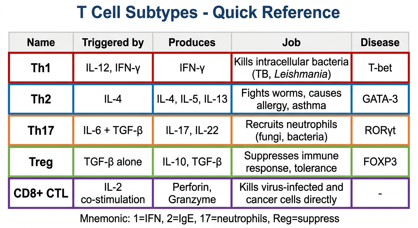
Create a summary flashcard of T cell subtypes and and continue with part 3
Medical study flashcard titled "T Cell Subtypes - Quick Reference" with a clean white background and colored sections. Show a table with 5 rows for Th1, Th2, Th17, Treg, and CD8+ CTL. Each row has columns: Name, Triggered by, Produces, Job, Disease. Th1 row in red: IL-12/IFN-γ, produces IFN-γ, kills intracellular bacteria TB Leishmania, T-bet. Th2 row in blue: IL-4, produces IL-4 IL-5 IL-13, fights worms causes allergy asthma, GATA-3. Th17 row in orange: IL-6+TGF-β, produces IL-17 IL-22, recruits neutrophils fungi bacteria, RORγt. Treg row in green: TGF-β alone, produces IL-10 TGF-β, suppresses immune response tolerance, FOXP3. CD8 CTL row in purple: IL-2 co-stimulation, produces Perforin Granzyme, kills virus-infected and cancer cells directly. Include a small mnemonic at bottom: "1=IFN, 2=IgE, 17=neutrophils, Reg=suppress". Professional medical flashcard style, clear readable fonts, color coded borders per row.
🛡️ IMMUNOLOGY - PART 3
B Cells, Antibodies & Complement
What Are B Cells?
How Does a B Cell Get Activated?
Step 1 - B Cell Sees Antigen
- B cell's receptor (BCR = surface IgM) grabs antigen floating in blood/lymph
- B cell chops up the antigen and presents it on MHC Class II
Step 2 - T Cell Comes to Help
- A CD4+ Tfh cell (T follicular helper - a special Th subtype) recognizes the peptide on MHC II
- CD40L (on T cell) locks onto CD40 (on B cell) → "YOU HAVE PERMISSION TO FIRE"
- T cell also releases cytokines (IL-4, IL-21) to further boost B cell
Step 3 - B Cell Activates
- B cell proliferates
- Enters the Germinal Center (a special zone inside lymph node)
- Magic happens there...
The Germinal Center - Where Antibodies Get UPGRADED
1. Somatic Hypermutation 🔬
- The B cell's antibody gene randomly mutates
- Creates B cells with slightly different antibodies
- Some mutations make the antibody bind the enemy better
- Some make it bind worse
2. Affinity Maturation 🏆
- B cells that make better-fitting antibodies → survive and multiply
- B cells that make worse antibodies → die (apoptosis)
- Result: Over time, antibodies get stronger and stronger
Analogy: Like a sporting tryout. Each round, only the best players survive. After several rounds, you have an elite team.
3. Class Switch Recombination 🔄
- B cell starts making IgM (basic antibody)
- Gets instructions from T cell cytokines to switch to a better antibody class
- IL-4 → switch to IgE or IgG
- TGF-β → switch to IgA
- Same antigen specificity, different antibody class
The 5 Antibody Classes - LEARN THESE COLD
🥇 IgG - The Main Fighter
- Most abundant antibody in blood
- Only antibody that crosses the placenta → gives baby protection for first 6 months
- Opsonization (coats bacteria so macrophages can eat them)
- Neutralization (blocks viruses from entering cells)
- Activates complement
- 4 subclasses: IgG1, IgG2, IgG3, IgG4
🥈 IgM - The First Responder
- First antibody produced in any infection
- Pentamer (5 Y-shapes joined together) → very large
- Best complement activator (classical pathway)
- Does NOT cross placenta
- Found on surface of naive B cells (IgM + IgD together)
Exam tip: If a question says "first antibody in infection" = IgM. Rising IgM = CURRENT infection.
🥉 IgA - The Mucosal Guardian
- Found in secretions: saliva, tears, breast milk, gut, respiratory tract
- Dimer in secretions (two Y-shapes joined)
- Protected by a secretory component (prevents digestion by enzymes)
- First line of defense at mucosal surfaces
- Passed from mother to baby in breast milk (passive immunity)
Analogy: IgA is the border patrol stationed at all entry points (mouth, nose, gut).
IgE - The Allergy Antibody
- Lowest concentration in blood normally
- Binds to mast cells and basophils via Fc receptor
- When antigen cross-links IgE on mast cell → DEGRANULATION → histamine → allergic reaction
- Also important for anti-parasitic (helminth) immunity
High IgE = think: allergy, asthma, parasites, Hyper-IgE syndrome (Job syndrome)
IgD - The Mystery One
- Found on surface of naive B cells alongside IgM
- Function not fully understood
- Not a major exam focus - just know it's a B cell surface marker
Quick Antibody Summary Table:
| Antibody | Special Feature | Crosses Placenta? | Where Found |
|---|---|---|---|
| IgG | Most abundant, opsonization | ✅ YES (only one) | Blood |
| IgM | First produced, pentamer, best complement | ❌ No | Blood |
| IgA | Mucosal immunity, dimer, in secretions | ❌ No | Saliva, gut, breast milk |
| IgE | Allergy + parasites, lowest level | ❌ No | On mast cells |
| IgD | Naive B cell marker | ❌ No | B cell surface |
How Do Antibodies Actually Fight?
1. Neutralization 🛡️
- Antibody physically blocks the virus/toxin from binding to your cell
- Like putting a cap on a key so it can't open the lock
2. Opsonization 🎯
- IgG coats the bacteria
- Macrophages and neutrophils have Fc receptors - they grab the IgG tail
- Phagocytose and destroy the bacteria
- Like putting a "EAT ME" sticker on bacteria
3. Complement Activation 💣
- IgM or IgG on bacteria → activates complement → bacteria gets blown up
4. ADCC (Antibody-Dependent Cell Cytotoxicity)
- IgG coats virus-infected cell
- NK cells grab the IgG tail via their CD16 receptor
- NK cell kills the infected cell
5. Mast Cell Degranulation
- IgE on mast cells → antigen cross-links → histamine release → allergy
COMPLEMENT - The Chemical Bomb System
3 Ways to Start the Cascade (3 Pathways):
| Pathway | Started by | First Step |
|---|---|---|
| Classical | IgM or IgG bound to bacteria | C1q recognizes antibody |
| Lectin | Mannose sugars on bacteria surface | MBL (Mannose Binding Lectin) |
| Alternative | Spontaneous C3 breakdown + bacterial surfaces | C3 hydrolysis |
Mnemonic: "CALM" = Classical-Antibody, Lectin-Mannose, Alternative-spontaneous
All 3 pathways converge at C3:
Classical ──┐
Lectin ─────┼──→ C3 convertase → C3 splits into C3a + C3b
Alternative ┘
                    ↓                    ↓
                   C3a              C3b coats bacteria
              (anaphylatoxin)        (opsonin = "eat me" tag)
                    ↓
              Mast cell degranulation
              (inflammation)
                    ↓
              C5 → C5a + C5b
                    ↓        ↓
              C5a =      C5b + C6+C7+C8+C9
         most powerful         ↓
         chemotactic      MAC (Membrane
           factor       Attack Complex)
                              ↓
                    Punches holes in bacteria
                    → bacteria bursts and dies
Key Complement Products - HIGH YIELD:
| Product | What it does |
|---|---|
| C3b | Opsonin - coats bacteria so phagocytes can eat them |
| C3a | Anaphylatoxin - causes mast cell degranulation, inflammation |
| C5a | Most powerful anaphylatoxin + best chemotactic factor (calls neutrophils) |
| MAC (C5b-9) | Membrane Attack Complex - kills Gram-negative bacteria, Neisseria |
Complement Deficiency Diseases:
| Deficiency | Disease |
|---|---|
| C1, C2, C4 | SLE-like autoimmune disease (immune complexes accumulate) |
| C3 | Most severe - recurrent infections with all encapsulated bacteria |
| C5-C9 (terminal) | Recurrent Neisseria infections ONLY (meningitis, gonorrhea) |
| C1-inhibitor | Hereditary Angioedema - recurrent episodes of swelling |
| DAF / CD55 | PNH (Paroxysmal Nocturnal Hemoglobinuria) - own RBCs destroyed |
Exam favourite: Patient with recurrent Neisseria meningitidis = C5-C9 deficiency
T-Dependent vs T-Independent Antigens
| Feature | T-Dependent | T-Independent |
|---|---|---|
| Antigen type | Proteins | Polysaccharides, lipids |
| Needs T cell help? | ✅ Yes (CD40L-CD40) | ❌ No |
| Class switching? | ✅ Yes (IgG, IgA, IgE) | ❌ No - only IgM |
| Memory cells? | ✅ Yes | ❌ No (weak memory) |
| Example | Tetanus toxoid vaccine | Pneumococcal polysaccharide vaccine |
This is why polysaccharide vaccines (like PPSV23) don't work well in infants under 2 - their T-independent response is weak. That's why we use conjugate vaccines (polysaccharide + protein carrier = now T-dependent) like PCV13.
Part 3 Summary - 5 Things to Remember
| # | Concept | One Line |
|---|---|---|
| 1 | Germinal center | Somatic hypermutation + Affinity maturation + Class switching |
| 2 | IgG | Most abundant, crosses placenta, opsonization |
| 3 | IgM | First produced, pentamer, best complement activator |
| 4 | IgA | Mucosal/secretory immunity, in breast milk |
| 5 | C5-C9 deficiency | Only susceptible to Neisseria infections |
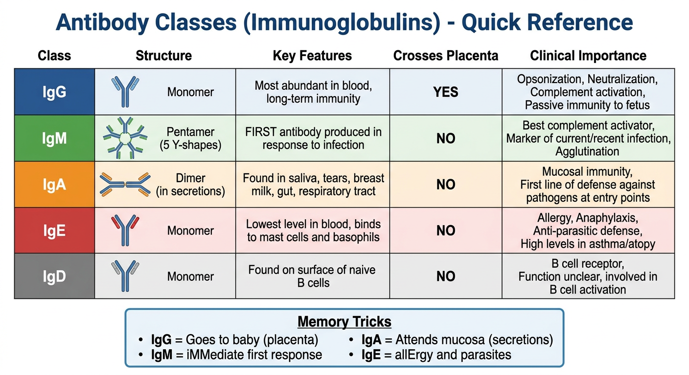
Create another flashcard for Antibody classes also continue with part 4
Medical study flashcard titled "Antibody Classes (Immunoglobulins) - Quick Reference" with clean white background. Show 5 color-coded rows for IgG, IgM, IgA, IgE, IgD. Each row has columns: Class, Structure, Key Features, Crosses Placenta, Clinical Importance. IgG row in dark blue: monomer, most abundant in blood, YES crosses placenta, opsonization neutralization complement activation. IgM row in green: pentamer (5 Y-shapes), FIRST antibody produced, NO, best complement activator current infection marker. IgA row in orange: dimer in secretions, found in saliva tears breast milk gut, NO, mucosal immunity first line defense. IgE row in red: monomer, lowest level in blood binds mast cells, NO, allergy anaphylaxis anti-parasite high in asthma. IgD row in grey: monomer, naive B cell surface marker, NO, function unclear. Bottom section: memory tricks box - "IgG = Goes to baby (placenta)", "IgM = iMMediate first", "IgA = Attends mucosa", "IgE = allErgy". Professional clean medical flashcard design, bold headers, easy to read fonts.
🛡️ IMMUNOLOGY - PART 4
Tolerance, Autoimmunity, Transplant & Tumor Immunity
SECTION 1: Immunologic Tolerance
What is Tolerance?
How Does the Body Prevent Attacking Itself?
Central Tolerance (in Thymus & Bone Marrow)
- Remember negative selection? T cells that bind self-antigens TOO strongly → killed
- Special gene called AIRE makes the thymus display proteins from other organs (thyroid, pancreas, eye)
- T cells that react to these → deleted before they leave
Analogy: Like a background check BEFORE someone joins the army. Anyone with criminal intent gets rejected at the gate.
Peripheral Tolerance (in Tissues - Second Line of Defense)
| Mechanism | How it Works | Analogy |
|---|---|---|
| Anergy | T cell sees antigen (Signal 1) but NO co-stimulation (no Signal 2) → permanently switched off | Car with ignition key but no fuel |
| Treg suppression | FOXP3+ Tregs release IL-10/TGF-β → silence nearby T cells | Internal affairs officer watching other officers |
| Deletion | Activated T cells express Fas → FasL on other cells triggers death → removes T cells after response | Army discharges soldiers after war ends |
| Ignorance | Some self-antigens are hidden in immune-privileged sites (eye, brain, testis) → T cells never see them | VIP area with no public access |
SECTION 2: Autoimmunity
Why Does Autoimmunity Happen?
-
Molecular mimicry - microbial antigen looks similar to self-antigen
- Example: GAS M protein looks like cardiac myosin → Rheumatic fever
- Example: Campylobacter antigen looks like nerve myelin → Guillain-Barré syndrome
-
Bystander activation - tissue damage releases hidden self-antigens → immune system sees them for first time
-
AIRE deficiency - self-reactive T cells escape thymus
-
Loss of Tregs (FOXP3 mutation) → no suppression → IPEX syndrome
Major Autoimmune Diseases - HIGH YIELD TABLE:
| Disease | What's Attacked | Type of Hypersensitivity | Key Autoantibody |
|---|---|---|---|
| SLE (Lupus) | Multiple organs, kidneys | Type III (immune complex) | Anti-dsDNA, Anti-Sm |
| Rheumatoid Arthritis | Joints (synovium) | Type III + IV | Rheumatoid Factor (RF), Anti-CCP |
| Type 1 Diabetes | Pancreatic β cells | Type IV (CTL kills β cells) | Anti-islet cell |
| Graves' Disease | Thyroid (TSH receptor) | Type II (stimulating Ab) | Anti-TSH receptor |
| Hashimoto's Thyroiditis | Thyroid tissue | Type IV | Anti-TPO, Anti-thyroglobulin |
| Myasthenia Gravis | ACh receptor at NMJ | Type II (blocking Ab) | Anti-AChR |
| Goodpasture's | Kidney + Lung basement membrane | Type II | Anti-GBM |
| Multiple Sclerosis | Myelin in CNS | Type IV | Anti-MBP |
Mnemonic for SLE antibodies: "DNA So Many" = dsDNA, Sm (Smith antigen)
SECTION 3: Transplantation Immunology
Why Does the Body Reject Transplants?
Types of Grafts:
| Type | From → To | Rejected? |
|---|---|---|
| Autograft | You → You (e.g. skin graft) | ❌ Never |
| Isograft | Identical twin → Twin | ❌ Never |
| Allograft | Same species, different person | ✅ Yes (if not controlled) |
| Xenograft | Different species (e.g. pig) | ✅ Vigorously |
HLA Matching
- HLA-DR (Class II) - mismatch here = worst rejection
- HLA-A, HLA-B (Class I)
Types of Rejection - EXTREMELY HIGH YIELD:
| Type | When | Mechanism | Features |
|---|---|---|---|
| Hyperacute | Minutes to hours after transplant | Pre-formed antibodies (ABO blood group mismatch OR prior sensitization) | Immediate thrombosis, graft turns black, must be removed NOW |
| Acute | Days to weeks | T cells (CD4+ and CD8+) recognize foreign HLA | Treatable with immunosuppressants |
| Chronic | Months to years | Slow antibody + T cell damage → vessel wall thickening, fibrosis | Gradual function loss, NOT reversible |
Hyperacute = antibody already made BEFORE transplant (like if patient had previous transplant or pregnancy)
Immunosuppression - How We Prevent Rejection:
| Drug | Mechanism | Side Effects |
|---|---|---|
| Cyclosporine / Tacrolimus | Block calcineurin → no NFAT → no IL-2 → no T cell activation | Nephrotoxicity, hypertension |
| Mycophenolate (MMF) | Blocks purine synthesis → lymphocytes can't divide | GI upset, bone marrow suppression |
| Prednisone (steroids) | Broad anti-inflammatory | Weight gain, diabetes, osteoporosis |
| Azathioprine | Blocks DNA synthesis | Bone marrow suppression |
Graft vs Host Disease (GVHD) - Unique to Bone Marrow Transplant
Happens in bone marrow transplant because you're transplanting IMMUNE CELLS (T cells) into the patient.
- Skin rash
- Diarrhea
- Liver dysfunction (jaundice)
- Looks like autoimmune disease
- Dry eyes, dry mouth, skin fibrosis, joint stiffness
- Donor T cells also attack remaining cancer cells in the host
- This is actually therapeutic - helps cure leukemia
SECTION 4: Tumor Immunology
Does the Immune System Fight Cancer?
- Immunocompromised patients (HIV, transplant patients on immunosuppressants) have MUCH higher cancer rates
- Cancers have immune cell infiltrates (immune cells surrounding them)
Tumor Antigens (What the Immune System Recognizes):
| Type | Example |
|---|---|
| Mutated proteins | Mutated p53, Ras - only in cancer cells |
| Oncofetal antigens | AFP (liver cancer), CEA (colon cancer) - proteins normally only in fetus |
| Viral antigens | HPV → cervical cancer; EBV → Burkitt lymphoma; HBV/HCV → liver cancer |
| Overexpressed proteins | HER2/neu (breast cancer) |
How the Immune System Fights Cancer:
| Cell | How It Kills Tumors |
|---|---|
| CD8+ CTL | Main killer - recognizes mutated proteins on MHC Class I → perforin + granzyme |
| NK cells | Kill tumor cells that LOST MHC Class I (virus trick - NK catches it) |
| Macrophages (M1) | Phagocytose and kill tumor cells |
How Cancer Escapes the Immune System:
This is what makes cancer so dangerous.
| Trick | How |
|---|---|
| Lose MHC Class I | CD8+ T cells can't recognize them (no peptide presented) |
| Express PD-L1 | Plugs into PD-1 on T cells → T cell shuts down and dies |
| Secrete TGF-β + IL-10 | Creates immunosuppressive microenvironment around tumor |
| Recruit Tregs | Tregs suppress anti-tumor T cells |
| Antigen loss | Stop expressing the antigen that immune cells were targeting |
Cancer Immunotherapy - The Revolution:
| Drug | Blocks | Cancer Types |
|---|---|---|
| Pembrolizumab / Nivolumab | Anti-PD-1 | Melanoma, lung, many others |
| Atezolizumab | Anti-PD-L1 | Bladder, lung |
| Ipilimumab | Anti-CTLA-4 | Melanoma |
How they work: Remove the brakes → T cells stay active → kill tumor cells Side effect: Autoimmune reactions (immune system over-activated)
- Take patient's own T cells
- Engineer them in lab to target specific tumor antigen (e.g. CD19 on B cell lymphoma)
- Infuse back → super-powered T cells hunt and kill tumor
- Rituximab → Anti-CD20 → kills B cell lymphomas
- Trastuzumab (Herceptin) → Anti-HER2 → breast cancer
- Cetuximab → Anti-EGFR → colorectal cancer
Part 4 Summary - 5 Things to Remember
| # | Concept | One Line |
|---|---|---|
| 1 | AIRE | Shows self-antigens in thymus → negative selection. Mutation → APS-1 |
| 2 | Anergy | Signal 1 without Signal 2 = T cell permanently switched off |
| 3 | Rejection types | Hyperacute (mins, pre-formed Ab) → Acute (weeks, T cells) → Chronic (months, irreversible) |
| 4 | GVHD | Donor T cells attack host - happens in bone marrow transplant |
| 5 | PD-L1 on tumors | Shuts down T cells → immune evasion. Anti-PD-1 drugs block this |
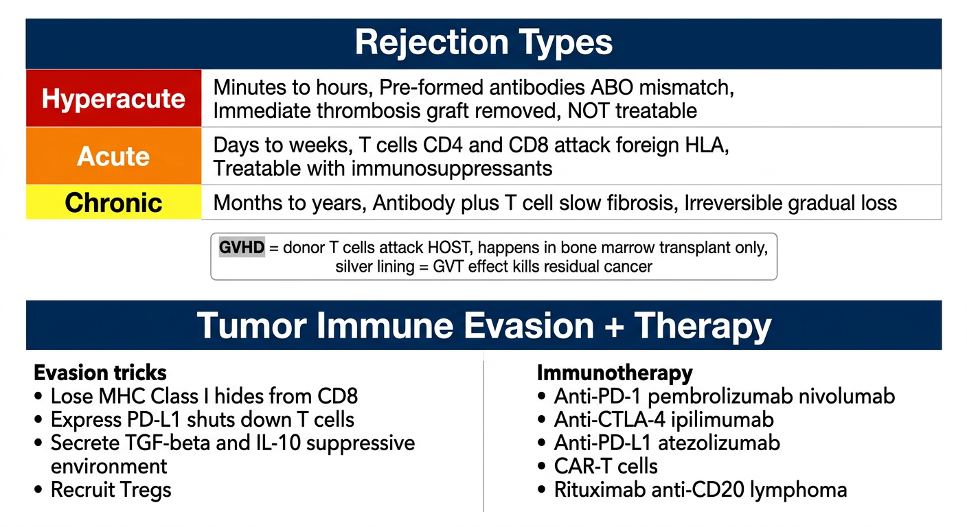
Create a flashcard for part 4 and continue with pa r 5
Medical study flashcard titled "Transplant Rejection + Tumor Immunity - Quick Reference" with clean white background. Two sections. TOP SECTION titled "Rejection Types" with 3 color coded rows: Hyperacute row in red - Minutes to hours, Pre-formed antibodies ABO mismatch, Immediate thrombosis graft removed, NOT treatable. Acute row in orange - Days to weeks, T cells CD4 and CD8 attack foreign HLA, Treatable with immunosuppressants. Chronic row in yellow - Months to years, Antibody plus T cell slow fibrosis, Irreversible gradual loss. Small box: GVHD = donor T cells attack HOST, happens in bone marrow transplant only, silver lining = GVT effect kills residual cancer. BOTTOM SECTION titled "Tumor Immune Evasion + Therapy" with two columns. Left column Evasion tricks: Lose MHC Class I hides from CD8, Express PD-L1 shuts down T cells, Secrete TGF-beta and IL-10 suppressive environment, Recruit Tregs. Right column Immunotherapy: Anti-PD-1 pembrolizumab nivolumab, Anti-CTLA-4 ipilimumab, Anti-PD-L1 atezolizumab, CAR-T cells, Rituximab anti-CD20 lymphoma. Clean professional medical flashcard, bold section headers, easy to read.
🛡️ IMMUNOLOGY - PART 5 (FINAL)
Hypersensitivity + Immunodeficiencies
⚠️ Most Exam-Heavy Topics of All
HYPERSENSITIVITY
What is Hypersensitivity?
Analogy: An overzealous security guard who not only stops the thief but also smashes the entire shop.
TYPE I - Immediate Hypersensitivity (Allergy)
⚡ Seconds to Minutes
The Simple Story:
Allergen enters body (pollen, peanut, bee sting)
        ↓
Th2 cells activated → IL-4 produced
        ↓
B cells switch to IgE
        ↓
IgE binds to Fc receptors on MAST CELLS and BASOPHILS
        ↓
Person is now "sensitized" - nothing happens yet
Same allergen enters again
        ↓
Allergen cross-links IgE on mast cell surface
        ↓
DEGRANULATION - mast cell explodes its granules
        ↓
Histamine + Leukotrienes + Prostaglandins released
        ↓
Vasodilation + Bronchoconstriction + Itching + Swelling
Mediators Released:
| Mediator | Type | Effect |
|---|---|---|
| Histamine | Preformed (instant) | Vasodilation, itching, bronchoconstriction |
| Tryptase | Preformed | Marker of mast cell activation (used in diagnosis) |
| Leukotrienes C4, D4, E4 | Newly formed (slower) | Powerful bronchoconstriction, mucus |
| Prostaglandin D2 | Newly formed | Bronchoconstriction |
| IL-4, IL-5 | Cytokines (late) | Sustain Th2, activate eosinophils |
Clinical Examples:
- Anaphylaxis - systemic, life-threatening (bee sting, peanut, penicillin)
- Asthma - bronchospasm
- Allergic rhinitis - hay fever
- Urticaria - hives
- Atopic dermatitis - eczema
- Food allergy
Treatment of Anaphylaxis:
- Epinephrine IM (FIRST AND MOST IMPORTANT - reverses everything)
- Antihistamines (H1 blockers)
- Corticosteroids
- Bronchodilators (salbutamol)
Exam tip: Anaphylaxis treatment = EPINEPHRINE FIRST. Always.
TYPE II - Cytotoxic Hypersensitivity
⏱️ Hours
The Simple Story:
- Complement activation → MAC punches holes → cell lyses
- Phagocytosis → IgG coats cell → macrophages eat it (opsonization gone wrong)
- ADCC → NK cells kill IgG-coated cells
- Instead of destroying the cell, antibody changes its function
- Example: Ab STIMULATES receptor → overactivation
- Example: Ab BLOCKS receptor → no function
Clinical Examples:
| Disease | What Happens | Antibody |
|---|---|---|
| ABO transfusion reaction | Wrong blood type → IgM attacks donor RBCs → lysis | Anti-A or Anti-B IgM |
| Hemolytic Disease of Newborn (HDN) | Rh- mother, Rh+ baby → maternal IgG crosses placenta → destroys fetal RBCs | Anti-Rh (IgG) |
| Autoimmune hemolytic anemia | IgG attacks own RBCs | Anti-RBC IgG |
| Graves Disease | IgG stimulates TSH receptor → excess thyroid hormone | Anti-TSH receptor (stimulating) |
| Myasthenia Gravis | IgG blocks ACh receptor at NMJ → muscle weakness | Anti-AChR (blocking) |
| Goodpasture Syndrome | IgG attacks basement membrane of kidney + lung | Anti-GBM IgG |
| Pemphigus Vulgaris | IgG attacks desmoglein (skin cell junction) → blisters | Anti-desmoglein |
Trick to remember Graves vs Myasthenia:
- Graves = Gas pedal stuck (stimulating = hyperthyroid, overactive)
- Myasthenia = Muscle paralysis (blocking = no signal = weak muscles)
TYPE III - Immune Complex Hypersensitivity
⏱️ 6-12 Hours
The Simple Story:
Analogy: Imagine police (antibodies) chasing criminals (antigens) through a city. They crash into each other (immune complex) right in the middle of a hospital (kidney). The explosion damages the hospital, not just the criminal.
Antigen + Antibody → Immune Complex formed
        ↓
Complex deposits in tissues (kidney, joints, vessels)
        ↓
Complement activated → C3a, C5a produced
        ↓
Neutrophils recruited → release enzymes
        ↓
TISSUE DAMAGE (not the pathogen - the bystander!)
Clinical Examples:
| Disease | Where Complex Deposits | Features |
|---|---|---|
| SLE (Lupus) | Kidney, skin, joints | Butterfly rash, nephritis, arthritis, low complement |
| Post-streptococcal GN | Kidney glomeruli | Hematuria, proteinuria, 2-3 weeks after Strep throat |
| Serum sickness | Vessels, joints, kidney | Fever, rash, arthritis 7-10 days after foreign protein injection |
| Arthus reaction | Local skin | Local swelling 4-8 hours after injection |
| Farmer's lung | Lung | Inhaled fungal spores → immune complexes in lung |
TYPE IV - Delayed Hypersensitivity
⏱️ 48-72 HOURS (slowest of all)
The Simple Story:
- Find the antigen-specific T cell
- Activate it
- T cell makes cytokines
- Macrophages accumulate
- THEN damage occurs
The 4 Subtypes of Type IV:
| Subtype | Cells Involved | Example |
|---|---|---|
| IVa | Th1 + macrophages | PPD (TB skin test), granuloma in TB |
| IVb | Th2 + eosinophils | Chronic allergic asthma |
| IVc | CD8+ CTL | Contact dermatitis (poison ivy, nickel, hair dye) |
| IVd | Th17 + neutrophils | Pustular psoriasis |
Clinical Examples:
| Condition | What Happens |
|---|---|
| PPD / Mantoux test (TB) | Inject TB antigen in skin → Th1 cells arrive → induration (hardening) at 48-72 hrs = positive |
| Contact dermatitis | Nickel earrings, poison ivy, hair dye, latex → CTLs → itchy rash after 48-72 hrs |
| Graft rejection (acute) | T cells attack foreign HLA on transplant |
| Type 1 Diabetes | CTLs kill pancreatic β cells |
| Multiple Sclerosis | T cells attack myelin |
| Tuberculosis granuloma | Th1 → IFN-γ → macrophages fuse → giant cells → wall off bacteria |
THE MASTER HYPERSENSITIVITY TABLE:
| Type I | Type II | Type III | Type IV | |
|---|---|---|---|---|
| Mediator | IgE | IgG/IgM (cell surface) | IgG (immune complex) | T cells |
| Timing | Seconds-minutes | Hours | 6-12 hours | 48-72 hours |
| Complement? | ❌ No | ✅ Yes | ✅ Yes | ❌ No |
| Mast cells? | ✅ Yes | ❌ No | ❌ No | ❌ No |
| Classic example | Anaphylaxis | Hemolytic anemia | SLE, PSGN | PPD test, Contact dermatitis |
| Nickname | Anaphylactic | Cytotoxic | Immune Complex | Delayed/Cell-mediated |
Mnemonic: "ACID"
- Anaphylactic (I)
- Cytotoxic (II)
- Immune complex (III)
- Delayed (IV)
IMMUNODEFICIENCIES
Primary (Born With It) vs Secondary (Acquired Later)
PRIMARY IMMUNODEFICIENCIES
Grouped by WHAT is missing:
GROUP 1: No B Cells / No Antibodies
- Gene: BTK (Bruton's Tyrosine Kinase) mutation
- Only in boys (X-linked)
- B cells don't mature → NO B cells → NO antibodies
- Fine at birth (has maternal IgG via placenta)
- Gets sick at 6 months when maternal IgG disappears
- Infections: Encapsulated bacteria (S.pneumo, H.flu, Neisseria)
- Treatment: IV immunoglobulin (IVIG) every month
- Most common primary immunodeficiency overall
- Only IgA missing, others normal
- Usually asymptomatic (most people don't know they have it)
- Some get recurrent sinus/chest infections, Giardia
- Danger: Give blood transfusion → anaphylaxis (recipient's anti-IgA antibodies react)
- Low IgG, IgA, IgM
- B cells present but don't mature into plasma cells
- Presents in adults (20-30 years old)
- Recurrent sinopulmonary infections, autoimmunity, lymphoma risk
GROUP 2: No T Cells
- Chromosome 22q11 deletion
- Thymus absent or small → no T cells
- Classic triad:
- Cardiac defects (conotruncal - Tetralogy of Fallot, truncus arteriosus)
- Abnormal facies
- Thymic hypoplasia (no T cells)
- Cleft palate
- Hypocalcemia (parathyroid glands absent → tetany/seizures in newborn)
- Mnemonic: CATCH-22 (Cardiac, Abnormal face, Thymic, Cleft palate, Hypocalcaemia - chromosome 22)
- Infections: Viral and fungal (need T cells to fight these)
GROUP 3: No T cells AND No B cells (SCID)
- The most severe of all - no T cells AND no B cells
- Most common cause: ADA deficiency (Adenosine Deaminase enzyme) → toxic metabolites kill lymphocytes
- Also: RAG1/RAG2 mutations → no V(D)J recombination
- Presents in first weeks-months of life
- Any infection = potentially fatal (viral, bacterial, fungal)
- Classic: "Bubble boy" (David Vetter - lived in sterile plastic bubble)
- Treatment: Bone marrow transplant (curative!)
- Danger: Do NOT give live vaccines → they will cause disease
GROUP 4: Phagocyte Defects
- NADPH oxidase gene mutation
- Neutrophils can engulf bacteria but CANNOT kill them (no respiratory burst, no ROS)
- Infections specifically with catalase-positive organisms (these destroy their own H2O2 so neutrophil can't use it against them)
- Staph aureus, Aspergillus, Serratia, Nocardia, Burkholderia
- Granulomas form because bacteria can't be killed → body walls them off
- Diagnosis: DHR test (Dihydrorhodamine) or NBT test (Nitroblue Tetrazolium) - no color change
- Treatment: Prophylactic trimethoprim-sulfamethoxazole + IFN-γ
- LYST gene mutation → lysosomes can't fuse properly
- Neutrophils have giant granules (visible on blood smear!)
- Partial albinism (melanocytes affected too)
- Recurrent pyogenic infections
- Nerve problems
- CD18 (integrin) mutation → neutrophils can't stick to vessel walls → can't get to infection site
- Hallmark: Delayed separation of umbilical cord (>3 weeks after birth)
- Infections without pus formation (no neutrophils arrive)
- Very high circulating neutrophil count (can't leave blood)
GROUP 5: Complement Deficiencies
| Deficiency | Disease |
|---|---|
| C1q, C2, C4 | SLE-like disease (immune complexes accumulate - not cleared) |
| C3 | Most severe - recurrent infections with ALL bacteria |
| C5-C9 (terminal) | ONLY Neisseria infections (meningitis, gonorrhea) |
| C1-inhibitor | Hereditary Angioedema - episodes of severe swelling (face, larynx - dangerous!) |
Quick Summary Table - Primary Immunodeficiencies:
| Disease | Defect | Bugs | Key Clue |
|---|---|---|---|
| Bruton's | BTK, no B cells | Encapsulated bacteria | Boys, starts at 6 months |
| Selective IgA | No IgA | Giardia, sinus/chest | Most common, anaphylaxis with transfusion |
| CVID | No plasma cells | Encapsulated bacteria | Adults, lymphoma risk |
| DiGeorge | No thymus, no T cells | Viral, fungal | CATCH-22, hypocalcemia, heart defects |
| SCID | No T + B cells | Everything | Bubble boy, ADA deficiency, BMT cures |
| CGD | No NADPH oxidase | Staph, Aspergillus, Serratia | Catalase +ve organisms, granulomas |
| Chediak-Higashi | LYST, bad lysosomes | Pyogenic bacteria | Giant granules, albinism |
| LAD | CD18, bad adhesion | Pyogenic bacteria | Delayed cord separation, no pus |
| C5-C9 deficiency | No MAC | Neisseria only | Recurrent meningococcal disease |
| Wiskott-Aldrich | WASp protein | Encapsulated + opportunistic | Triad: Thrombocytopenia + Eczema + Immunodeficiency |
| Ataxia-Telangiectasia | ATM kinase, DNA repair | Sinopulmonary | Cerebellar wobble + spider veins on eyes |
| Hyper-IgE (Job's) | STAT3 | Staph aureus | Very high IgE, cold abscesses (no redness!) |
SECONDARY IMMUNODEFICIENCIES (Acquired)
| Cause | What's Lost | Example Infections |
|---|---|---|
| HIV/AIDS | CD4+ T cells | Opportunistic: PCP, CMV, Toxoplasma, Cryptococcus |
| Corticosteroids | Broad suppression | Any |
| Chemotherapy | Neutrophils | Bacterial, fungal |
| Splenectomy | Opsonization lost | Encapsulated bacteria (pneumococcal sepsis) |
| Malnutrition | Global suppression | Any |
| Diabetes | Neutrophil function | Skin infections, Mucormycosis |
PART 5 Summary - 6 Things to Remember
| # | Concept | One Line |
|---|---|---|
| 1 | Type I | IgE + mast cell + histamine = seconds. Treat anaphylaxis with EPINEPHRINE |
| 2 | Type II | IgG/IgM against cells. Graves (stimulating) vs Myasthenia (blocking) |
| 3 | Type III | Immune complexes in tissues. Low complement. SLE, PSGN |
| 4 | Type IV | T cells only. 48-72 hrs. PPD test, contact dermatitis |
| 5 | SCID | No T + B cells, ADA deficiency, BMT is cure, NO live vaccines |
| 6 | C5-C9 deficiency | Only Neisseria infections. Always in exams |
🏁 ALL 5 PARTS OF IMMUNOLOGY COMPLETE!
- ✅ Part 1 - Innate vs Adaptive, Receptors, MHC, Lymphocyte development
- ✅ Part 2 - T cell activation (2-signal rule), CD4+ subtypes, CD8+ killing
- ✅ Part 3 - B cells, Antibody classes, Complement pathways
- ✅ Part 4 - Tolerance, Autoimmunity, Transplant, Tumor immunity
- ✅ Part 5 - Hypersensitivity Types I-IV + All immunodeficiencies
Make more flashcards for exam prep like it should inculde everything important
Medical exam flashcard titled "Hypersensitivity Types I-IV Master Table" on white background. Large clear table with 5 columns: Feature, Type I Anaphylactic, Type II Cytotoxic, Type III Immune Complex, Type IV Delayed. Rows: Mediator - IgE / IgG IgM against cells / IgG immune complexes / T cells no antibody. Timing - Seconds-minutes / Hours / 6-12 hours / 48-72 hours. Complement - No / Yes / Yes / No. Mast cells - Yes / No / No / No. Key examples - Anaphylaxis Asthma Hay fever / Graves Myasthenia Hemolytic anemia Goodpasture / SLE PSGN Serum sickness / PPD test Contact dermatitis Type1 DM MS. Treatment - Epinephrine antihistamines / Remove antibody steroids / Steroids / Steroids avoid antigen. Lab clue - High IgE / Direct Coombs positive / Low complement C3 C4 / Positive skin test 48-72hr. Bottom box in yellow: MNEMONIC ACID = Anaphylactic Cytotoxic Immune-complex Delayed. Color code each type column: Type I pink, Type II blue, Type III green, Type IV purple. Professional medical flashcard style.
Medical exam flashcard titled "Primary Immunodeficiencies - High Yield" on white background. Two column layout. Left column lists diseases with colored boxes: Bruton's XLA - BTK mutation, no B cells, boys only, starts 6 months, IVIG treatment, encapsulated bacteria. DiGeorge - 22q11 deletion, no thymus no T cells, CATCH-22 mnemonic Cardiac Abnormal face Thymic Cleft palate Hypocalcemia chromosome 22, viral fungal infections. SCID - No T and B cells, ADA deficiency, bubble boy, BMT cures, NO live vaccines. Right column: CGD - NADPH oxidase defect, catalase positive organisms Staph Aspergillus Serratia, NBT test negative, granulomas. Wiskott-Aldrich - WASp mutation, TRIAD Thrombocytopenia plus Eczema plus Immunodeficiency. Selective IgA - Most common primary immunodeficiency, anaphylaxis with blood transfusion. LAD - CD18 defect, delayed umbilical cord separation, no pus. Hyper IgE Job syndrome - STAT3, cold abscesses no redness, very high IgE. Bottom row: Complement deficiencies - C1q C2 C4 = SLE-like, C3 = all bacteria, C5-C9 = Neisseria only, C1-inh = Hereditary Angioedema. Clean professional design bold text color coded rows.
Medical exam flashcard titled "MHC Class I vs II + T Cell Activation" on white background. TOP SECTION: Two side by side colored boxes. Blue box MHC Class I: Found on ALL nucleated cells, Presents to CD8+ T cells Killers, Source = inside cell endogenous viruses tumors, Peptide 8-9 amino acids, HLA-A HLA-B HLA-C, CD8 binds alpha-3 domain. Red box MHC Class II: Found on APCs only dendritic cells macrophages B cells, Presents to CD4+ T cells Helpers, Source = outside cell exogenous bacteria, Peptide 10-20 amino acids, HLA-DR HLA-DP HLA-DQ, CD4 binds beta-2 domain. Memory trick box: CD8 times 8 = Class I. CD4 times 4 = Class II. MIDDLE SECTION titled Two Signal Rule: Signal 1 = TCR binds peptide-MHC antigen specific. Signal 2 = CD28 binds B7 co-stimulation. Both needed = ACTIVATION. Signal 1 alone = ANERGY permanently off. BOTTOM SECTION: Checkpoints table - CTLA-4 competes with CD28 for B7 = brakes on T cell, PD-1 binds PD-L1 on tumor = shuts down T cell, Cancer drugs Anti-PD-1 pembrolizumab Anti-CTLA-4 ipilimumab remove these brakes. Professional clean medical flashcard.
Medical exam flashcard titled "Complement Pathways + Deficiency Diseases" on white background. TOP SECTION: Three pathway boxes side by side. Box 1 Classical pathway - Triggered by IgM or IgG bound to antigen, starts with C1q. Box 2 Lectin pathway - Triggered by Mannose on bacteria surface, starts with MBL. Box 3 Alternative pathway - Spontaneous C3 hydrolysis on bacterial surface. All three arrows pointing down to center box: ALL CONVERGE AT C3. C3 splits to C3a anaphylatoxin mast cell degranulation inflammation AND C3b opsonin coats bacteria for phagocytosis. Then C5 splits to C5a most powerful chemotactic factor calls neutrophils AND C5b leads to C5b+C6+C7+C8+C9 = MAC Membrane Attack Complex = punches holes in bacteria lysis. BOTTOM SECTION: Deficiency Disease table - C1q C2 C4 deficiency causes SLE-like disease. C3 deficiency causes severe recurrent all bacterial infections. C5-C9 deficiency causes Neisseria meningitidis and gonorrhea ONLY. C1-inhibitor deficiency causes Hereditary Angioedema episodes of swelling. DAF CD55 deficiency causes PNH Paroxysmal Nocturnal Hemoglobinuria. Bold color coded professional flashcard design.
Medical exam flashcard titled "Key Cytokines You Must Know" on clean white background. Table with 3 columns: Cytokine, Main Source, Key Function. Rows color coded by function group. Inflammatory group in red: IL-1 from Macrophages - fever acute inflammation. TNF-alpha from Macrophages - fever inflammation cachexia septic shock. IL-6 from Macrophages - fever acute phase proteins CRP. IL-8 CXCL8 from Macrophages - neutrophil chemotaxis. T cell group in blue: IL-2 from T cells - T cell proliferation master growth factor blocked by cyclosporine. IL-4 from Th2 - IgE class switching Th2 differentiation. IL-5 from Th2 - eosinophil activation. IL-12 from Macrophages DCs - Th1 differentiation. IL-17 from Th17 - neutrophil recruitment. IFN-gamma from Th1 NK cells - macrophage activation kills intracellular bacteria. Anti-inflammatory group in green: IL-10 from Tregs Macrophages - suppresses immune response. TGF-beta from Tregs - immunosuppression Treg induction class switch to IgA. Antiviral group in orange: IFN-alpha IFN-beta from virus infected cells - antiviral state in neighboring cells. Bottom mnemonic box: IL-2 multiply, IL-4 IgE allergy, IL-5 eosinophils worms, IL-12 Th1, IFN-gamma activate macrophages. Clean professional design.
Medical exam flashcard titled "Autoimmune Diseases Quick Reference" on white background. Large table with 4 columns: Disease, What is Attacked, Hypersensitivity Type, Key Autoantibody. Rows: SLE Lupus - Multiple organs kidneys skin - Type III immune complex - Anti-dsDNA Anti-Sm butterfly rash. Rheumatoid Arthritis - Joint synovium - Type III plus IV - Rheumatoid Factor RF Anti-CCP. Type 1 Diabetes - Pancreatic beta cells - Type IV CTL - Anti-islet cell GAD65. Graves Disease - TSH receptor thyroid - Type II stimulating - Anti-TSH receptor hyperthyroid. Hashimoto Thyroiditis - Thyroid tissue - Type IV - Anti-TPO Anti-thyroglobulin hypothyroid. Myasthenia Gravis - ACh receptor NMJ - Type II blocking - Anti-AChR muscle weakness. Goodpasture Syndrome - Kidney plus lung basement membrane - Type II - Anti-GBM hematuria hemoptysis. Multiple Sclerosis - Myelin CNS - Type IV - Anti-MBP demyelination. Pemphigus Vulgaris - Skin desmoglein - Type II - Anti-desmoglein blisters. Bottom box: Graves = Gas pedal stuck stimulating = hyperthyroid. Myasthenia = Muscles paralyzed blocking = weak. SLE = Low complement anti-dsDNA. Professional color coded medical flashcard.
400, message: Got more than 4194304 bytes when reading: b'data: {"candidates": [{"content": {"role": "model","parts": [{"inlineData": {"mimeType": "image/png"...'.
We encountered an error while processing your request. Please try again or reach out to us for help.
Create and sens 1 by one
An error occurred (ValidationException) when calling the ConverseStream operation: Expected toolResult blocks at messages.64.content for the following Ids: tooluse_M4rqP3v9olpafrlUvWgtFH, tooluse_M4pYJDUcXes8pcAQX4Wgu5
We encountered an error while processing your request. Please try again or reach out to us for help.
Create and send images 1 by 1
An error occurred (ValidationException) when calling the ConverseStream operation: Expected toolResult blocks at messages.64.content for the following Ids: tooluse_M4rqP3v9olpafrlUvWgtFH, tooluse_M4pYJDUcXes8pcAQX4Wgu5
We encountered an error while processing your request. Please try again or reach out to us for help.
HERPES (CMV,EBV,HSV) Ebola what virus Parasites (atealst know their name add manifest) zika virus complication Parvob19 Papiloma( manifest) Contaminated water bacteria Pox (cowpox) Hepatitis c , b and d manifest Prions Polio vaccine immunity Hypersensitivity Which cell activate Cd8 , th1 , th2 Giant cell related heterophile antibody
An error occurred (ValidationException) when calling the ConverseStream operation: Expected toolResult blocks at messages.64.content for the following Ids: tooluse_M4rqP3v9olpafrlUvWgtFH, tooluse_M4pYJDUcXes8pcAQX4Wgu5
We encountered an error while processing your request. Please try again or reach out to us for help.