
Chyne stroke respiration case based question for 15 marks mbbs physiology 1st year
Reading File
Now let me search the medical library for Cheyne-Stokes respiration content.
Finding Sources
Finding Sources
Excellent! Now let me read the most relevant sections - Ganong's (physiology textbook for 1st year MBBS) and Guyton's:
Reading File
Reading File
Finding Sources
Now I have rich content from Guyton and Ganong. Let me also fetch the Cheyne-Stokes diagram from Guyton:
Loading Image
Reading File
I now have all the content needed from Guyton and Ganong to write a comprehensive 15-mark case-based question. Let me compose the answer.
Cheyne-Stokes Respiration - 15 Marks Case-Based Question (MBBS 1st Year Physiology)
CASE VIGNETTE
A 62-year-old male with a known history of congestive heart failure (CHF) is brought to the emergency department by his wife. She reports that for the past week, while he sleeps, she has noticed his breathing becoming progressively deeper and faster, then gradually slowing down and stopping entirely for about 15-20 seconds, after which the cycle repeats. The patient complains of waking up suddenly at night feeling breathless, excessive daytime sleepiness, and morning headaches. On examination, his pulse is weak and irregular, and he appears pale and slightly confused.
QUESTIONS
(a) Name the breathing pattern being described. Draw and label its graphical representation. (3 marks)
(b) Explain the physiological mechanism responsible for this pattern of breathing in this patient. (5 marks)
(c) What are the other clinical conditions in which this breathing pattern is seen? (3 marks)
(d) How does this breathing pattern differ from normal respiratory regulation? What are the consequences for the patient? (4 marks)
MODEL ANSWER
(a) Name and Diagram (3 marks)
The breathing pattern is Cheyne-Stokes Respiration (CSR), also called periodic breathing.
It is a cyclical pattern of breathing characterized by a crescendo-decrescendo (waxing and waning) increase in depth and rate of respiration, followed by a period of central apnea (complete cessation of breathing), lasting approximately 40-60 seconds per cycle.
Diagram:
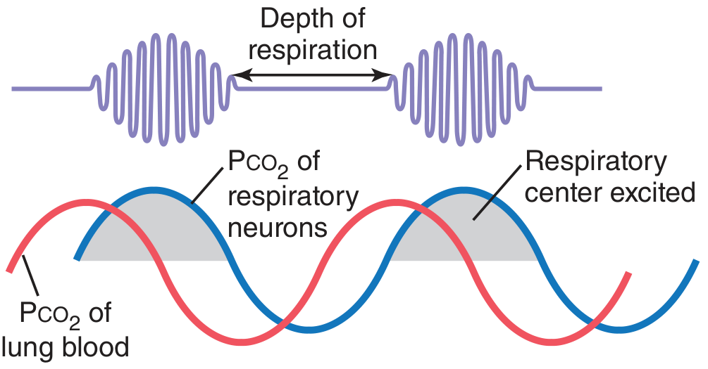
Fig: Cheyne-Stokes breathing. The red line shows PCO2 of pulmonary blood; the blue line shows PCO2 at the respiratory neurons. Note that the depth of respiration follows the PCO2 at the brain, not the lung. (Guyton and Hall, Fig 42.12)
Label your diagram to show:
- Phase of apnea
- Phase of hyperpnea (crescendo phase)
- Phase of declining ventilation (decrescendo phase)
- PCO2 in pulmonary blood (changes first)
- PCO2 at respiratory center (changes with a delay)
(b) Mechanism of Cheyne-Stokes Respiration in CHF (5 marks)
The underlying mechanism involves a delayed negative feedback loop between the lungs and the brainstem respiratory center. In this patient with CHF, two factors operate:
Step-by-step mechanism:
-
Slow circulation (prolonged lung-to-brain circulation time): In CHF, cardiac output is reduced. Blood flow from the lungs to the brainstem chemoreceptors is significantly delayed (the normal lung-to-brain transit time is prolonged).
-
Hyperpnea phase: The respiratory center in the medulla responds to a rise in PCO2 / fall in PO2, driving vigorous hyperventilation. This rapidly lowers the PCO2 of pulmonary blood and raises PO2.
-
The delay problem: Because circulation is slow, this low-PCO2 blood takes several extra seconds to reach the brain and medullary chemoreceptors. During this delay, the lungs continue to blow off CO2, lowering PCO2 even further than intended.
-
Apnea phase: When the blood with very low PCO2 finally reaches the brain, the respiratory center is strongly inhibited, producing central apnea (cessation of breathing). During apnea, metabolic CO2 begins to accumulate in the blood again.
-
Cycle repeats: When the now high-PCO2 blood reaches the brain after another delay, the respiratory center is re-excited, and vigorous breathing restarts. The negative feedback loop overshoots in both directions because of the abnormal delay, producing the characteristic cyclic pattern.
In Ganong's words: "The respiratory control system oscillates because the negative feedback loop from lungs to brain is abnormally long." (Ganong's Review of Medical Physiology, 26th ed.)
Additionally: Some CHF patients also show increased CO2 sensitivity - a small rise in PCO2 causes a disproportionately large ventilatory response (10-20 fold instead of normal 2-3 fold), which further amplifies the oscillation.
Key point: Depth of respiration corresponds to PCO2 at the respiratory neurons (brain), NOT to PCO2 at the pulmonary blood. Changes in pulmonary blood PCO2 occur first, but the effect on respiration is delayed.
(c) Other Conditions Causing Cheyne-Stokes Respiration (3 marks)
| Category | Conditions |
|---|---|
| Cardiac | Congestive heart failure (most common), severe mitral stenosis |
| Renal | Uremia (renal failure) - metabolic cause |
| Neurological | Stroke, traumatic brain injury, brain tumors, increased intracranial pressure, bilateral supramedullary dysfunction |
| Metabolic | Severe hypoxemia (e.g., high altitude - above 2700 m) |
| Physiological | During normal sleep in some elderly individuals, premature neonates |
| Drug-induced | Opioid/narcotic overdose (depresses respiratory center) |
- Ganong's: "It is seen most commonly in patients with heart failure and uremia, but it occurs also in patients with brain disease and during sleep in some normal individuals."
- In neurological disorders, the mechanism is increased gain (hypersensitivity) of the respiratory center due to supramedullary dysfunction, rather than delayed circulation.
(d) Difference from Normal Respiratory Regulation and Consequences (4 marks)
Normal respiratory regulation:
- Medullary chemoreceptors respond to PCO2/H+ changes and peripheral chemoreceptors (carotid/aortic bodies) respond to PO2 falls.
- Under normal conditions, the feedback loop is well-damped. Blood and tissue have large buffering capacities for CO2/O2, so the respiratory response is smooth and proportionate.
- Normal ventilatory response to a 3 mmHg rise in PCO2 = 2-3 fold increase in ventilation.
How Cheyne-Stokes differs:
| Feature | Normal Regulation | Cheyne-Stokes |
|---|---|---|
| Feedback loop | Short, well-damped | Prolonged, oscillating |
| Ventilatory response | Proportionate | Exaggerated (10-20 fold) |
| PCO2 at brain | Reflects lung PCO2 quickly | Delayed by slow circulation |
| Breathing pattern | Regular, even | Cyclic waxing-waning with apnea |
| CO2 buffering | Effective (large CO2 stores in blood) | Overwhelmed by slow circulation |
Consequences for this patient:
- Nocturnal hypoxemia - Apneic phases cause oxygen desaturation, leading to morning headaches, confusion, and fatigue.
- Sleep fragmentation - Patients typically arouse at the peak of hyperpnea, causing excessive daytime sleepiness and poor quality of life.
- Increased sympathetic activity - Repeated hypoxemia activates the sympathetic nervous system, worsening cardiac function and potentially triggering arrhythmias.
- CO2 fluctuations - Swings in PCO2 cause fluctuations in cerebral blood flow, which can produce altered mental states and EEG changes.
- Prognostic significance - In CHF patients, the presence of CSR is associated with increased mortality. It is not merely a symptom but a marker of disease severity.
SUMMARY TABLE FOR QUICK REVISION
| Feature | Detail |
|---|---|
| Type of pattern | Crescendo-decrescendo hyperpnea + central apnea |
| Cycle duration | ~40-60 seconds |
| Primary mechanism in CHF | Prolonged lung-to-brain circulation time |
| Mechanism in brain disease | Increased gain/sensitivity of respiratory center |
| Key difference from normal | Abnormally long negative feedback loop |
| Consequences | Hypoxemia, arousals, sympathetic activation, poor prognosis |
| Treatment principle | Treat underlying cause (CHF - optimize cardiac output; CPAP may help) |
Sources: Guyton and Hall Textbook of Medical Physiology, 14th ed., Ch. 42 (p. 539); Ganong's Review of Medical Physiology, 26th ed., Ch. on Regulation of Respiration.
This is a shared conversation. Sign in to Orris to start your own chat.