

Explain this to me in simplest language possible
🔴 TIER 1 - Study These FIRST (Most Important)
| Topic | What it means simply |
|---|---|
| Cardiac Output | How much blood the heart pumps per minute, and how to measure it |
| Cardiac Cycle | The steps the heart goes through with each beat (squeeze → relax) |
| Shock | When blood supply fails - types and how the body tries to fix it |
| Circulatory Shock | Same as above but more detail on stages and treatment |
| O2-Hb Dissociation Curve | How oxygen attaches to and releases from blood cells |
| Hypoxia | When the body doesn't get enough oxygen |
| Surfactant | A liquid in lungs that stops them from collapsing; if missing → baby lung disease |
| GFR | How fast kidneys filter blood |
| Peptic Ulcer | Stomach ulcer - causes and the physiology behind it |
| HCl Secretion | How the stomach produces acid |
| Countercurrent Multiplier | How kidneys concentrate urine (a kidney mechanism) |
🟡 TIER 2 - Study These SECOND (Good Backup)
- Blood Pressure - how the body controls BP (baroreceptors, RAAS system)
- Hemostasis + Hemophilia A - how blood clots; what goes wrong in Hemophilia
- ECG - reading heart electrical signals
- Venous Return - blood flowing back to the heart
- Digestion topics - swallowing phases, gastric emptying, gut hormones, fat absorption
- CO2 transport - how CO2 is carried in blood
- High-altitude acclimatization - how the body adjusts at high altitude
- Lung compliance - how easily lungs can stretch
🟢 TIER 3 - Study if Time Permits (Bonus)
- GFR regulation, kidney in acid-base balance, bladder physiology
- Breathing control, Ondine's curse (rare breathing disorder)
- VO2 max, oxygen debt, chemoreceptors
💡 "Explain Why" - Short Answer Questions to Prepare
- Why can yogurt be tolerated but not milk in lactose intolerance?
- Why does protein leak into urine in nephritis?
- Why do we breathe faster during exercise?
- Why is coronary blood flow highest during heart relaxation (diastole)?
- Why are blankets avoided in shock patients?
- Why can't the heart go into sustained spasm (tetany)?
- Why are vaccines less effective in immunocompromised people?
- Why does lack of surfactant cause IRDS (infant respiratory distress)?
📌 Bottom Line
Study Tier 1 first, Tier 2 second. Tier 3 is a bonus if you have extra time.
Can you explain these topic briefly but in simple language
🔴 TIER 1 Topics - Simple Explanations
1. 🫀 Cardiac Output
CO = Heart Rate × Stroke Volume (e.g., 72 beats/min × 70 mL/beat = ~5 litres/min)
| Factor | Simple Meaning |
|---|---|
| Heart Rate | How fast the heart beats |
| Preload | How much blood fills the heart before it contracts (stretch) |
| Afterload | The resistance the heart must push against (like blood pressure) |
| Contractility | How strongly the heart muscle squeezes |
2. 🔄 Cardiac Cycle
- Atrial Systole - Atria contract, top-up the ventricles with a last bit of blood (~20%)
- Isovolumetric Contraction - Ventricles start squeezing. All valves closed. Volume stays same, pressure builds.
- Rapid Ejection - Aortic valve opens. Blood shoots out into aorta.
- Reduced Ejection - Blood flow slows down.
- Isovolumetric Relaxation - Ventricles relax. All valves closed again. Pressure falls.
- Rapid Ventricular Filling - Mitral valve opens. Blood rushes from atria into ventricles.
- Reduced Filling - Flow slows, passive filling continues.
- P wave = atria contracting
- QRS complex = ventricles contracting
- T wave = ventricles relaxing
- S1 ("Lub") = Mitral and tricuspid valves closing (start of systole)
- S2 ("Dub") = Aortic and pulmonary valves closing (end of systole)
3. ⚡ Shock (Types + Hypovolemic + Compensatory Mechanisms)
| Type | Simple Cause | Example |
|---|---|---|
| Hypovolemic | Not enough blood/fluid in body | Heavy bleeding, severe dehydration |
| Cardiogenic | Heart is too weak to pump | Heart attack |
| Distributive | Blood vessels too dilated, BP drops | Sepsis, anaphylaxis, neurogenic |
| Obstructive | Blood flow physically blocked | Pulmonary embolism, cardiac tamponade |
- Initial (non-progressive): Body compensates successfully
- Progressive: Compensation failing, organs start suffering
- Irreversible: Permanent organ damage, death likely
- Heart beats faster (tachycardia)
- Blood vessels constrict (cool, pale skin)
- Kidneys retain water (reduced urine)
- Adrenaline released (fight-or-flight response)
- Blood diverted from skin/gut to brain and heart
4. 🩸 Circulatory Shock (Classify, Stages, Management)
- Hypovolemic: Give IV fluids, stop bleeding
- Cardiogenic: Support the heart (dopamine, dobutamine), treat the cause
- Septic/Distributive: Antibiotics + IV fluids + vasopressors (noradrenaline)
- Obstructive: Remove the obstruction (e.g., drain tamponade, clot-bust PE)
5. 📈 O2-Hb Dissociation Curve (Bohr Effect, 2,3-BPG)
- High affinity = Hb grips oxygen tightly → oxygen stays in blood (not released to tissues)
- Low affinity = Hb releases oxygen easily → tissues get more oxygen
6. 😮💨 Hypoxia (Types + Hypoxic Hypoxia)
| Type | Cause | Simple Example |
|---|---|---|
| Hypoxic hypoxia | Low O2 in blood (low PaO2) | High altitude, lung disease |
| Anemic hypoxia | Not enough hemoglobin to carry O2 | Severe anemia, CO poisoning |
| Stagnant/Ischemic hypoxia | Blood flow too slow | Heart failure, shock |
| Histotoxic hypoxia | Cells can't use the O2 delivered | Cyanide poisoning |
- Most common type
- Low atmospheric O2 OR lungs can't get O2 into blood
- Causes: high altitude, pneumonia, COPD, drowning
- Response: breathe faster, heart pumps harder, make more red blood cells (if chronic)
7. 🫁 Surfactant (RDS, Hyaline Membrane Disease)
- Reduces surface tension in alveoli
- Stops alveoli from collapsing after each breath out
- Makes breathing much easier - lungs don't have to work hard to re-inflate
- Premature babies lack surfactant
- Alveoli collapse with each breath
- Baby has to work extremely hard to breathe
- This is IRDS (Infant Respiratory Distress Syndrome) = also called Hyaline Membrane Disease
- Pink "hyaline membranes" form in alveoli (dead cells + fluid)
- Treatment: Give artificial surfactant, oxygen, breathing support; give mother steroids before preterm birth to speed up lung maturity
8. 🧫 GFR (Factors + Tubuloglomerular Feedback)
- Low protein diet (less oncotic pressure)
- High blood pressure
- Dilation of afferent arteriole (coming in)
- Constriction of afferent arteriole (e.g., in shock)
- Low blood pressure
- Obstruction in urinary tract
It's the kidney's own self-regulation system.
- If GFR goes too HIGH → more fluid/salt reaches the macula densa (a sensor in the kidney tubule)
- Macula densa senses this → sends signal to constrict the afferent arteriole → GFR comes back down
- If GFR too LOW → opposite happens → arteriole dilates → GFR goes up
- This keeps filtration stable automatically - like a thermostat for the kidney!
9. 🔴 Peptic Ulcer (Etiology + Physiological Basis)
- H. pylori infection (most common - a bacteria that lives in stomach)
- NSAIDs (painkillers like ibuprofen damage the stomach lining)
- Excess acid (Zollinger-Ellison syndrome)
- Stress (serious illness, burns → "stress ulcer")
- Normally, stomach has protective factors: mucus layer, bicarbonate, blood flow, tight cell junctions
- When acid/pepsin overpower the protective factors → ulcer forms
- H. pylori breaks down the mucus layer directly
- NSAIDs block prostaglandins (which normally stimulate mucus production)
10. 🧪 HCl Secretion by Parietal Cells
| Stimulator | Source |
|---|---|
| Acetylcholine | Vagus nerve (sight/smell/thought of food) |
| Gastrin | G-cells in stomach antrum (when food arrives) |
| Histamine | Enterochromaffin-like (ECL) cells (responds to gastrin) |
- CO2 + H2O → H2CO3 (carbonic acid) via carbonic anhydrase
- H2CO3 → H⁺ + HCO3⁻
- H⁺ pumped into stomach by H⁺/K⁺ ATPase pump (the "proton pump")
- Cl⁻ follows H⁺ → making HCl
11. 🌊 Countercurrent Multiplier Mechanism
- Descending limb: Permeable to water → water leaves → fluid inside becomes more concentrated
- Ascending limb: Pumps out NaCl but is impermeable to water → salt leaves, but water stays → fluid becomes dilute
- ADH present → water pulled out → concentrated urine (small amount)
- No ADH → water stays → dilute urine (diabetes insipidus)
🟡 TIER 2 Topics - Quick Summaries
| Topic | Simple Explanation |
|---|---|
| Blood Pressure (Baroreceptor, RAAS) | Baroreceptors = pressure sensors in arteries that signal the brain to adjust BP. RAAS = hormonal system (Renin→Angiotensin→Aldosterone) that raises BP by retaining salt and water. |
| Hemostasis + Hemophilia A | Hemostasis = stopping bleeding. Steps: vessel spasm → platelet plug → clotting cascade → fibrin clot. Hemophilia A = missing Factor VIII → clotting cascade fails → uncontrolled bleeding. |
| Countercurrent mechanism in kidney | Same as above - the loop of Henle mechanism for concentrating urine. |
| ECG (Unipolar/Bipolar, Lead II) | ECG records electrical activity of the heart. Bipolar leads (I, II, III) measure between two points. Unipolar leads (aVR, aVL, aVF; V1-V6) measure from one point. Lead II is standard for rhythm strips. |
| Venous Return | Blood returning from body to right side of heart. Helped by: muscle pump (leg muscles), respiratory pump (breathing), venous valves, and heart suction. |
| Factors affecting Stroke Volume | Preload (↑ = ↑ SV), Afterload (↑ = ↓ SV), Contractility (↑ = ↑ SV) - these are the key three. |
| Phases of Deglutition | Swallowing has 3 phases: Oral (voluntary, tongue pushes food back), Pharyngeal (reflex, food passes throat safely), Esophageal (peristalsis waves push food down). |
| Regulation of Gastric Emptying | Stomach empties slowly (1-4 hours). Slowed by: fats, acids, high osmolality (via CCK). Speeded by: gastrin, stomach distension. |
| GIT Hormones (Gastrin, CCK) | Gastrin (from G-cells) = stimulates acid + gastric motility. CCK (from duodenum) = stimulates pancreatic enzymes + bile release, slows gastric emptying. |
| Fat Absorption | Fats broken down by lipase → absorbed as monoglycerides + fatty acids → packaged into micelles (with bile salts) → enter intestinal cells → packaged into chylomicrons → enter lymphatics (not blood directly). |
| CO2 Transport + Haldane Effect | CO2 transported as: bicarbonate (70%), attached to Hb as carbaminoHb (23%), dissolved (7%). Haldane effect: oxygenated blood carries less CO2 (and vice versa). |
| High-Altitude Acclimatization | Body adapts to low O2: breathe faster → blow off CO2 → kidneys compensate by excreting bicarbonate. Kidneys make more EPO → more red blood cells. 2,3-BPG increases → more O2 released to tissues. |
| Lung Compliance | How easily lungs stretch. High compliance = lungs expand easily (emphysema). Low compliance = stiff lungs, hard to breathe (pulmonary fibrosis). Surfactant increases compliance. |
💡 "Explain Why" - Answers in Simple Language
| Question | Simple Answer |
|---|---|
| Yogurt OK, milk not in lactose intolerance? | Lactose intolerance = no lactase enzyme → can't digest milk sugar. Yogurt contains bacteria that have already pre-digested the lactose, so very little remains to cause symptoms. |
| Albuminuria in nephritis? | Normally kidneys filter blood but protein (albumin) is too big to pass through. In nephritis, glomeruli are inflamed/damaged → their filter becomes "leaky" → protein spills into urine. |
| Tachypnea during exercise? | Muscles produce more CO2 → CO2 dissolved in blood → slightly acidic → chemoreceptors detect this → signal brain to breathe faster to blow off CO2. |
| Coronary flow maximal in diastole? | During systole (heart squeezing), heart muscle is so tightly contracted that it squeezes its own vessels shut. Only during diastole (relaxation) do the coronary vessels open up fully and get adequate blood flow. |
| Blanket avoided in shock? | Shock causes vasoconstriction in the skin (skin becomes cold/pale) to redirect blood to vital organs. A blanket causes vasodilation in skin → blood pools in skin → even less blood available for vital organs → shock worsens. |
| Cardiac muscle can't be tetanized? | Tetany = sustained muscle contraction. Cardiac muscle has a very long refractory period (the heart cannot be re-stimulated while it's still contracting). This is a safety feature - if the heart could tetanize, it would stop pumping blood. |
| Vaccines less effective in immunocompromised? | Vaccines work by training the immune system to recognize a pathogen. If the immune system is weakened (chemotherapy, HIV, steroids), it can't mount a strong enough response to "learn" from the vaccine. |
| Surfactant deficiency causes IRDS? | Without surfactant → high surface tension in alveoli → alveoli collapse after each breath → baby must work enormously hard to re-inflate collapsed lungs → respiratory failure. |
📌 Study Tip: For each Tier 1 topic, know: (1) the basic definition, (2) the mechanism, and (3) one clinical example. That's usually enough to answer exam questions!That covers every single topic from the guide! Here's a quick recap of what was covered:
- Cardiac Output, Cardiac Cycle, Shock, Circulatory Shock
- O2-Hb Dissociation Curve (Bohr effect, 2,3-BPG)
- Hypoxia (all 4 types), Surfactant + IRDS
- GFR + Tubuloglomerular Feedback
- Peptic Ulcer, HCl secretion, Countercurrent Multiplier
Peptic ulsar
peptic ulcer stomach diagram pathophysiology
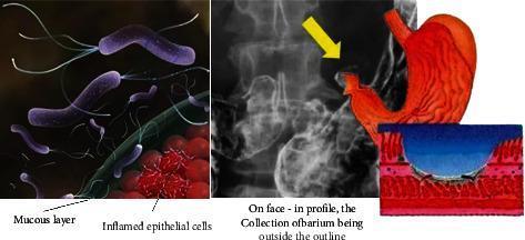
This composite educational illustration details the pathogenesis and clinical imaging of a peptic ulcer. The left panel features a pathophysiology diagram showing purple, flagellated Helicobacter pylori bacteria penetrating a green mucous layer to reach red, inflamed epithelial cells, representing the primary infectious cause of gastric irritation. The center-right panel integrates diagnostic and anatomical content, featuring a grayscale barium swallow X-ray alongside a red anatomical diagram of the stomach. A yellow arrow on the X-ray identifies a barium collection outside the normal gastric outline, characteristic of an ulcer crater (ulcer niche). A supplementary cross-sectional illustration provides a microscopic view of the ulcer, depicting a blue-shaded crater penetrating through the mucosal and submucosal layers into the underlying tissue. This graphic is intended for medical students to understand the transition from microbial infection to macroscopic tissue damage and its subsequent appearance in radiological imaging.
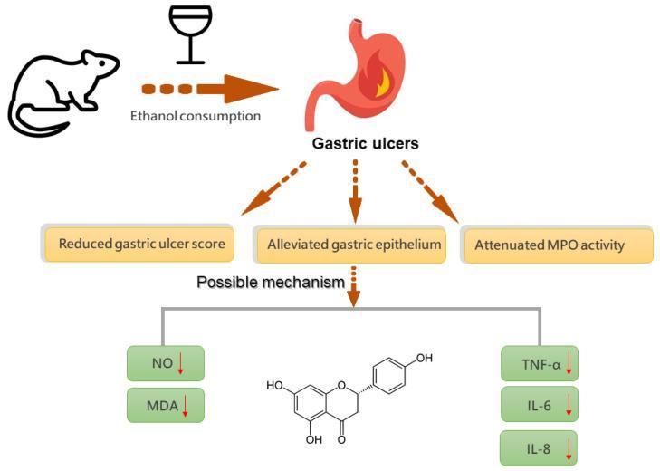
This pathophysiology diagram illustrates the gastroprotective mechanism of naringenin against ethanol-induced gastric ulcers. The diagram starts with an animal model (rat/mouse) consuming ethanol, depicted by a wine glass icon, leading to the development of 'Gastric ulcers' in a stomach graphic showing inflammation/burning. Three primary outcomes are highlighted following intervention: reduced gastric ulcer score, alleviated gastric epithelium damage, and attenuated myeloperoxidase (MPO) activity. The lower portion of the infographic details the 'Possible mechanism' involving naringenin, whose chemical structure is centrally displayed. The mechanism indicates a reduction (marked with red downward arrows) of several biomarkers: oxidative stress markers Nitric Oxide (NO) and Malondialdehyde (MDA), as well as pro-inflammatory cytokines Tumor Necrosis Factor-alpha (TNF-α), Interleukin-6 (IL-6), and Interleukin-8 (IL-8). This educational summary emphasizes the anti-inflammatory and antioxidant role of flavonoids in treating mucosal injury.
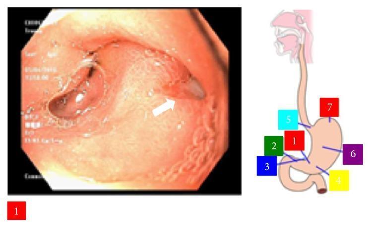
This composite educational image features an upper gastrointestinal endoscopy view of the stomach and an anatomical diagram of the upper digestive tract. The endoscopic image displays the gastric antrum, specifically highlighting a deep, nonbleeding penetrating ulcer on the posterior wall. The ulcer presents as a well-circumscribed lesion with a dark, necrotic-appearing base, approximately 7mm in diameter. A white arrow points to a small amount of extruding material adjacent to the ulcer edge. The surrounding gastric mucosa shows signs of inflammation, characterized by erythema and distorted, thickened rugal folds indicative of localized edema. To the right, a schematic illustration of the esophagus and stomach includes color-coded numerical labels (1 through 7) marking key anatomical regions: the cardia, fundus, body, antrum, and pylorus. This visual is intended to teach clinical findings of peptic ulcer disease and the endoscopic appearance of penetrating gastric lesions, relevant to gastroenterology and internal medicine.
🔴 Peptic Ulcer Disease - Complete Simple Guide
🤔 What is a Peptic Ulcer?
- Stomach → Gastric ulcer
- First part of small intestine (duodenum) → Duodenal ulcer (most common!)
- Rarely the esophagus
⚔️ The Battle Inside Your Stomach
| 🛡️ PROTECTIVE FORCES | ⚔️ DAMAGING FORCES |
|---|---|
| Mucus layer (gel coating) | Stomach acid (HCl) |
| Bicarbonate (HCO3⁻) trapped in mucus | Pepsin (digestive enzyme) |
| Prostaglandins (maintain the barrier) | H. pylori bacteria |
| Good mucosal blood flow | NSAIDs (painkillers) |
| Growth factors | Smoking, alcohol, stress |
🦠 Causes (Etiology)
1. H. pylori Infection (Most Common - 70%+ of cases)
- A gram-negative bacterium that lives in the stomach
- Normally, the acidic stomach kills bacteria - but H. pylori survives by producing urease enzyme
- Urease converts urea → ammonia (NH3) → ammonia makes the local area alkaline → bacteria survive
- H. pylori then attaches to the stomach lining and releases toxins (CagA) that destroy the mucus layer
- Without the mucus shield, acid burns through the stomach wall → ulcer
Diagnostic test: Patient drinks 13C-labelled urea → if H. pylori is present, urease breaks it down → 13CO2 exhaled → measured in a breath test (urea breath test)
2. NSAIDs (Ibuprofen, Aspirin, Diclofenac)
- NSAIDs block prostaglandins
- Prostaglandins normally tell the stomach to make mucus and bicarbonate
- No prostaglandins → no mucus → acid erodes the lining
- This is why you're told to take ibuprofen WITH food!
3. Excess Acid (Zollinger-Ellison Syndrome)
- A tumor called a gastrinoma produces gastrin non-stop
- Gastrin tells parietal cells to make massive amounts of acid
- So much acid that ulcers form in the stomach, duodenum, AND even the jejunum
4. Other Causes
| Factor | Why it Causes Ulcers |
|---|---|
| Smoking | Reduces mucosal blood flow, impairs healing |
| Alcohol | Directly damages the mucosal lining |
| Stress (severe illness, burns) | Reduces blood flow to stomach, reduces protective mucus |
| Steroids | Suppress prostaglandin synthesis |
| Chronic kidney/liver disease | Hypercalcemia → stimulates more gastrin → more acid |
📍 Where Do Ulcers Form?
Esophagus (rare) ← acid reflux
    ↓
Stomach:
  - Lesser curvature of antrum (gastric ulcer)
  - Near pylorus (very common)
    ↓
Duodenum (first part) ← MOST COMMON SITE
🔬 Gastric Ulcer vs Duodenal Ulcer
| Feature | Gastric Ulcer | Duodenal Ulcer |
|---|---|---|
| Main cause | Weak protective barrier (H. pylori, NSAIDs) | Excess acid production |
| Acid levels | Normal or LOW | HIGH |
| Pain timing | During or right after eating (food stimulates acid) | 2-3 hours after eating, at night (empty stomach) |
| Food effect on pain | Food WORSENS pain | Food RELIEVES pain (temporarily buffers acid) |
| H. pylori | ~70% cases | ~95% cases |
| Cancer risk | Yes (small risk) | Virtually none |
😣 Symptoms
- Burning/gnawing pain in upper abdomen (epigastric region)
- Pain worse at night (duodenal ulcer classic)
- Nausea, bloating, belching
- Loss of appetite
- In severe cases: vomiting blood (hematemesis) or black tarry stools (melena = blood in stool)
⚠️ Complications
| Complication | What Happens |
|---|---|
| Bleeding | Ulcer erodes a blood vessel → blood in stools or vomit |
| Perforation | Ulcer eats all the way through the stomach wall → contents spill into abdomen → emergency! |
| Pyloric stenosis | Repeated ulcers near pylorus → scarring → narrowing → food can't pass → vomiting |
| Penetration | Ulcer goes into nearby organ (pancreas) |
💊 Treatment
| Treatment | How it Works |
|---|---|
| PPI (Omeprazole, Pantoprazole) | Blocks the H⁺/K⁺-ATPase pump in parietal cells → drastically reduces acid production |
| H2 blockers (Ranitidine) | Block histamine receptors on parietal cells → less acid |
| Antacids (Magnesium/Aluminium hydroxide) | Directly neutralise acid already in stomach |
| Triple therapy for H. pylori | PPI + Amoxicillin + Clarithromycin for 7-14 days → kills the bacteria |
| Misoprostol | A prostaglandin analogue - used with NSAIDs to protect stomach lining |
| Surgery | For perforation or severe bleeding - rare now with PPIs |
🧠 Simple Summary to Remember
"Ulcer = Acid wins the battle against the mucosal defense"
- H. pylori BREAKS the defense (destroys mucus)
- NSAIDs WEAKEN the defense (block prostaglandins)
- Excess acid OVERWHELMS the defense (Zollinger-Ellison)
- Treatment = REDUCE acid (PPI) + KILL the bacteria (antibiotics)
explain why section all 8
💡 "EXPLAIN WHY" - All 8 Questions Answered
1. 🥛 Why is Yogurt Tolerated but NOT Milk in Lactose Intolerance?
The Problem:
What happens when they drink milk:
- Lactose passes undigested into the large intestine
- Gut bacteria ferment it → produce gas (CO2, H2, methane)
- Result: bloating, cramps, diarrhea, flatulence
Why yogurt is fine:
- Yogurt is made by fermenting milk with live bacteria (Lactobacillus, Streptococcus thermophilus)
- These bacteria pre-digest the lactose during fermentation - most of it is already gone
- The remaining lactase-producing bacteria in yogurt also help digest whatever lactose is left inside the gut
- So yogurt arrives in the intestine with very little lactose left → no symptoms!
Simple Memory:
🥛 Milk = lactose intact → gut bacteria ferment it → gas + pain 🍶 Yogurt = bacteria already digested the lactose → almost none left → no problem
2. 🫘 Why Does Albuminuria Occur in Nephritis?
Normal kidney filtration:
- A size barrier - large proteins like albumin (molecular weight ~69,000 Da) are TOO BIG to pass
- A charge barrier - the membrane is negatively charged, and albumin is also negatively charged, so it gets repelled (like charges repel)
What happens in nephritis:
- Nephritis = inflammation of the glomeruli (caused by immune complex deposition, autoimmune attack, infections like strep)
- Inflammation damages the filtration membrane:
- The pores enlarge (size barrier breaks down)
- The negative charge is lost (charge barrier breaks down)
- Now albumin - which was too big and too charged to pass - slips right through
- Result: albumin spills into urine = albuminuria/proteinuria
Simple memory:
Normal filter = fine mesh + negative charge → blocks albumin Nephritis = mesh torn + charge lost → albumin leaks through → albuminuria
3. 🏃 Why Does Tachypnea (Fast Breathing) Occur During Exercise?
What happens during exercise:
- Muscles work hard → need more oxygen and produce more CO2 and lactic acid
- CO2 dissolves in blood → forms carbonic acid → blood pH drops (becomes more acidic)
How the body detects this:
- Central chemoreceptors (in the brainstem medulla) detect ↑ CO2 and ↑ H⁺ in blood
- Peripheral chemoreceptors (carotid and aortic bodies) also detect ↑ CO2 and ↓ O2
- These send signals to the respiratory centre in the brain
The response:
- Respiratory centre sends signals to breathing muscles
- You breathe faster and deeper (tachypnea + hyperpnea)
- This blows off the excess CO2 → blood pH returns to normal
Also:
- Body temperature rises during exercise → also stimulates faster breathing
- Proprioceptors in moving limbs send early signals even before CO2 builds up
Simple memory:
Exercise → more CO2 → blood gets acidic → chemoreceptors detect it → brain says "breathe faster!" → CO2 blown off → acid-base balanced again
4. ❤️ Why is Coronary Blood Flow MAXIMAL During Diastole?
The paradox:
Why coronary flow falls during systole:
- The heart muscle squeezes incredibly hard (pressure inside left ventricle reaches ~120 mmHg)
- This contraction physically compresses the coronary vessels running through the muscle wall
- The intramuscular coronary arteries get squeezed shut from outside
- Blood flow is blocked during systole - the heart temporarily stops feeding itself!
Why coronary flow is maximal during diastole:
- When the heart relaxes (diastole), the muscle compression is released
- Coronary vessels open up freely
- The aorta (which was filled during systole) now acts like a pressure reservoir and pushes blood through the open coronary vessels
- Blood rushes in → maximum coronary filling happens during diastole
Clinical importance:
- In tachycardia (fast heart rate), diastole gets shorter → less time for coronary filling → heart can become ischemic even without a blockage
- This is why heart rate control is important in coronary artery disease
Simple memory:
Systole = heart squeezing = coronary vessels crushed = NO flow Diastole = heart relaxing = coronary vessels open = MAXIMUM flow
5. 🧥 Why is a Blanket AVOIDED in Shock?
First - why does skin feel cold in shock?
- Massive sympathetic stimulation → vasoconstriction of skin and peripheral blood vessels
- Blood is redirected (shunted) away from skin → to vital organs (heart, brain, kidneys)
- Skin becomes cold, pale, clammy - this is actually a life-saving mechanism
Why a blanket is harmful:
- A warm blanket heats the skin → causes vasodilation of peripheral skin vessels
- Now blood flows back into the skin → pools in peripheral vessels
- Less blood returns to the heart (reduced venous return)
- Cardiac output drops further
- The already-failing vital organ perfusion gets even worse
- Shock is worsened, not helped
Exception:
- In neurogenic shock (spinal cord injury), the patient loses the ability to vasoconstrict skin → truly loses heat → a blanket may be needed to prevent hypothermia
- In septic shock (early/warm phase), skin may already be warm - here blanket is also unnecessary
Simple memory:
Normal shock response = vasoconstrict skin → blood to vital organs Blanket → vasodilates skin → blood pools in skin → less to vital organs → shock worsens ❌ No blanket in shock!
6. 💓 Why Can't Cardiac Muscle Be TETANIZED?
What is tetany (tetanization)?
Why cardiac muscle is different:
Why this is a brilliant design:
- If the heart could tetanize → it would freeze in a sustained contraction → blood would stop pumping → instant death
- The long refractory period ensures the heart must relax (diastole) between every beat → blood fills in → pumping continues
Simple memory:
Skeletal muscle: short action potential → can be re-stimulated during contraction → tetany possible Cardiac muscle: LONG action potential = long refractory period → no re-stimulation during contraction → tetany impossible → heart must relax → life continues!
7. 💉 Why Are Vaccines Less Effective in Immunocompromised Patients?
How vaccines normally work:
- Vaccine introduces a harmless antigen (killed pathogen, weakened virus, or just a protein/mRNA)
- The immune system mounts a response:
- Antigen-presenting cells (dendritic cells) pick up the antigen
- B cells are activated → produce antibodies
- T cells (helper + cytotoxic) are activated
- The immune system forms memory B cells and T cells
- Next time the real pathogen arrives → rapid, powerful response = immunity
What happens in immunocompromised patients?
- HIV/AIDS (destroys CD4+ T helper cells)
- Chemotherapy (kills all rapidly dividing cells including immune cells)
- Organ transplant drugs (immunosuppressants to prevent rejection)
- High-dose steroids, severe malnutrition
- B cell and T cell numbers/function are reduced
- Antigen-presenting cells may not work properly
- The immune system cannot mount a strong enough response to the vaccine antigen
- Few or no memory cells are formed
- Antibody levels (titres) produced are low or short-lived
Result:
Additional concern:
- Live attenuated vaccines (MMR, BCG, oral polio) can actually cause disease in severely immunocompromised patients because the weakened pathogen in the vaccine can replicate uncontrolled
- These vaccines are contraindicated in severely immunocompromised patients
Simple memory:
Vaccine = lesson for the immune system Immunocompromised = immune system too weak to learn the lesson Result = poor antibody production + no memory cells = vaccine doesn't work well
8. 👶 Why Does Surfactant Deficiency Cause IRDS?
What is surfactant?
What does surfactant do?
- Alveoli stay open after each breath out
- Less effort needed to inflate the lungs with each new breath
- Small alveoli don't collapse into large ones (by Laplace's Law: P = 2T/r - small radius = higher pressure to stay open unless surface tension is lowered)
What happens without surfactant (premature baby)?
- Alveoli collapse completely after each breath out (atelectasis)
- With every new breath, the baby must use enormous effort to re-inflate completely collapsed alveoli
- Like trying to blow up a brand new balloon from flat, every single breath
- Baby's breathing muscles exhaust rapidly
- Oxygen levels fall → CO2 rises → respiratory failure
- Collapsed alveoli fill with fluid + dead cells → form pink hyaline membranes (hence "Hyaline Membrane Disease")
Signs of IRDS:
- Rapid breathing (tachypnea) within hours of birth
- Grunting (baby trying to keep alveoli open with its own voice)
- Nasal flaring
- Chest retractions (skin pulls inward between ribs with each breath)
- Cyanosis (bluish color)
Treatment:
- Artificial surfactant given directly into the windpipe (trachea) at birth
- CPAP (continuous positive airway pressure) keeps alveoli open
- Antenatal steroids (given to mother before preterm delivery) speed up surfactant production in the baby
Simple memory:
Surfactant = keeps alveoli open (reduces surface tension) No surfactant = alveoli collapse after every breath Every new breath = trying to inflate a fully collapsed balloon = exhausting Baby tires out → respiratory failure = IRDS
📋 Quick Summary Table
| Question | One-Line Answer |
|---|---|
| Yogurt vs milk | Bacteria in yogurt pre-digest the lactose → no lactose left to cause symptoms |
| Albuminuria in nephritis | Inflamed glomerulus loses size + charge barrier → albumin leaks into urine |
| Tachypnea in exercise | ↑CO2 + ↓pH → chemoreceptors stimulate respiratory centre → fast breathing |
| Coronary flow in diastole | Systolic contraction crushes coronary vessels → diastolic relaxation opens them |
| Blanket avoided in shock | Blanket → vasodilates skin → blood pools peripherally → less to vital organs |
| Cardiac muscle can't tetanize | Long action potential = long refractory period → can't receive new stimulus during contraction |
| Vaccines less effective | Weak immune system can't make enough antibodies or memory cells from vaccine |
| Surfactant deficiency → IRDS | No surfactant → high surface tension → alveoli collapse every breath → exhaustion |