
Etiology and pathophysiology of cva
ischemic stroke cerebrovascular accident pathophysiology diagram
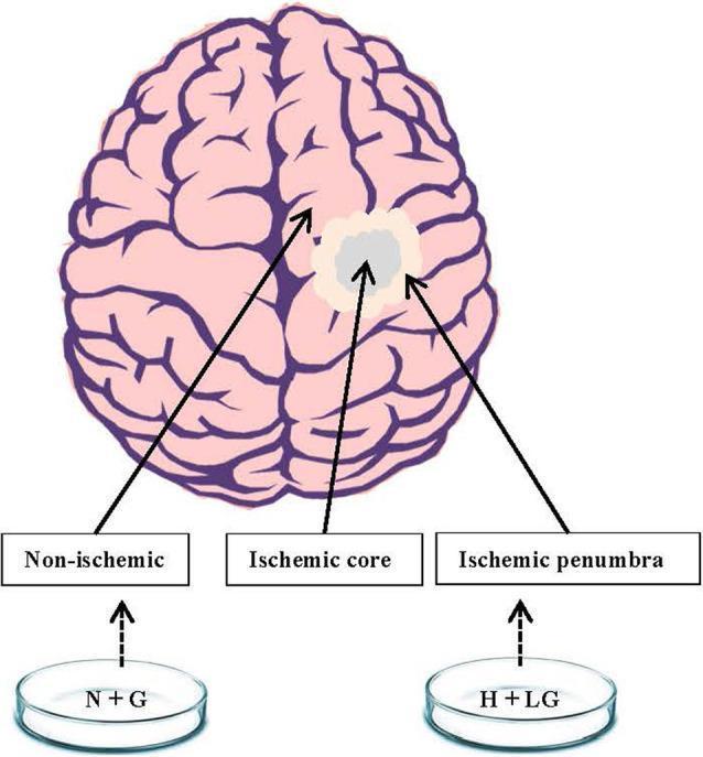
A schematic diagram illustrating the pathophysiology of ischemic stroke and corresponding in vitro experimental models. The central feature is a superior view of the human brain, depicted with pink-colored gyri and sulci. This illustration highlights three distinct clinical zones following a cerebrovascular accident: the 'Ischemic core' (indicated by a gray central circle representing irreversible tissue necrosis), the surrounding 'Ischemic penumbra' (shown as a white halo representing potentially salvageable tissue), and the remaining 'Non-ischemic' healthy regions (pink). Below the brain, the diagram links these clinical states to laboratory cell culture models. An 'N+G' (Normoxia + Glucose) petri dish is correlated with the non-ischemic region, serving as the experimental control. An 'H+LG' (Hypoxia + Low Glucose) petri dish is correlated with the ischemic penumbra, illustrating the experimental strategy to mimic penumbral conditions in HT22 cells to study neuroprotective mechanisms and cellular survival factors like HIF1α.
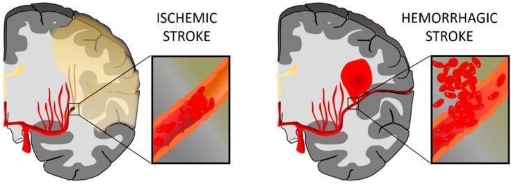
This medical illustration presents a side-by-side comparison of the two primary types of cerebrovascular accidents: Ischemic Stroke and Hemorrhagic Stroke. The diagram uses a coronal cross-section of the human brain and magnified insets to demonstrate the underlying pathophysiology. On the left, 'Ischemic Stroke' is depicted by a large yellowish-tan region in the cerebral cortex, representing infarcted tissue resulting from hypoperfusion. The inset reveals an intraluminal thrombus (blood clot) obstructing an artery, preventing distal blood flow. On the right, 'Hemorrhagic Stroke' is characterized by a focal, bright red intraparenchymal hematoma. The corresponding inset shows a ruptured vessel with erythrocytes extravasating into the surrounding brain tissue. Both illustrations highlight the middle cerebral artery territory. This anatomical diagram serves as an educational tool to differentiate between occlusive and hemorrhagic mechanisms of brain injury, emphasizing the clinical significance of vascular integrity and patency in maintaining cerebral blood flow.
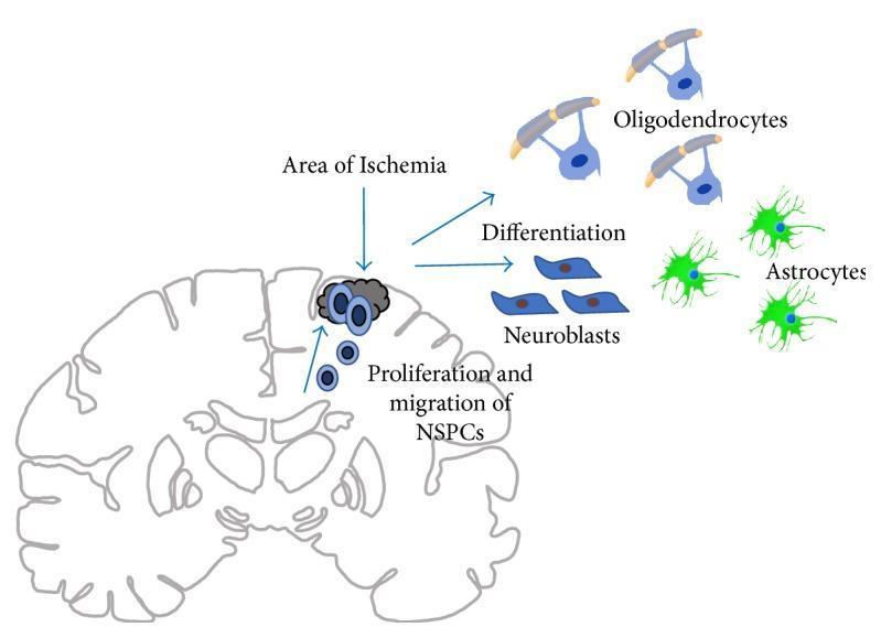
This pathophysiology diagram illustrates the endogenous response to ischemic stroke within a coronal section of the brain. The diagram focuses on the proliferation and migration of neural stem and progenitor cells (NSPCs) following injury. Small, round blue cells representing NSPCs are shown originating from the subventricular zone (SVZ) and migrating toward a dark gray-shaded 'Area of Ischemia' in the cerebral cortex. The visual logic demonstrates that once NSPCs reach or reside outside the SVZ in the context of ischemia, they undergo differentiation into three primary neural lineages. These are depicted as: 1) Oligodendrocytes (blue cells with complex processes wrapping around yellow axonal segments), 2) Astrocytes (bright green, star-shaped cells with multiple radiating processes), and 3) Neuroblasts (elongated, boat-shaped blue cells). The illustration highlights the brain's innate neurogenic potential and the specific cellular pathways involved in tissue repair and glial differentiation following a cerebral vascular accident.
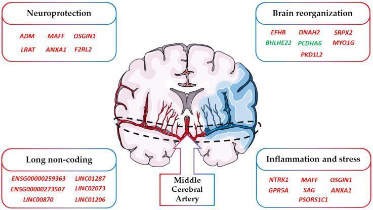
This infographic presents a pathophysiology diagram of an ischemic stroke and its associated molecular changes. At the center is a stylized horizontal cross-section of a human brain. The right hemisphere is shaded light blue to indicate an affected vascular territory, with an arrow highlighting the 'Middle Cerebral Artery' (MCA). Red vascular structures represent perfusion, while the blue-tinted vessels on the right correspond to the stroke zone. Surrounding the brain are four labeled boxes classifying Differentially Expressed Genes (DEGs) into clinical categories: 'Neuroprotection' (e.g., ADM, ANXA1), 'Brain reorganization' (e.g., EFHB, BHLHE22), 'Long non-coding' (e.g., LINC01287), and 'Inflammation and stress' (e.g., NTRK1, PSORS1C1). The text colors denote expression levels: red gene names signify upregulation (increased expression) and green gene names signify downregulation (decreased expression) following a stroke event. This visual aid is intended for medical education on the genomic response to cerebrovascular accidents and neuroinflammation.
Cerebrovascular Accident (CVA) - Etiology and Pathophysiology
Definition
Classification and Etiology
1. Ischemic Stroke (~85% of all strokes)
- Atherosclerotic plaques form at bifurcations and bends of major cerebral arteries (internal carotid, middle cerebral artery, basilar artery, vertebrals)
- Plaque rupture triggers in-situ thrombosis or artery-to-artery embolism
- Most commonly affects the MCA territory
- Emboli originate from cardiac sources and lodge in intracranial vessels
- Major cardiac sources:
- Atrial fibrillation (most common)
- Recent myocardial infarction with mural thrombus
- Dilated cardiomyopathy
- Valvular heart disease (rheumatic mitral stenosis, prosthetic valves)
- Infective endocarditis
- Patent foramen ovale (paradoxical embolism)
- Lipohyalinosis and microatheroma of small penetrating arteries (lenticulostriate, thalamoperforating arteries)
- Strongly associated with chronic hypertension and diabetes
- Produces small deep infarcts (lacunes), typically <1.5 cm
- No identifiable cause after workup (~30% of ischemic strokes)
- Arterial dissection (carotid or vertebral) - especially in younger patients, often from trauma or connective tissue disorders
- Vasculitis (e.g., Takayasu's arteritis affecting common carotid arteries)
- Hypercoagulable states (antiphospholipid antibody syndrome, protein C/S deficiency, factor V Leiden)
- Sickle cell disease (sickling in small vessels)
- Cocaine and amphetamine use (vasospasm)
- Oral contraceptive use (increases thrombotic risk)
2. Hemorrhagic Stroke (~15% of all strokes)
- Primary cause: Hypertension (most common) - causes lipohyalinosis and Charcot-Bouchard microaneurysms in penetrating arteries
- Cerebral amyloid angiopathy (lobar hemorrhages in elderly)
- Anticoagulation therapy (warfarin, DOACs)
- Arteriovenous malformations (AVMs)
- Hemorrhagic transformation of ischemic infarction
- Cocaine/sympathomimetics
- Ruptured saccular (berry) aneurysm (~85% of SAH)
- AVM rupture
- Trauma
Risk Factors
| Modifiable | Non-Modifiable |
|---|---|
| Hypertension (single most important) | Age (risk doubles per decade after 55) |
| Atrial fibrillation | Male sex |
| Diabetes mellitus | Family history/genetics |
| Hyperlipidemia | Prior stroke or TIA |
| Smoking | Race (Black > White) |
| Obesity / physical inactivity | |
| Excessive alcohol |
Pathophysiology
Ischemic Cascade
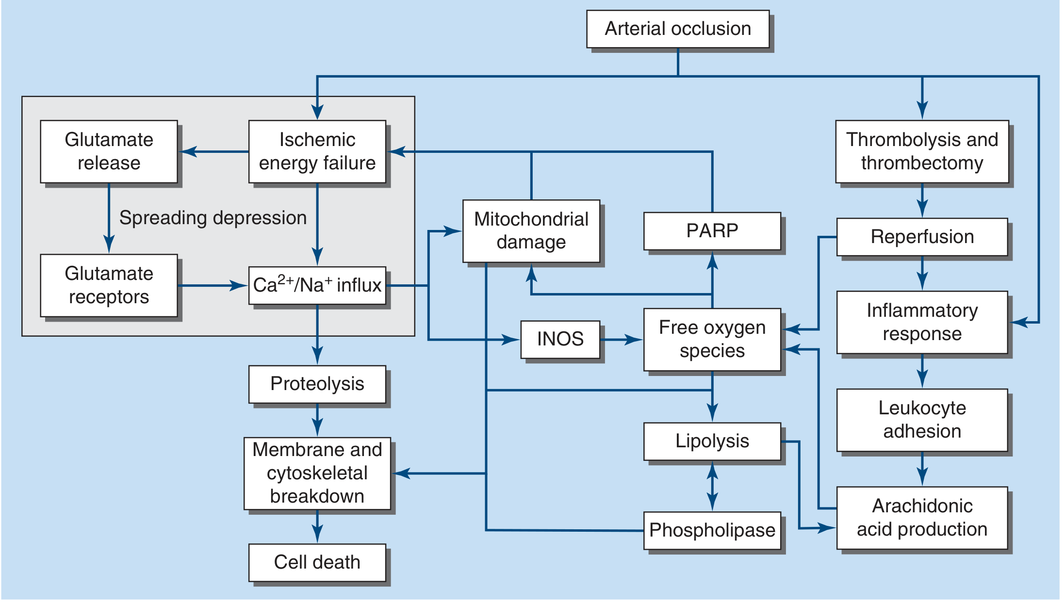
- Flow <16-18 mL/100g/min → neurons cease functioning (neurologic symptoms appear within seconds)
- Flow <10 mL/100g/min → infarction within ~1 hour
- Flow <20 mL/100g/min sustained → ischemia without infarction (reversible if restored)
- Reduction of O₂ and glucose delivery → reduced ATP generation
- Na⁺/K⁺-ATPase pump failure → cellular depolarization
- Intracellular Na⁺ and Ca²⁺ rise (ionic homeostasis lost)
- Cellular depolarization → massive glutamate release from synaptic terminals
- Glial glutamate reuptake fails
- Sustained extracellular glutamate activates NMDA and AMPA receptors
- Massive Ca²⁺ influx into neurons
- Ca²⁺ activates destructive enzymes: proteases, phospholipases, endonucleases
- Ca²⁺ overload damages mitochondria
- Inducible nitric oxide synthase (iNOS) is activated
- ROS (superoxide, hydrogen peroxide, peroxynitrite) damage DNA, lipid membranes, and proteins
- Poly ADP-ribose polymerase (PARP) activation depletes NAD⁺, worsening energy failure
- Lipolysis and phospholipase activation → arachidonic acid production → further inflammation
- Within hours: activation of microglia (brain-resident immune cells)
- Infiltration of peripheral leukocytes (neutrophils, macrophages)
- Leukocyte adhesion molecules upregulated on endothelium
- Release of cytokines (IL-1, TNF-α), matrix metalloproteinases (MMPs)
- MMP activation disrupts the blood-brain barrier → vasogenic edema
- This inflammatory response, while important for repair, also contributes to secondary injury
- Waves of cortical spreading depression emanate from the ischemic core
- Add metabolic stress to surrounding penumbral tissue
The Ischemic Penumbra
- Ischemic core: Irreversibly infarcted tissue with zero or near-zero blood flow. Neurons die within minutes.
- Ischemic penumbra: Surrounding zone of reduced perfusion (functionally silent but structurally viable). Can be saved if perfusion is restored promptly.
- The penumbra is the target of reperfusion therapy (tPA, thrombectomy)
- Restoration of flow reverses acidosis, clears glutamate and toxic ROS, and halts spreading depression
Aggravating Factors
- Fever dramatically worsens ischemic brain injury (increases metabolic demand)
- Hyperglycemia (glucose >11.1 mmol/L / 200 mg/dL) worsens outcomes - likely by increasing lactate production and ROS
- Hypotension can expand the infarct core by collapsing collateral flow into the penumbra
Hemorrhagic Pathophysiology
- Vessel rupture → hematoma formation → direct mechanical compression of adjacent structures
- Hematoma expansion (occurs in ~30-40% within the first hours)
- Perihematomal edema develops within hours to days (secondary injury)
- Raised intracranial pressure (ICP) → herniation
- In SAH: vasospasm (days 4-14) causes delayed cerebral ischemia in ~30% of patients; hemoglobin breakdown products irritate meningeal vessels
Blood-Brain Barrier Disruption
- Both ischemia and hemorrhage disrupt the BBB
- In ischemia: MMP-9 and cytokines degrade tight junctions
- Results in vasogenic edema (protein-rich fluid leaks into parenchyma) compounding cytotoxic edema (intracellular swelling from energy failure)
Summary Table
| Feature | Ischemic Stroke | Hemorrhagic Stroke |
|---|---|---|
| Frequency | ~85% | ~15% |
| Mechanism | Occlusion (thrombosis/embolism) | Vessel rupture |
| Major causes | Atherosclerosis, AF, lipohyalinosis | HTN, aneurysm, AVM, amyloid angiopathy |
| Onset | Sudden; maximal at onset (embolism) or stuttering (thrombosis) | Sudden; often with headache, vomiting, coma |
| CT early | Normal initially; hypodensity later | Immediate hyperdensity (white) |
| Core pathology | Excitotoxicity, energy failure, ROS | Mechanical compression, edema, ICP rise |
| Reversible zone | Penumbra (salvageable) | Perihematomal zone |
Make it simple for 5 marks question
Etiology and Pathophysiology of CVA (Stroke)
Definition
Etiology (Causes)
- Thrombosis - atherosclerosis of cerebral arteries (commonest cause overall)
- Embolism - clot from heart (atrial fibrillation, MI, valvular disease)
- Lacunar infarct - small vessel disease due to hypertension/diabetes
- Intracerebral hemorrhage - hypertension (most common cause)
- Subarachnoid hemorrhage - ruptured berry aneurysm
Pathophysiology
Key Concept: Ischemic Penumbra
- Core = irreversibly dead tissue
- Penumbra = surrounding viable but at-risk tissue
- Goal of treatment is to save the penumbra (via tPA or thrombectomy)
Generate a one-page study guide for stroke etiology and pathophysiology
mkdir -p /tmp/workspace/stroke-study-guide && ls /tmp/workspace/stroke-study-guide/
~/stroke-study-guide/generate.py
from reportlab.lib.pagesizes import A4
from reportlab.lib import colors
from reportlab.lib.units import mm
from reportlab.platypus import (
SimpleDocTemplate, Paragraph, Spacer, Table, TableStyle, HRFlowable
)
from reportlab.lib.styles import getSampleStyleSheet, ParagraphStyle
from reportlab.lib.enums import TA_CENTER, TA_LEFT
from reportlab.platypus import KeepTogether
OUTPUT = "/tmp/workspace/stroke-study-guide/Stroke_Etiology_Pathophysiology.pdf"
# ── Colour palette ────────────────────────────────────────────────────────────
NAVY = colors.HexColor("#1a3c5e")
TEAL = colors.HexColor("#0d7377")
ORANGE = colors.HexColor("#e87722")
LIGHTBLUE = colors.HexColor("#ddeeff")
LIGHTORANGE = colors.HexColor("#fff3e0")
LIGHTGREEN = colors.HexColor("#e8f5e9")
LIGHTGRAY = colors.HexColor("#f5f5f5")
WHITE = colors.white
RED = colors.HexColor("#c0392b")
GREEN = colors.HexColor("#27ae60")
doc = SimpleDocTemplate(
OUTPUT,
pagesize=A4,
leftMargin=12*mm, rightMargin=12*mm,
topMargin=10*mm, bottomMargin=8*mm,
)
W = A4[0] - 24*mm # usable width
styles = getSampleStyleSheet()
# Custom styles
TITLE = ParagraphStyle("title",
fontSize=18, fontName="Helvetica-Bold",
textColor=WHITE, alignment=TA_CENTER, spaceAfter=2)
SUBTITLE = ParagraphStyle("subtitle",
fontSize=9, fontName="Helvetica",
textColor=colors.HexColor("#cce0ff"), alignment=TA_CENTER, spaceAfter=0)
SEC = ParagraphStyle("sec",
fontSize=9, fontName="Helvetica-Bold",
textColor=WHITE, alignment=TA_LEFT, leading=11)
BODY = ParagraphStyle("body",
fontSize=7.8, fontName="Helvetica",
textColor=colors.black, leading=10.5, spaceAfter=1)
BODYBOLD = ParagraphStyle("bodybold",
fontSize=7.8, fontName="Helvetica-Bold",
textColor=NAVY, leading=10.5)
SMALL = ParagraphStyle("small",
fontSize=7.2, fontName="Helvetica",
textColor=colors.HexColor("#444444"), leading=9.5)
MNEMO = ParagraphStyle("mnemo",
fontSize=8.5, fontName="Helvetica-BoldOblique",
textColor=ORANGE, alignment=TA_CENTER, leading=12)
story = []
# ══════════════════════════════════════════════════════════════════════════════
# HEADER BANNER
# ══════════════════════════════════════════════════════════════════════════════
header_data = [[
Paragraph("STROKE (CVA) — Etiology & Pathophysiology", TITLE),
Paragraph("One-Page Study Guide | Based on Harrison's Principles of Internal Medicine 22E", SUBTITLE),
]]
header_table = Table([[
Paragraph("STROKE (CVA) — Etiology & Pathophysiology", TITLE),
]], colWidths=[W])
header_table.setStyle(TableStyle([
("BACKGROUND", (0,0), (-1,-1), NAVY),
("TOPPADDING", (0,0), (-1,-1), 7),
("BOTTOMPADDING", (0,0), (-1,-1), 4),
("LEFTPADDING", (0,0), (-1,-1), 6),
("RIGHTPADDING", (0,0), (-1,-1), 6),
("ROUNDEDCORNERS", [4]),
]))
story.append(header_table)
sub_table = Table([[
Paragraph("One-Page Study Guide · Harrison's Principles of Internal Medicine 22E", SUBTITLE),
]], colWidths=[W])
sub_table.setStyle(TableStyle([
("BACKGROUND", (0,0), (-1,-1), TEAL),
("TOPPADDING", (0,0), (-1,-1), 3),
("BOTTOMPADDING", (0,0), (-1,-1), 3),
("LEFTPADDING", (0,0), (-1,-1), 6),
]))
story.append(sub_table)
story.append(Spacer(1, 3*mm))
# ══════════════════════════════════════════════════════════════════════════════
# ROW 1 — Definition | Types at a glance
# ══════════════════════════════════════════════════════════════════════════════
def section_header(text, bg=TEAL):
t = Table([[Paragraph(text, SEC)]], colWidths=["100%"])
t.setStyle(TableStyle([
("BACKGROUND", (0,0), (-1,-1), bg),
("TOPPADDING", (0,0),(-1,-1), 3),
("BOTTOMPADDING",(0,0),(-1,-1), 3),
("LEFTPADDING",(0,0),(-1,-1), 5),
]))
return t
def box(content_rows, bg=LIGHTBLUE, col_width=None):
cw = [col_width] if col_width else ["100%"]
t = Table(content_rows, colWidths=cw)
t.setStyle(TableStyle([
("BACKGROUND", (0,0),(-1,-1), bg),
("TOPPADDING", (0,0),(-1,-1), 3),
("BOTTOMPADDING",(0,0),(-1,-1), 3),
("LEFTPADDING",(0,0),(-1,-1), 5),
("RIGHTPADDING",(0,0),(-1,-1), 5),
("GRID",(0,0),(-1,-1),0.3,colors.HexColor("#cccccc")),
]))
return t
# ── Definition block ─────────────────────────────────────────────────────────
def_text = [
[Paragraph("DEFINITION", SEC)],
[Paragraph(
"<b>CVA / Stroke</b> = Sudden onset of neurological deficit due to <b>vascular disruption</b> of brain blood supply. "
"2nd leading cause of death worldwide (7.1 million deaths/yr).", BODY)],
]
def_tbl = Table(def_text, colWidths=[W])
def_tbl.setStyle(TableStyle([
("BACKGROUND",(0,0),(0,0), NAVY),
("BACKGROUND",(0,1),(0,1), LIGHTBLUE),
("TOPPADDING",(0,0),(-1,-1),3),("BOTTOMPADDING",(0,0),(-1,-1),3),
("LEFTPADDING",(0,0),(-1,-1),5),("RIGHTPADDING",(0,0),(-1,-1),5),
("BOX",(0,0),(-1,-1),0.5, TEAL),
]))
story.append(def_tbl)
story.append(Spacer(1, 2*mm))
# ══════════════════════════════════════════════════════════════════════════════
# ROW 2 — Two columns: Ischemic | Hemorrhagic
# ══════════════════════════════════════════════════════════════════════════════
CW = (W - 3*mm) / 2 # each column width
# ── Ischemic column ───────────────────────────────────────────────────────────
isch_rows = [
[Paragraph("ISCHEMIC STROKE (85%)", SEC)],
[Paragraph("<b>1. Large-vessel atherothrombosis</b>", BODYBOLD)],
[Paragraph("Atherosclerotic plaque → thrombosis / artery-to-artery embolism<br/>"
"Common sites: ICA, MCA, basilar artery", BODY)],
[Paragraph("<b>2. Cardioembolic</b>", BODYBOLD)],
[Paragraph("Atrial fibrillation (most common), MI mural thrombus,<br/>"
"Valvular disease, Dilated cardiomyopathy, PFO", BODY)],
[Paragraph("<b>3. Small-vessel (Lacunar)</b>", BODYBOLD)],
[Paragraph("Lipohyalinosis of penetrating arteries → tiny deep infarcts<br/>"
"Driven by: Hypertension, Diabetes", BODY)],
[Paragraph("<b>4. Other / Cryptogenic</b>", BODYBOLD)],
[Paragraph("Arterial dissection, Vasculitis, Hypercoagulable states,<br/>"
"Sickle cell, OCP, Cocaine (~30% no cause found)", BODY)],
]
isch_tbl = Table(isch_rows, colWidths=[CW])
isch_tbl.setStyle(TableStyle([
("BACKGROUND",(0,0),(0,0), TEAL),
("BACKGROUND",(0,1),(-1,-1), LIGHTBLUE),
("TOPPADDING",(0,0),(-1,-1),2),("BOTTOMPADDING",(0,0),(-1,-1),2),
("LEFTPADDING",(0,0),(-1,-1),5),("RIGHTPADDING",(0,0),(-1,-1),5),
("BOX",(0,0),(-1,-1),0.5,TEAL),
]))
# ── Hemorrhagic column ────────────────────────────────────────────────────────
hem_rows = [
[Paragraph("HEMORRHAGIC STROKE (15%)", SEC)],
[Paragraph("<b>Intracerebral Hemorrhage (ICH)</b>", BODYBOLD)],
[Paragraph("• <b>Hypertension</b> → Charcot-Bouchard microaneurysms → rupture<br/>"
"• Cerebral amyloid angiopathy (lobar, elderly)<br/>"
"• Anticoagulants, AVM, Cocaine/sympathomimetics<br/>"
"• Hemorrhagic transformation of ischemic stroke", BODY)],
[Paragraph("<b>Subarachnoid Hemorrhage (SAH)</b>", BODYBOLD)],
[Paragraph("• Ruptured <b>berry (saccular) aneurysm</b> — 85% of SAH<br/>"
"• AVM rupture<br/>"
"• Classic: sudden 'thunderclap' worst headache of life", BODY)],
[Paragraph("<b>Pathophysiology of ICH</b>", BODYBOLD)],
[Paragraph("Hematoma → mechanical compression → perihematomal edema → "
"↑ICP → herniation. Vasospasm (days 4–14) causes delayed ischemia in SAH.", BODY)],
]
hem_tbl = Table(hem_rows, colWidths=[CW])
hem_tbl.setStyle(TableStyle([
("BACKGROUND",(0,0),(0,0), RED),
("BACKGROUND",(0,1),(-1,-1), colors.HexColor("#fdecea")),
("TOPPADDING",(0,0),(-1,-1),2),("BOTTOMPADDING",(0,0),(-1,-1),2),
("LEFTPADDING",(0,0),(-1,-1),5),("RIGHTPADDING",(0,0),(-1,-1),5),
("BOX",(0,0),(-1,-1),0.5, RED),
]))
two_col = Table([[isch_tbl, hem_tbl]], colWidths=[CW, CW], spaceBefore=0)
two_col.setStyle(TableStyle([
("VALIGN",(0,0),(-1,-1),"TOP"),
("LEFTPADDING",(0,0),(-1,-1),0),
("RIGHTPADDING",(0,0),(-1,-1),0),
("TOPPADDING",(0,0),(-1,-1),0),
("BOTTOMPADDING",(0,0),(-1,-1),0),
("INNERGRID",(0,0),(-1,-1),2,WHITE),
]))
story.append(two_col)
story.append(Spacer(1, 2*mm))
# ══════════════════════════════════════════════════════════════════════════════
# RISK FACTORS
# ══════════════════════════════════════════════════════════════════════════════
rf_data = [
[Paragraph("RISK FACTORS", SEC), ""],
[Paragraph("<b>Modifiable:</b> Hypertension (#1), Atrial fibrillation, Diabetes, "
"Hyperlipidemia, Smoking, Obesity, Alcohol, OCP", BODY),
Paragraph("<b>Non-modifiable:</b> Age (doubles/decade after 55), Male sex, "
"Family history, Prior stroke/TIA, Black ethnicity", BODY)],
]
rf_tbl = Table(rf_data, colWidths=[W/2, W/2])
rf_tbl.setStyle(TableStyle([
("BACKGROUND",(0,0),(1,0), ORANGE),
("BACKGROUND",(0,1),(1,1), LIGHTORANGE),
("SPAN",(0,0),(1,0)),
("TOPPADDING",(0,0),(-1,-1),2),("BOTTOMPADDING",(0,0),(-1,-1),2),
("LEFTPADDING",(0,0),(-1,-1),5),("RIGHTPADDING",(0,0),(-1,-1),5),
("BOX",(0,0),(-1,-1),0.5, ORANGE),
("LINEAFTER",(0,1),(0,1),0.3,colors.grey),
]))
story.append(rf_tbl)
story.append(Spacer(1, 2*mm))
# ══════════════════════════════════════════════════════════════════════════════
# ISCHEMIC PATHOPHYSIOLOGY CASCADE
# ══════════════════════════════════════════════════════════════════════════════
cascade_steps = [
("1", TEAL, "Arterial Occlusion",
"CBF drops → zero = necrosis in 4–10 min; <16 mL/100g/min → infarction within 1 hr"),
("2", colors.HexColor("#1565c0"), "Energy Failure",
"↓O₂ + ↓Glucose → ATP depleted → Na⁺/K⁺-ATPase fails → cellular depolarisation, ↑intracellular Na⁺ & Ca²⁺"),
("3", colors.HexColor("#6a1b9a"), "Excitotoxicity",
"Depolarisation → massive glutamate release → NMDA/AMPA receptor activation → Ca²⁺ floods into neuron"),
("4", colors.HexColor("#bf360c"), "ROS & Mitochondrial Damage",
"Ca²⁺ overload → activates proteases, phospholipases → ROS (superoxide, peroxynitrite) → iNOS, PARP activation → DNA & membrane damage"),
("5", colors.HexColor("#2e7d32"), "Inflammation",
"Microglia activate within hours → leukocyte infiltration → IL-1, TNF-α, MMP-9 → BBB breakdown → vasogenic oedema"),
("6", colors.HexColor("#4e342e"), "Cell Death / Oedema",
"Cytotoxic + vasogenic oedema → ↑ICP → herniation if severe. Spreading cortical depression adds metabolic stress to penumbra"),
]
casc_rows = [[Paragraph("ISCHEMIC PATHOPHYSIOLOGY CASCADE", SEC)]]
for num, col, title, detail in cascade_steps:
num_cell = Paragraph(f"<b>{num}</b>", ParagraphStyle("n", fontSize=10,
fontName="Helvetica-Bold", textColor=WHITE, alignment=TA_CENTER, leading=13))
title_cell = Paragraph(f"<b>{title}</b>", ParagraphStyle("t", fontSize=7.8,
fontName="Helvetica-Bold", textColor=col, leading=10))
detail_cell = Paragraph(detail, SMALL)
casc_rows.append([num_cell, title_cell, detail_cell])
casc_tbl = Table(casc_rows, colWidths=[W])
casc_tbl.setStyle(TableStyle([
("BACKGROUND",(0,0),(0,0), NAVY),
("TOPPADDING",(0,0),(-1,-1),0),("BOTTOMPADDING",(0,0),(-1,-1),0),
("LEFTPADDING",(0,0),(-1,-1),5),("RIGHTPADDING",(0,0),(-1,-1),5),
]))
# Rebuild as proper multi-column table
casc_data = [[Paragraph("ISCHEMIC PATHOPHYSIOLOGY CASCADE", SEC)]]
inner_rows = []
for num, col, title, detail in cascade_steps:
num_p = Paragraph(f"<b>{num}</b>",
ParagraphStyle("nn", fontSize=10, fontName="Helvetica-Bold",
textColor=WHITE, alignment=TA_CENTER, leading=13))
title_p = Paragraph(f"<b>{title}</b>",
ParagraphStyle("tt", fontSize=7.8, fontName="Helvetica-Bold",
textColor=col, leading=10))
detail_p = Paragraph(detail, SMALL)
inner_rows.append([num_p, title_p, detail_p])
inner_tbl = Table(inner_rows, colWidths=[8*mm, 42*mm, W - 50*mm])
step_bg = [
colors.HexColor("#0d7377"),
colors.HexColor("#1565c0"),
colors.HexColor("#6a1b9a"),
colors.HexColor("#bf360c"),
colors.HexColor("#2e7d32"),
colors.HexColor("#4e342e"),
]
for i, bg in enumerate(step_bg):
inner_tbl.setStyle(TableStyle([
("BACKGROUND", (0,i),(0,i), bg),
("BACKGROUND", (1,i),(2,i), LIGHTGRAY if i % 2 == 0 else WHITE),
("TOPPADDING",(0,i),(-1,i),3),("BOTTOMPADDING",(0,i),(-1,i),3),
("LEFTPADDING",(0,i),(0,i),2),("RIGHTPADDING",(0,i),(0,i),2),
("LEFTPADDING",(1,i),(-1,i),5),("RIGHTPADDING",(1,i),(-1,i),5),
("VALIGN",(0,i),(-1,i),"MIDDLE"),
("LINEBELOW",(0,i),(-1,i),0.3,colors.HexColor("#cccccc")),
]))
full_casc = Table([
[Paragraph("ISCHEMIC PATHOPHYSIOLOGY CASCADE", SEC)],
[inner_tbl],
], colWidths=[W])
full_casc.setStyle(TableStyle([
("BACKGROUND",(0,0),(0,0), NAVY),
("TOPPADDING",(0,0),(0,0),3),("BOTTOMPADDING",(0,0),(0,0),3),
("LEFTPADDING",(0,0),(-1,-1),5),("RIGHTPADDING",(0,0),(-1,-1),5),
("TOPPADDING",(0,1),(0,1),0),("BOTTOMPADDING",(0,1),(0,1),0),
("LEFTPADDING",(0,1),(0,1),0),("RIGHTPADDING",(0,1),(0,1),0),
("BOX",(0,0),(-1,-1),0.5,NAVY),
]))
story.append(full_casc)
story.append(Spacer(1, 2*mm))
# ══════════════════════════════════════════════════════════════════════════════
# ROW 3 — Penumbra | Aggravating Factors | Mnemonic
# ══════════════════════════════════════════════════════════════════════════════
C3 = (W - 4*mm) / 3
# Penumbra
pen_rows = [
[Paragraph("ISCHEMIC PENUMBRA", SEC)],
[Paragraph(
"<b>Core</b> — Irreversibly dead; CBF ≈ 0<br/>"
"<b>Penumbra</b> — Ischaemic, viable; CBF reduced<br/>"
"<b>Goal</b> — Save penumbra via tPA / thrombectomy<br/>"
"Reperfusion clears glutamate, reverses acidosis, "
"halts spreading depression", BODY)],
]
pen_tbl = Table(pen_rows, colWidths=[C3])
pen_tbl.setStyle(TableStyle([
("BACKGROUND",(0,0),(0,0), TEAL),
("BACKGROUND",(0,1),(0,1), LIGHTBLUE),
("TOPPADDING",(0,0),(-1,-1),3),("BOTTOMPADDING",(0,0),(-1,-1),3),
("LEFTPADDING",(0,0),(-1,-1),5),("RIGHTPADDING",(0,0),(-1,-1),5),
("BOX",(0,0),(-1,-1),0.5,TEAL),
]))
# Aggravating factors
agg_rows = [
[Paragraph("AGGRAVATING FACTORS", SEC)],
[Paragraph(
"<b>Fever</b> — dramatically worsens ischaemia;<br/>"
"treat aggressively<br/>"
"<b>Hyperglycaemia</b> >200 mg/dL — ↑lactate & ROS;<br/>"
"worsens outcome<br/>"
"<b>Hypotension</b> — collapses collateral flow<br/>"
"into penumbra → expands core<br/>"
"<b>Oedema</b> — cytotoxic + vasogenic;<br/>"
"peaks 48–72 hr", BODY)],
]
agg_tbl = Table(agg_rows, colWidths=[C3])
agg_tbl.setStyle(TableStyle([
("BACKGROUND",(0,0),(0,0), ORANGE),
("BACKGROUND",(0,1),(0,1), LIGHTORANGE),
("TOPPADDING",(0,0),(-1,-1),3),("BOTTOMPADDING",(0,0),(-1,-1),3),
("LEFTPADDING",(0,0),(-1,-1),5),("RIGHTPADDING",(0,0),(-1,-1),5),
("BOX",(0,0),(-1,-1),0.5,ORANGE),
]))
# Mnemonic
mne_rows = [
[Paragraph("MEMORY AID", SEC)],
[Paragraph(
"Ischaemic cascade:<br/><br/>"
"<b>E</b>nergy failure<br/>"
"<b>E</b>xcitotoxicity (glutamate/Ca²⁺)<br/>"
"<b>R</b>OS & mitochondrial damage<br/>"
"<b>I</b>nflammation (microglia)<br/>"
"<b>E</b>dema + Cell death<br/><br/>"
'"<i>Every Event Ruins Injured tissue Eventually</i>"',
MNEMO)],
]
mne_tbl = Table(mne_rows, colWidths=[C3])
mne_tbl.setStyle(TableStyle([
("BACKGROUND",(0,0),(0,0), colors.HexColor("#4a235a")),
("BACKGROUND",(0,1),(0,1), colors.HexColor("#f3e5f5")),
("TOPPADDING",(0,0),(-1,-1),3),("BOTTOMPADDING",(0,0),(-1,-1),3),
("LEFTPADDING",(0,0),(-1,-1),5),("RIGHTPADDING",(0,0),(-1,-1),5),
("BOX",(0,0),(-1,-1),0.5,colors.HexColor("#4a235a")),
]))
bottom_row = Table([[pen_tbl, agg_tbl, mne_tbl]], colWidths=[C3, C3, C3])
bottom_row.setStyle(TableStyle([
("VALIGN",(0,0),(-1,-1),"TOP"),
("LEFTPADDING",(0,0),(-1,-1),0),("RIGHTPADDING",(0,0),(-1,-1),0),
("TOPPADDING",(0,0),(-1,-1),0),("BOTTOMPADDING",(0,0),(-1,-1),0),
("INNERGRID",(0,0),(-1,-1),2,WHITE),
]))
story.append(bottom_row)
story.append(Spacer(1, 2*mm))
# ══════════════════════════════════════════════════════════════════════════════
# QUICK COMPARISON TABLE
# ══════════════════════════════════════════════════════════════════════════════
cmp_header_style = ParagraphStyle("ch", fontSize=7.5, fontName="Helvetica-Bold",
textColor=WHITE, alignment=TA_CENTER, leading=10)
cmp_body_style = ParagraphStyle("cb", fontSize=7.2, fontName="Helvetica",
textColor=colors.black, alignment=TA_CENTER, leading=9.5)
cmp_label_style = ParagraphStyle("cl", fontSize=7.5, fontName="Helvetica-Bold",
textColor=NAVY, alignment=TA_LEFT, leading=10)
cmp_data = [
[Paragraph("FEATURE", cmp_header_style),
Paragraph("ISCHEMIC STROKE", cmp_header_style),
Paragraph("HEMORRHAGIC STROKE", cmp_header_style)],
[Paragraph("Frequency", cmp_label_style),
Paragraph("~85%", cmp_body_style), Paragraph("~15%", cmp_body_style)],
[Paragraph("Mechanism", cmp_label_style),
Paragraph("Occlusion (thrombosis / embolism)", cmp_body_style),
Paragraph("Vessel rupture", cmp_body_style)],
[Paragraph("Main Causes", cmp_label_style),
Paragraph("Atherosclerosis, AF, Lipohyalinosis", cmp_body_style),
Paragraph("HTN, Aneurysm, AVM, Amyloid angiopathy", cmp_body_style)],
[Paragraph("CT Finding", cmp_label_style),
Paragraph("Normal early; hypodensity later", cmp_body_style),
Paragraph("Immediate hyperdensity (white)", cmp_body_style)],
[Paragraph("Core Pathology", cmp_label_style),
Paragraph("Excitotoxicity, Energy failure, ROS", cmp_body_style),
Paragraph("Mechanical compression, Oedema, ↑ICP", cmp_body_style)],
[Paragraph("Treatment Target", cmp_label_style),
Paragraph("Save penumbra (tPA / thrombectomy)", cmp_body_style),
Paragraph("Stop bleeding, reduce ICP", cmp_body_style)],
]
col_w = [36*mm, (W-36*mm)/2, (W-36*mm)/2]
cmp_tbl = Table(cmp_data, colWidths=col_w)
cmp_tbl.setStyle(TableStyle([
("BACKGROUND", (0,0), (-1,0), NAVY),
("BACKGROUND", (0,1), (-1,1), LIGHTBLUE),
("BACKGROUND", (0,2), (-1,2), WHITE),
("BACKGROUND", (0,3), (-1,3), LIGHTBLUE),
("BACKGROUND", (0,4), (-1,4), WHITE),
("BACKGROUND", (0,5), (-1,5), LIGHTBLUE),
("BACKGROUND", (0,6), (-1,6), WHITE),
("GRID", (0,0), (-1,-1), 0.3, colors.HexColor("#aaaaaa")),
("TOPPADDING", (0,0),(-1,-1), 2),
("BOTTOMPADDING",(0,0),(-1,-1), 2),
("LEFTPADDING", (0,0),(-1,-1), 4),
("RIGHTPADDING",(0,0),(-1,-1), 4),
("VALIGN", (0,0),(-1,-1), "MIDDLE"),
("BOX", (0,0),(-1,-1), 0.8, NAVY),
]))
story.append(cmp_tbl)
# ══════════════════════════════════════════════════════════════════════════════
# FOOTER
# ══════════════════════════════════════════════════════════════════════════════
story.append(Spacer(1, 2*mm))
footer = Table([[
Paragraph("Source: Harrison's Principles of Internal Medicine 22E (2025), Chapters 437–438 | "
"Orris Medical Study Guides", SMALL)
]], colWidths=[W])
footer.setStyle(TableStyle([
("BACKGROUND",(0,0),(-1,-1), colors.HexColor("#eceff1")),
("TOPPADDING",(0,0),(-1,-1),2),("BOTTOMPADDING",(0,0),(-1,-1),2),
("LEFTPADDING",(0,0),(-1,-1),5),
("BOX",(0,0),(-1,-1),0.3,colors.grey),
]))
story.append(footer)
# ── Build ─────────────────────────────────────────────────────────────────────
doc.build(story)
print(f"PDF saved to: {OUTPUT}")
cd /tmp/workspace/stroke-study-guide && python generate.py 2>&1
ls -lh /tmp/workspace/stroke-study-guide/
Stroke Etiology Pathophysiology
PDF Document · PDF
- Header banner - title and source (Harrison's 22E)
- Definition - concise CVA definition with epidemiology
- Two-column etiology block - Ischemic (85%) with all 4 subtypes vs. Hemorrhagic (15%) with ICH and SAH causes
- Risk factors bar - modifiable vs. non-modifiable side by side
- Ischemic pathophysiology cascade - 6 colour-coded steps from arterial occlusion to cell death, with detailed notes for each step
- Bottom row - Ischemic Penumbra explained + Aggravating Factors (fever, hyperglycaemia, hypotension) + Memory mnemonic "Every Event Ruins Injured tissue Eventually"
- Quick comparison table - Ischemic vs. Hemorrhagic stroke across 6 features
- Footer - source citation
Clinical features
stroke clinical features arterial territory brain infarction
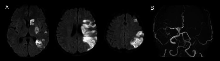
This composite diagnostic image features brain MRI findings from a patient with an acute ischemic stroke. Panel A consists of three axial slices of Diffusion-Weighted Imaging (DWI), which demonstrate acute cerebral infarctions in the left middle cerebral artery (MCA) territory. These infarctions are characterized by areas of restricted diffusion (hyperintensities). Visible pathology includes scattered embolic-pattern infarcts in the deep white matter and basal ganglia, as well as a large, confluent cortical infarct involving the left parietal lobe. Panel B shows a coronal view from a Magnetic Resonance Angiogram (MRA) of the intracranial circulation. The image reveals a significant obstruction or occlusion of the left internal carotid artery (ICA). Notable vascular findings include the absence of the normal arterial signal on the left side and the presence of fine, tortuous collateral vessels attempting to reperfuse the ischemic territory. This clinical imaging set highlights the relationship between proximal large vessel occlusion and subsequent multi-territory cortical and subcortical infarction, characteristic of thromboembolic events.
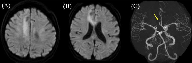
This composite diagnostic image illustrates an acute ischemic stroke within the right anterior cerebral artery (ACA) territory using MRI and MRA modalities. Panels (A) and (B) are axial Diffusion-Weighted Imaging (DWI) scans of the brain. (A) shows a distinct area of high signal intensity (hyperintensity) in the right frontal lobe convexity, indicating restricted water diffusion characteristic of acute infarction. (B) displays similar hyperintensity at a lower axial level, involving the corpus callosum and medial frontal cortex. Panel (C) presents a Magnetic Resonance Angiogram (MRA) of the intracranial vasculature. A yellow arrow points to the right ACA, which appears truncated and lacks normal distal signal, confirming an occlusion. The combined imaging demonstrates a large territory infarction secondary to proximal arterial blockage. This resource is suitable for medical education regarding radiological signs of ischemic stroke, neuroanatomy of the ACA, and the clinical application of DWI and MRA in cerebrovascular emergencies.
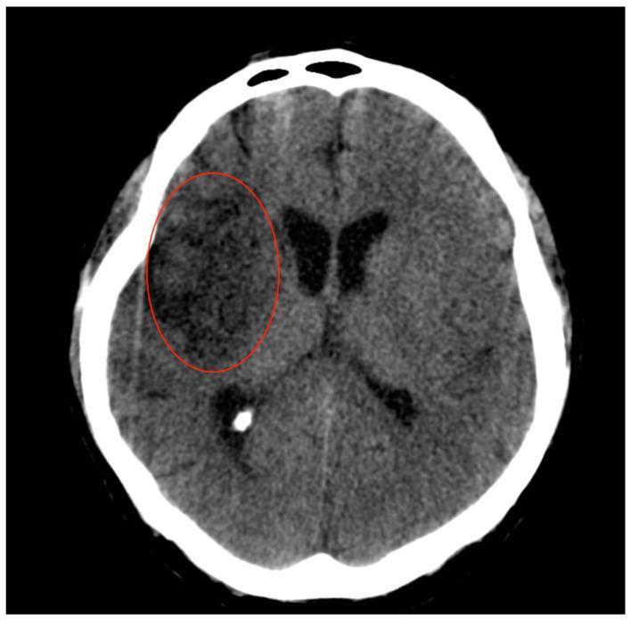
This diagnostic image is an axial non-contrast Computed Tomography (CT) scan of the brain, demonstrating a subacute ischemic infarct. A red oval highlights a large, well-demarcated area of hypodensity within the right hemisphere, specifically involving the fronto-temporal-insular regions. The hypodensity extends to include deep gray matter structures, notably the lentiform nucleus and the right caudate nucleus, consistent with a vascular territory supplied by the middle cerebral artery (MCA). The darker shade of gray within the highlighted region compared to the contralateral parenchyma indicates cytotoxic edema and tissue necrosis following ischemia. Key educational features include the loss of gray-white matter differentiation and the involvement of the lateral lenticulostriate arterial territory. This image illustrates the progression of cerebral infarction 7 days post-mechanical thrombectomy, serving as a clinical example of established ischemic stroke evolution and large vessel territory involvement in neurology and radiology education.
Clinical Features of CVA (Stroke)
General / Universal Features
| Feature | Detail |
|---|---|
| Onset | Abrupt; maximal at onset (embolism) or stuttering/stepwise (thrombosis) |
| Consciousness | Preserved in most ischemic strokes; depressed in large MCA/brainstem/hemorrhagic |
| FAST signs | Facial drooping, Arm weakness, Speech difficulty, Time to call help |
By Arterial Territory
1. Middle Cerebral Artery (MCA) - Most Common
- Contralateral hemiplegia (face + arm > leg)
- Contralateral hemianesthesia (all sensory modalities)
- Homonymous hemianopia (contralateral visual field loss)
- Gaze deviation toward the side of the lesion (day 1-2)
- Global aphasia (both production and comprehension of language lost)
- Dysarthria
- Hemiplegia + hemianesthesia + hemianopia
- Anosognosia (unawareness of the deficit)
- Constructional apraxia
- Hemispatial neglect (ignores the contralateral side)
| Branch Occluded | Features |
|---|---|
| Superior division (dominant) | Broca's (non-fluent) aphasia + arm/face weakness - leg spared |
| Inferior division (dominant) | Wernicke's (fluent) aphasia + upper quadrantanopia, no weakness |
| Inferior division (non-dominant) | Hemineglect, spatial agnosia, no weakness |
| Lenticulostriate (penetrating) | Lacunar syndromes: pure motor, pure sensory, or clumsy-hand dysarthria |
2. Anterior Cerebral Artery (ACA)
- Contralateral leg weakness (arm and face relatively spared - opposite of MCA)
- Abulia - slowness in verbal and motor responses; apathy
- Urinary incontinence
- Gait apraxia
- Grasp reflex, sucking reflex (primitive reflexes)
- If bilateral ACA occlusion: paraplegia + profound abulia + urinary incontinence
3. Posterior Cerebral Artery (PCA)
- Contralateral homonymous hemianopia (with macular sparing often)
- Cortical blindness (bilateral occipital involvement)
- Memory impairment (hippocampal involvement)
- Visual agnosia, prosopagnosia (can't recognise faces)
- Topographic disorientation
- Unformed visual hallucinations
- Thalamic syndrome - contralateral loss of all sensory modalities + spontaneous pain/dysesthesia (thalamic pain)
- Choreoathetosis, intention tremor
- Weber's syndrome - ipsilateral CN III palsy + contralateral hemiplegia (midbrain involvement)
- Claude's syndrome - ipsilateral CN III palsy + contralateral ataxia
4. Internal Carotid Artery (ICA)
- Symptoms combine MCA + ACA territory features
- Amaurosis fugax - transient monocular blindness ("curtain coming down") - hallmark of ipsilateral carotid disease (via ophthalmic artery)
- Carotid bruit on auscultation in stenosis
- If massive: entire hemisphere infarction with stupor/coma
5. Posterior Circulation - Vertebrobasilar
- Bilateral motor and sensory deficits (crossed signs)
- Locked-in syndrome (complete paralysis except eye movements)
- Coma, pin-point pupils, loss of brainstem reflexes
- Gaze palsies, internuclear ophthalmoplegia
| Sign | Structure |
|---|---|
| Ipsilateral facial pain/numbness | V nucleus/tract |
| Ipsilateral Horner's syndrome | Sympathetic fibres |
| Ipsilateral ataxia/falling | Cerebellar peduncle |
| Dysphagia, hoarseness | IX, X nuclei |
| Contralateral loss of pain & temperature (body) | Spinothalamic tract |
- No hemiplegia (corticospinal tract spared) - characteristic!
- Ipsilateral cerebellar ataxia, nausea, vomiting
- Contralateral loss of pain and temperature (body + face)
- Ipsilateral Horner's syndrome
6. Small-Vessel (Lacunar) Syndromes
| Syndrome | Location | Features |
|---|---|---|
| Pure motor hemiplegia | Posterior limb, internal capsule / pons | Face + arm + leg weakness equally |
| Pure sensory stroke | Thalamus (VPL nucleus) | Hemisensory loss of all modalities |
| Sensorimotor stroke | Capsule + thalamus | Motor + sensory combined |
| Clumsy hand - dysarthria | Genu of internal capsule / pons | Facial weakness + clumsiness + slurred speech |
| Ataxic hemiparesis | Pons / internal capsule | Ipsilateral ataxia + contralateral hemiparesis |
Hemorrhagic Stroke - Clinical Features
Intracerebral Hemorrhage (ICH)
- Sudden severe headache
- Rapid progression over minutes to hours (unlike ischemic which is maximal at onset)
- Nausea and vomiting (raised ICP)
- Altered consciousness / coma
- Hypertension (usually markedly elevated BP at presentation)
- Features depend on location:
| Location | Features |
|---|---|
| Putamen (most common) | Contralateral hemiplegia, hemisensory loss, homonymous hemianopia, gaze deviation |
| Thalamus | Hemisensory loss, eyes deviated downward and inward |
| Pons | Quadriplegia, pin-point pupils, hyperthermia, coma |
| Cerebellum | Ataxia, vomiting, gaze palsy - NO hemiplegia; may need urgent surgery |
Subarachnoid Hemorrhage (SAH)
- "Thunderclap" headache - sudden, worst headache of life
- Neck stiffness (meningism) - Kernig's and Brudzinski's signs
- Photophobia, phonophobia
- Vomiting
- Loss of consciousness at onset (in severe cases)
- Subhyaloid (preretinal) haemorrhages on fundoscopy - pathognomonic
- Delayed complication: vasospasm (days 4-14) → delayed ischemia
Differentiating Ischemic vs. Hemorrhagic at Bedside
| Feature | Ischemic | Hemorrhagic |
|---|---|---|
| Onset | Sudden, maximal at onset | Sudden, progressive over minutes |
| Headache | Mild or absent | Severe ("thunderclap" in SAH) |
| Vomiting | Uncommon | Common |
| BP | Variable | Markedly elevated |
| Consciousness | Usually preserved | Often impaired |
| Progression | Stable or improving | Worsening |
| CT | Hypodensity (later) | Hyperdensity (immediate) |
Warning Signs Before Stroke
- TIA (Transient Ischaemic Attack) - same symptoms as stroke but resolve within 24 hours (usually <1 hour); high risk of completed stroke within 48 hours
- Amaurosis fugax - transient monocular blindness (ICA disease)
- Recurrent stereotyped episodes suggest thrombotic mechanism
Nursing management
stroke nursing care assessment neurological monitoring
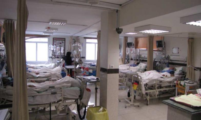
Clinical photograph of a specialized acute stroke unit within a hospital setting. The image demonstrates a multi-bed ward layout specifically designed for the management of neurological emergencies. Visible equipment includes adjustable medical hospital beds, bedside physiological monitors for continuous hemodynamic and vital sign tracking, and IV infusion poles with multiple fluid bags. The environment is equipped with bedside privacy curtains, wall-mounted medical gas outlets, and rolling crash or utility carts for emergency intervention supplies. A ceiling-mounted surveillance camera is visible, indicative of the high-acuity monitoring required for acute stroke patients. Natural light from windows and recessed fluorescent panels provide illumination. The unit serves as the clinical environment for managing patients undergoing treatments such as intravenous thrombolysis or pre-/post-procedure care for mechanical thrombectomy.
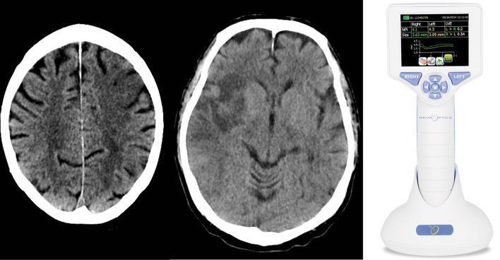
This composite educational image illustrates the clinical assessment of stroke patients using neuroimaging and automated pupillometry. On the left and center are two axial non-contrast CT scans of the brain. The left image demonstrates a hypodense area in the left prefrontal region, consistent with an ischemic infarction in the prefrontal eye field. The center image shows a larger area of hypodensity involving the right insular cortex and surrounding white matter, characteristic of a strategic stroke in an autonomic control center. On the right, a clinical photograph displays a handheld automated pupillometer (NeurOptics NPi-200). The device's digital interface displays quantitative data for both eyes, including the Neurological Pupil index (NPi) and pupil size in millimeters, alongside a pupillary light reflex waveform. This visual combination demonstrates the integration of anatomical diagnostic imaging with functional neurological monitoring in acute stroke management.
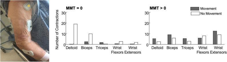
This composite image consists of a clinical photograph and a data visualization chart focused on electromyography (EMG) monitoring in stroke rehabilitation. The clinical photograph on the left shows the paretic arm of a patient with several wireless BioStamp sensors attached to monitor muscle activity. Sensors are visible over the deltoid, biceps, triceps, wrist flexors, and wrist extensors. The skin exhibits signs of atrophy and wrinkling, common in acute stroke or geriatric populations. The right side contains two bar graphs comparing the median number of muscle contractions per 30 minutes in patients with a Manual Muscle Test (MMT) score of 0 (flaccid paralysis) versus those with MMT > 0. The graphs categorize contractions based on whether they occurred during 'Movement' (grey bars) or 'No Movement' (white bars). In the MMT = 0 group, proximal muscles like the deltoid show the highest frequency of contractions without movement. In the MMT > 0 group, contractions are more evenly distributed across all muscle groups, with a higher prevalence during active movement. This educational resource illustrates the utility of surface EMG in detecting subclinical muscle activity for prognostic assessment during neurological recovery.
Nursing Management of CVA (Stroke)
Phases of Nursing Management
| Phase | Timeframe | Priority |
|---|---|---|
| Acute | First 24-72 hours | Stabilise, prevent death, acute treatment |
| Subacute | Days 3-14 | Prevent complications, begin mobilisation |
| Rehabilitation | Weeks to months | Restore function, patient/family education |
1. IMMEDIATE / ACUTE NURSING CARE
A. Airway, Breathing, Circulation (ABCs) - First Priority
- Assess airway - patients with decreased consciousness or bulbar palsy are at risk of aspiration
- Position patient in lateral (recovery) position if consciousness is impaired
- Administer supplemental oxygen if SpO₂ <94%
- Assist with intubation if GCS <8 or airway unprotectable
- Monitor respiratory rate, depth, SpO₂ continuously
- Keep suction equipment at bedside at all times
B. Neurological Assessment - Ongoing
- Glasgow Coma Scale (GCS) - consciousness level
- National Institutes of Health Stroke Scale (NIHSS) - standardised stroke severity
- Pupillary size and reaction (fixed/dilated = herniation emergency)
- Motor strength in all four limbs
- Facial symmetry
- Speech (fluency, comprehension)
- Any sudden deterioration = immediate physician notification
C. Vital Sign Monitoring
| Parameter | Target / Action |
|---|---|
| Blood pressure | Do NOT lower unless >220/120 mmHg (ischemic) or >180/105 (hemorrhagic) - hypotension collapses penumbral flow |
| Temperature | Keep <37.5°C - fever dramatically worsens ischemia; use antipyretics + surface cooling |
| Blood glucose | Maintain 60-180 mg/dL; correct hypoglycemia immediately; avoid hyperglycemia (worsens outcome) |
| Heart rate/rhythm | Continuous cardiac monitoring - detect AF, arrhythmias |
| ICP signs | Watch for Cushing's triad (hypertension + bradycardia + irregular breathing) |
D. IV Access and Medications
- Establish large-bore IV access (2 lines preferred)
- Collect bloods: CBC, coagulation, electrolytes, glucose, lipids, ECG
- Assist with IV tPA administration if eligible (within 4.5 hours, no hemorrhage):
- Confirm eligibility checklist (BP, glucose, CT result, time of onset)
- Monitor for bleeding complications during and after infusion (headache, BP spike, neurological worsening = stop infusion, urgent CT)
- BP must be <185/110 before tPA and <180/105 for 24 hours after
- Prepare patient for thrombectomy if indicated (large vessel occlusion)
2. PREVENTING COMPLICATIONS (Nursing Priority List)
A. Aspiration Pneumonia (Most Common Early Complication)
- Nil by mouth (NBM) until formal swallow assessment by speech therapist
- Perform bedside swallow screen (water swallow test) before any oral intake
- If dysphagia confirmed → nasogastric (NG) tube feeding
- Keep head of bed elevated 30-45° during and 30 min after feeds
- Oral hygiene every 4-6 hours (suction secretions if needed)
- Monitor for signs: fever, cough, reduced SpO₂, abnormal chest sounds
B. Deep Vein Thrombosis (DVT) and Pulmonary Embolism
- Apply pneumatic compression stockings (proven benefit, safe alternative to heparin)
- Subcutaneous heparin (LMWH or UFH) as prescribed
- Begin early passive limb exercises as soon as haemodynamically stable
- Progress to active assisted exercises when possible
- Monitor calves for warmth, swelling, Homan's sign
- Early mobilisation is key
C. Pressure Ulcers (Decubitus Ulcers)
- Perform Braden Scale risk assessment on admission
- Reposition every 2 hours (log-roll technique for paralysed patients)
- Use pressure-relieving mattress
- Keep skin clean and dry; barrier creams for moisture protection
- Inspect bony prominences (sacrum, heels, elbows) every shift
D. Urinary Complications
- Catheterise only if urinary retention or unconscious (avoid routine catheterisation - UTI risk)
- If catheter in situ: strict catheter care, maintain closed drainage system
- Begin bladder retraining as soon as patient is alert
- Monitor fluid balance (input/output chart)
- In continent patients with neglect: prompted voiding, bedpan/urinal at intervals
E. Cerebral Oedema (peaks Day 2-3, can last 10 days)
- Monitor for signs of rising ICP: decreasing GCS, BP rising, HR falling, unequal pupils
- Head of bed elevated 30°
- Avoid hypotonic fluids (worsen oedema)
- Administer IV mannitol or hypertonic saline as prescribed (osmotic therapy)
- Fluid restriction as ordered
- Prepare for decompressive hemicraniectomy if massive MCA infarction with malignant oedema
F. Seizures
- Observe for focal or generalised seizures (5-10% of stroke patients)
- Institute seizure precautions: padded bed rails, suction at bedside, IV access
- Administer antiepileptics as prescribed if seizures occur
- Do NOT give prophylactic antiepileptics routinely
G. Nutritional Support
- Nutritional assessment within 24-48 hours (MUST score or NRS-2002)
- Begin enteral feeding (NG tube) if unable to swallow safely
- High-protein diet once oral feeding established
- Refer to dietitian for individualised plan
- Monitor weight weekly
3. POSITIONING AND MOBILISATION
| Position | Purpose |
|---|---|
| HOB 30° elevation | Reduces ICP, prevents aspiration |
| Affected limbs in neutral alignment | Prevents contractures and subluxation |
| Shoulder protection | Paralysed shoulder subluxes easily - use sling when upright |
| Side-lying (unaffected side) | Reduces skin breakdown on paralysed side |
- Passive range-of-motion exercises (Day 1-2)
- Sitting up in bed → edge of bed (Day 2-3)
- Transfer to chair → standing with assist (Day 3-5)
- Ambulation with physiotherapist (once stable)
4. REHABILITATION NURSING
Multidisciplinary Team (MDT) Roles
| Team Member | Role |
|---|---|
| Nurse | Coordinates care, monitors progress, education |
| Physiotherapist | Mobility, balance, gait retraining |
| Occupational therapist | ADLs (dressing, bathing), home modifications |
| Speech therapist | Dysphagia, aphasia, communication aids |
| Dietitian | Nutritional needs |
| Social worker | Discharge planning, community support |
| Psychologist | Depression, adjustment, cognitive rehab |
Nursing Goals in Rehabilitation
- Activities of daily living (ADLs): Assist and progressively reduce help as ability improves (use unaffected side initially, train affected side)
- Communication: Speak slowly and clearly; use simple sentences; allow extra time; use gesture/picture boards for aphasic patients
- Constrained movement therapy: Immobilise unaffected limb to force use of affected side - proven to improve hemiparesis even years after stroke
- Bowel programme: Dietary fibre, adequate hydration, scheduled toileting
- Psychological support: Screen for post-stroke depression (occurs in 30-40%); SSRI may be prescribed; active listening and therapeutic communication
5. PATIENT AND FAMILY EDUCATION
| Topic | Key Teaching Points |
|---|---|
| Stroke recognition | FAST acronym: Face, Arm, Speech, Time - call emergency services immediately |
| Medications | Antiplatelets (aspirin, clopidogrel), anticoagulants (AF patients), statins, antihypertensives - emphasise compliance |
| Risk factor control | BP <130/80, glucose control, stop smoking, limit alcohol, exercise |
| Diet | Low salt, low fat, Mediterranean-style diet; texture-modified if dysphagia persists |
| Follow-up | Regular outpatient neurology/GP review; carotid Doppler monitoring |
| Home safety | Grab rails, non-slip mats, raised toilet seat, wheelchair access if needed |
| Recurrence warning | Any new sudden neurological symptoms = emergency; do not wait |
| Caregiver support | Educate family in safe transfer, exercises, communication strategies |
6. NURSING DIAGNOSES (NANDA Framework)
| Nursing Diagnosis | Related to |
|---|---|
| Ineffective cerebral tissue perfusion | Cerebrovascular occlusion/haemorrhage |
| Impaired physical mobility | Hemiplegia/paresis |
| Impaired verbal communication | Aphasia/dysarthria |
| Risk for aspiration | Dysphagia, impaired swallow reflex |
| Risk for impaired skin integrity | Immobility, incontinence |
| Self-care deficit (bathing, dressing, feeding) | Neuromuscular impairment |
| Risk for falls | Hemiparesis, ataxia, neglect |
| Anxiety/Fear | Sudden disability, uncertain prognosis |
| Caregiver role strain | Complex care needs at home |
7. MONITORING FOR DETERIORATION
- Sudden drop in GCS (>2 points)
- New or worsening neurological deficit
- Severe headache (haemorrhagic transformation)
- BP spike after tPA
- Signs of herniation: unequal pupils, Cushing's triad
- Persistent fever unresponsive to antipyretics
- Respiratory distress or oxygen desaturation