
Pathogenesis of parkinson's disease
Reading File
Finding Sources
Searching PubMed
"Parkinson disease" AND pathogenesis
Reading File
Finding Sources
Reading File
Reading File
Reading File
Reading File
Searching Images
Lewy body substantia nigra Parkinson disease histology
Pathogenesis of Parkinson's Disease
Parkinson disease (PD) is a neurodegenerative disorder driven by progressive loss of pigmented dopaminergic neurons, most prominently in the substantia nigra pars compacta, with a hallmark pathological signature of misfolded α-synuclein aggregation.
1. Core neurodegenerative process
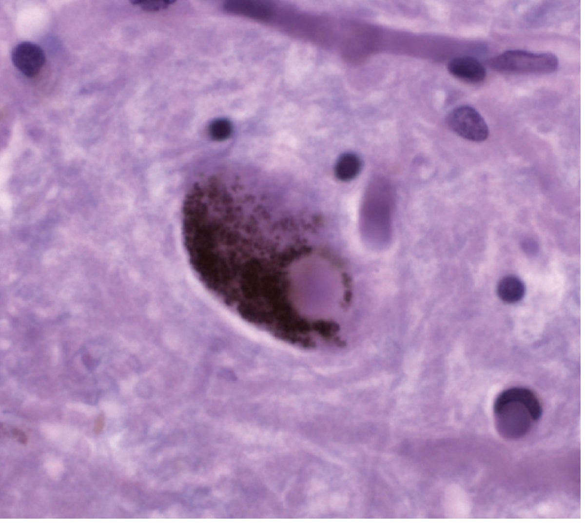
- Selective neuronal loss: The most constant pathological finding is depletion of pigmented dopaminergic neurons in the substantia nigra (and other pigmented brainstem nuclei such as the locus ceruleus and dorsal motor nucleus of the vagus), with replacement gliosis. In PD patients, pigmented neuron counts fall to roughly 30% or less of age-matched controls (versus a normal age-related decline from ~425,000 to ~200,000 by age 80) - Adams and Victor's Principles of Neurology, 12th Ed.
- Nigrostriatal pathway disruption: These neurons project from the substantia nigra to the striatum and normally regulate motor output via basal ganglia direct/indirect circuitry. Their loss produces abnormal firing in these circuits, generating the cardinal motor features (tremor, rigidity, bradykinesia, postural instability) - Bradley and Daroff's Neurology in Clinical Practice.
- Tyrosine hydroxylase (the rate-limiting enzyme in dopamine synthesis) declines correspondingly with neuronal loss.
2. Lewy bodies and α-synuclein
- The defining microscopic lesion is the Lewy body: an eosinophilic, cytoplasmic inclusion with a halo, found in surviving pigmented neurons. Ultrastructurally, Lewy bodies are composed of aggregated α-synuclein filaments along with neurofilament proteins and ubiquitin. Dystrophic neurites containing aggregated α-synuclein ("Lewy neurites") also occur - Robbins & Kumar Basic Pathology.
- α-synuclein is a normal presynaptic protein involved in synaptic vesicle trafficking and neurotransmission. In PD it misfolds, aggregates, and propagates in a prion-like fashion between neurons.
- Braak staging hypothesis: Braak and Braak proposed that synuclein pathology begins outside the substantia nigra - first in the dorsal glossopharyngeal-vagal nucleus and anterior olfactory structures - and ascends to the midbrain only later. This would explain prodromal non-motor features (anosmia, constipation, REM sleep behavior disorder) preceding motor onset, and is supported indirectly by epidemiological data showing reduced PD risk after truncal vagotomy (suggesting the vagus is an entry route for gut-to-brain synuclein spread). This staging model remains debated - Adams and Victor's Principles of Neurology.
3. Protein clearance and organelle dysfunction
- Genetic and mechanistic evidence points to failure of protein and organelle quality control:
- Synuclein aggregates are normally cleared via autophagy; defects in autophagic-lysosomal degradation are thought to be pathogenic.
- Parkin and related gene products function in mitochondrial quality control and endosomal trafficking; their dysfunction impairs clearance of damaged mitochondria and proteins.
- Mitochondrial abnormalities and resultant oxidative stress are considered central contributors to nigral neuronal death.
- Robbins & Kumar Basic Pathology, p. 853-854
4. Genetic contributions
- Most PD is sporadic, but recognized monogenic and risk-associated causes include:
- Point mutations and gene duplications of SNCA (α-synuclein) - autosomal dominant PD.
- LRRK2 gain-of-function mutations - the most common cause of autosomal dominant PD (mechanism still unclear).
- Heterozygous mutations in GBA (glucocerebrosidase, the Gaucher disease gene) - a lysosomal enzyme whose deficiency is a significant PD risk factor, again implicating impaired lysosomal turnover.
- Robbins & Kumar Basic Pathology, p. 854
5. Environmental/toxin models
- The neurotoxin MPTP (accidentally self-administered by drug users) causes an irreversible parkinsonian syndrome via selective destruction of nigral dopaminergic neurons. It is converted by monoamine oxidase to the toxic metabolite MPP+, which concentrates in melanin-containing nigral neurons and destroys them. This remains a useful experimental model of nigral degeneration and dopaminergic biochemistry, though it does not reproduce Lewy body pathology or the full clinical picture of naturally occurring PD.
- Epidemiological associations with pesticide/organophosphate exposure in agrarian regions and industrialized settings have been noted, but no toxin, heavy metal, or infection has been proven causal. Iron deposition in the substantia nigra has also been implicated in the degenerative process.
- Adams and Victor's Principles of Neurology, 12th Ed.
6. Beyond the nigrostriatal system
- Neuronal loss also occurs in the dorsal motor nucleus of the vagus and reticular formation (contributing to sleep disorders that can precede motor symptoms), and cerebral cortical involvement contributes to dementia. When dementia develops within one year of motor onset, it is classified as Lewy body dementia rather than PD with dementia - Robbins & Kumar Basic Pathology.
Recent evidence to note
A recent systematic review/meta-analysis (PMID: 39577101, 2025) examined the role of Th17 immune cells in PD pathogenesis, adding an emerging neuroinflammatory dimension not covered in detail in the textbook sources above - suggesting adaptive immune dysregulation may contribute alongside protein aggregation and mitochondrial dysfunction. Another 2025 meta-analysis (PMID: 40827506) explored epigenetic mechanisms (SETD1A expression) linking educational attainment to PD risk via genome-wide association and Mendelian randomization data. These represent active areas of research that extend, but do not contradict, the classical pathogenic framework above.
Pathogenesis of parkinsonism
Finding Sources
Finding Sources
Searching PubMed
parkinsonism AND pathogenesis
Reading File
Reading File
Reading File
Reading File
Reading File
Pathogenesis of Parkinsonism
Parkinsonism is a clinical syndrome, not a single disease. It is defined by four cardinal signs - tremor, rigidity, akinesia/bradykinesia, and postural instability - that result whenever the dopaminergic nigrostriatal pathway or its downstream basal ganglia circuitry is disrupted, regardless of the underlying cause (Goldman-Cecil Medicine, Ch. 378). Parkinson disease (PD) is the most common cause, but the same clinical picture arises from several distinct pathogenic mechanisms.
1. Shared final pathway
Whatever the etiology, most parkinsonian syndromes converge on loss of dopaminergic input to the striatum (either from destruction of substantia nigra neurons or from blockade of dopamine receptors), which disinhibits the basal ganglia's inhibitory output circuitry (via the direct/indirect pathways through the globus pallidus internus and subthalamic nucleus), producing bradykinesia and rigidity - Bradley and Daroff's Neurology in Clinical Practice.
2. Idiopathic Parkinson disease (most common cause)
- Progressive loss of pigmented dopaminergic neurons in the substantia nigra pars compacta, with Lewy bodies (α-synuclein aggregates) as the pathological hallmark.
- Proposed mechanisms: α-synuclein misfolding/aggregation, mitochondrial dysfunction and complex I inhibition, oxidative stress, impaired autophagic/lysosomal clearance, excitotoxicity, neuroinflammation, apoptosis, and loss of trophic support.
- Genetic causes account for only ~10% of cases (SNCA, LRRK2 - autosomal dominant; parkin, PINK1, DJ1 - autosomal recessive early-onset; GBA as a risk-modifying gene). The remainder is thought to reflect gene-environment interaction.
- Goldman-Cecil Medicine, Ch. 378, p. 3729-3733 (covered in detail in the previous answer on PD pathogenesis)
3. Drug-induced parkinsonism (2nd most common cause overall)
- Caused by dopamine receptor-blocking drugs (typical and atypical antipsychotics, antidopaminergic antiemetics) or drugs that deplete presynaptic dopamine stores (reserpine, tetrabenazine, deutetrabenazine, valbenazine).
- Mechanism is postsynaptic D2-receptor blockade rather than neuronal loss - this is why it is usually symmetric with more postural/action tremor than classic rest tremor, and is often (though not always) reversible over weeks to months after the offending drug is stopped.
- Risk correlates with a drug's D2-receptor binding affinity (risperidone/ziprasidone > olanzapine > quetiapine > clozapine). Occasionally symptoms persist, suggesting the drug "unmasked" latent subclinical PD.
- Bradley and Daroff's Neurology in Clinical Practice, Ch. 96
4. Vascular parkinsonism (2nd most common form in movement disorder clinics, ~8%)
- Associated with subcortical small-vessel/lacunar cerebrovascular disease, with relative preservation of substantia nigra dopaminergic cells - a key pathogenic distinction from PD.
- Risk factors: diabetes, chronic hypertension, hyperlipidemia.
- Produces "lower-body parkinsonism" (broad-based shuffling gait, start/freezing hesitation) with prominent postural instability, pyramidal signs, and dementia, but little tremor.
- Presynaptic striatal dopamine transporter imaging (SPECT) is typically normal, unlike in PD - reflecting that neurons themselves are not primarily lost, but their connectivity/white matter is disrupted.
- Bradley and Daroff's Neurology in Clinical Practice, Ch. 96
5. Toxin-induced parkinsonism
- MPTP: converted by monoamine oxidase-B to MPP+, which is selectively taken up by nigral dopaminergic neurons via the dopamine reuptake transporter and inhibits mitochondrial complex I, blocking oxidative phosphorylation and causing cell death. This remains the classic experimental/toxicologic model implicating oxidative stress and mitochondrial dysfunction in nigral degeneration, though it produces neuronal loss without Lewy bodies. MAO-B inhibitors (selegiline) block this conversion and prevent toxicity.
- Manganese: causes levodopa-unresponsive, symmetric parkinsonism with dystonic features; PET shows normal presynaptic dopamine function, indicating primarily postsynaptic/striatal pathology rather than nigral neuronal loss.
- Carbon monoxide: causes bilateral globus pallidus necrosis and white-matter injury.
- Katzung's Basic and Clinical Pharmacology, 16th Ed.; Bradley and Daroff's Neurology in Clinical Practice
6. Atypical parkinsonian syndromes (heredodegenerative/multisystem)
These involve more widespread neurodegeneration beyond the nigrostriatal pathway, explaining poorer levodopa response and additional features:
- Multiple system atrophy (MSA): combined degeneration of basal ganglia, cerebellum, locus ceruleus, inferior olivary nucleus, and pyramidal tracts, with glial cytoplasmic α-synuclein inclusions and prominent autonomic failure.
- Progressive supranuclear palsy (PSP): tauopathy affecting brainstem and basal ganglia, causing symmetric parkinsonism with vertical gaze palsy and early falls.
- Corticobasal degeneration (CBD): asymmetric tauopathy affecting cortex and basal ganglia.
- Diffuse Lewy body disease: cortical and subcortical Lewy body spread producing parkinsonism with early dementia/hallucinations.
- Katzung's Basic and Clinical Pharmacology; Bradley and Daroff's Neurology in Clinical Practice
7. Other/secondary causes
- Postencephalitic parkinsonism: neurofibrillary tangles in nigral cells without Lewy bodies, following viral encephalitis (historically von Economo's encephalitis).
- Structural lesions: distortion of the midbrain from hydrocephalus, over-drainage of CSF, or incipient transtentorial herniation can produce symptomatic parkinsonism by direct mechanical disruption of nigrostriatal pathways - Adams and Victor's Principles of Neurology.
- Genetic/metabolic: mitochondrial diseases (e.g., POLG1 mutations), Wilson disease, Fahr disease (basal ganglia calcification).
- Guadeloupean parkinsonism: an atypical, levodopa-unresponsive form linked to dietary exposure to mitochondrial complex I toxins in tropical plants (soursop/graviola).
Summary comparison
| Cause | Core mechanism | Key pathologic clue |
|---|---|---|
| Idiopathic PD | Nigral neuron loss, α-synuclein aggregation | Lewy bodies, asymmetric onset, rest tremor, levodopa-responsive |
| Drug-induced | Postsynaptic D2 blockade or presynaptic dopamine depletion | Symmetric, reversible, normal DAT imaging |
| Vascular | Subcortical ischemic small-vessel disease | Lower-body gait pattern, normal DAT imaging, preserved SN |
| Toxin (MPTP/Mn/CO) | Mitochondrial complex I inhibition or striatal/pallidal necrosis | Acute exposure history, no Lewy bodies |
| Atypical (MSA/PSP/CBD) | Multisystem neurodegeneration (synucleinopathy or tauopathy) | Poor levodopa response, additional neurologic signs |
Note: My literature search for very recent evidence returned papers specific to Parkinson disease pathogenesis (Th17 immune involvement, PMID 39577101; epigenetic/SETD1A mechanisms, PMID 40827506; PD-diabetes links, PMID 41241609) rather than parkinsonism-as-a-syndrome broadly - these support and extend the PD-specific mechanisms above but don't materially change the classification of parkinsonism's differential causes described here.
This is a shared conversation. Sign in to Orris to start your own chat.