
Period delaying medicine
period delaying medicine norethisterone how to use dose side effects 2025
Period-Delaying Medicines
1. Norethisterone (Most Common Option)
- Dose: 5 mg (1 tablet) three times a day
- Start taking it 3 days before your expected period
- Continue until you want your period to begin
- Can be used for up to 17-27 days (per most prescribing guidance)
- Your period will usually arrive 2-3 days after stopping
- It is not a contraceptive - it will not prevent pregnancy
- Requires a prescription in most countries
- Not suitable if you have a history of blood clots, liver disease, or are pregnant
- Nausea
- Breast tenderness
- Bloating
- Headaches
- Mood changes
- Spotting/breakthrough bleeding
2. Combined Oral Contraceptive Pill (COC)
3. Medroxyprogesterone Acetate (Provera)
Comparison at a Glance
| Feature | Norethisterone | COC Pill | Medroxyprogesterone |
|---|---|---|---|
| Prescription needed | Yes | Yes | Yes |
| Delay duration | Up to ~27 days | Indefinitely (run packs back-to-back) | Short-term |
| Contraceptive effect | No | Yes | No |
| Suitability for non-pill users | Yes | No (new start needs time) | Yes |
Who Should Not Use These Medicines
- Pregnant or possibly pregnant women
- Those with a history of blood clots (DVT/PE)
- History of breast or hormone-sensitive cancer
- Severe liver disease
- Unexplained vaginal bleeding
- Those with migraine with aura (COC is especially contraindicated)
Disclaimer: These medicines require a doctor's prescription. Always consult a healthcare professional before starting, as suitability depends on your medical history, current medications, and individual circumstances. Do not self-medicate.
Fibroadenosis
fibroadenosis breast clinical features pathology treatment
Fibroadenosis (Benign Breast Disease / ANDI)
Nomenclature
ANDI - Aberrations of Normal Development and Involution
- Bailey and Love's Short Practice of Surgery, 28th Ed., p. 940
- S Das Manual on Clinical Surgery, 13th Ed., p. 432
Definition
Epidemiology
- Affects women between 20-50 years (reproductive age)
- Incidence falls sharply after menopause (unlike breast carcinoma)
- More common in:
- Nulliparous women (spinsters, childless married women)
- Women who have not breastfed their children
- Higher incidence in western countries (England, Wales)
Aetiology
- Cyclical hormonal imbalances
- Oral contraceptives and HRT
- Diet and smoking
- High caffeine intake (proposed, not strongly proven)
Pathology
| Phase | Age | Changes |
|---|---|---|
| Lobule development | 15-25 years | Lobular proliferation → fibroadenoma |
| Cyclical changes | 15-50 years | Adenosis, cysts, epithelial hyperplasia |
| Involution | 35-55 years | Stromal involution → cyst formation |
Microscopic Features:
- Adenosis - increase in number of acini per lobule
- Cyst formation - micro- and macrocysts (kinking/narrowing of ductules causes secretion accumulation)
- Epithelial hyperplasia - >2 cell layers lining ducts/acini
- With atypia: Atypical Ductal Hyperplasia (ADH) or Atypical Lobular Hyperplasia (ALH)
- Papillomatosis
- Fibrosis
- Lymphocytic infiltration
Key: If ADH involves >2 ducts or the lesion is >2 mm, the term DCIS is used. - Bailey & Love, p. 940
Clinical Features
Symptoms
- Breast pain (Mastalgia) - the dominant symptom
- Cyclical (premenstrual): starts around day 14, worsens until day 27-28, relieved by onset of menses
- Both breasts usually involved; may radiate to the upper arm
- Non-cyclical: persistent, well-localised, not related to the cycle
- Nodularity / Lump - often multiple lumps, bilateral, upper and outer quadrant predominance
- Lumps become larger and more tender premenstrually
- Size fluctuates with the cycle - this almost excludes carcinoma
- Nipple discharge - greenish or serous
Signs
- Multiple, firm, rubbery nodules - best felt by fingers and thumb (not palmar surfaces)
- Usually bilateral, upper outer quadrant
- Nodules are not fixed to skin or pectoralis fascia
- Cysts - smooth, round, variable consistency; very tense cysts may feel hard and non-fluctuant
- Axillary lymph nodes: occasionally slightly enlarged and tender
- "Blue-domed cyst of Bloodgood" - appearance of a solitary breast cyst
Investigations
Triple Assessment (the cornerstone):
- Clinical examination
- Imaging
- Mammography (women >35 years)
- Ultrasound (younger women with dense breasts)
- Fine Needle Aspiration Cytology (FNAC)
- Aspirate from cyst: if non-blood-stained, no residual lump after aspiration, cyst does not refill, and cytology is benign → benign diagnosis confirmed
Criteria for a benign cyst on aspiration:
- Aspirate is not blood-stained
- No residual lump after aspiration
- Cyst does not refill
- Cytology shows no malignant cells
Cancer Risk
- Simple fibroadenosis: no significantly increased risk
- Epithelial hyperplasia without atypia: RR ~1.5-2x
- Atypical ductal/lobular hyperplasia: RR ~4-5x
- Papillomatosis (5+ papillomas): RR ~3x
- Positive family history of breast cancer further increases lifetime risk
Treatment
1. Reassurance and Explanation
- Most important step - the patient must be reassured that the condition is benign
- Explain the cyclical nature of symptoms
2. Mastalgia Management
- Well-fitted, supportive bra (worn 24 hours/day if needed)
- NSAIDs / analgesics
- Evening primrose oil (gamma-linolenic acid) - first-line non-hormonal option
- Danazol (anti-gonadotropin) - effective but androgenic side effects (acne, hirsutism, voice changes)
- Tamoxifen (anti-oestrogen) - effective; used in low doses (10-20 mg/day)
- Ormeloxifene (selective oestrogen receptor modulator)
- Bromocriptine (dopamine agonist / prolactin inhibitor) - less used now due to side effects
- GnRH analogues - for severe refractory cases
3. Cyst Management
- Aspiration - both diagnostic and therapeutic
- If cyst recurs after 2-3 aspirations or cytology is atypical → surgical excision
4. Surgical Treatment
- Dominant, discrete lump that is uncertain after triple assessment
- Atypia on cytology/histology
- Patient preference
Differential Diagnosis
| Feature | Fibroadenosis | Carcinoma | Fibroadenoma |
|---|---|---|---|
| Age | 20-50 years | >40 years | 15-30 years |
| Lumps | Multiple, bilateral | Usually solitary | Single, solitary |
| Cyclical variation | Yes - hallmark | No | No |
| Skin fixity | Absent | Often present | Absent |
| Axillary nodes | Occasionally tender | Firm, enlarged | Not enlarged |
| After menopause | Incidence falls | Incidence rises | Rare |
| Mobility | Mobile | Fixed/tethered | Highly mobile ("breast mouse") |
Key Points to Remember
- Fibroadenosis = ANDI = benign mammary dysplasia - all refer to the same spectrum
- It is the most common cause of a breast lump in reproductive-aged women
- The cyclical fluctuation in lump size is almost pathognomonic and effectively excludes carcinoma
- Triple assessment (clinic + imaging + FNAC) is mandatory before labelling any breast lump as benign
- Epithelial hyperplasia with atypia carries a significantly elevated cancer risk and warrants close follow-up
Sources: Bailey and Love's Short Practice of Surgery, 28th Ed., pp. 939-944; S Das Manual on Clinical Surgery, 13th Ed., pp. 432-434
Treatment protocol for viral hepatitis A.. with sgot 880 and sgpt 850 bilirubin 13.2
viral hepatitis A treatment protocol high transaminases jaundice management 2024
hepatitis A severe jaundice bilirubin 13 SGOT 880 SGPT 850 hospitalization criteria fulminant
https://emedicine.medscape.com/article/177484-treatment
Viral Hepatitis A - Treatment Protocol
First: Interpreting This Patient's Values
| Parameter | Patient's Value | Interpretation |
|---|---|---|
| SGOT (AST) | 880 U/L | Severely elevated (~22x ULN) |
| SGPT (ALT) | 850 U/L | Severely elevated (~21x ULN) |
| Bilirubin (Total) | 13.2 mg/dL | Markedly elevated (jaundice confirmed) |
- Prothrombin time / INR (most important prognostic marker)
- Hepatic encephalopathy (confusion, flapping tremor)
- Serum albumin
- Blood glucose (hypoglycemia = poor sign)
- Serum creatinine
Core Principle
There is NO specific antiviral treatment for Hepatitis A. HAV infection is self-limited. Management is entirely supportive. - Goldman-Cecil Medicine, p. 1568
Treatment Protocol
Step 1 - Confirm Diagnosis
- Anti-HAV IgM (ELISA) - peaks in the second month; diagnostic for acute infection
- IgM anti-HAV disappears after 4-12 months; IgG persists for life
- HAV RNA PCR in stool (not routinely necessary)
- Exclude Hep B (HBsAg, anti-HBc IgM) and Hep C (anti-HCV)
Step 2 - Assess Severity and Decide Admission
- Bilirubin >10 mg/dL ✓ (patient has 13.2)
- Inability to maintain oral intake (nausea, vomiting)
- Signs of dehydration
- PT/INR prolonged (>1.5)
- Any encephalopathy
- Age >50 years or underlying chronic liver disease
- Immunocompromised state
"In severe cases, patients may need to be hospitalized. If liver function is deteriorating, patients may need to be assessed for liver transplantation." - Goldman-Cecil Medicine, p. 1568
"About 3-8% of FHF cases are caused by HAV; however, only 1-2% of HAV infections in adults lead to FHF." - Medscape/Emedicine
Step 3 - Supportive In-Hospital Care
A. Rest and Activity
- Bed rest during the acute symptomatic phase
- Return to work/school only after fever and jaundice have cleared
- Restrict physical exertion until symptoms resolve
B. Diet and Nutrition
- High-calorie, high-carbohydrate, low-fat diet
- Small, frequent meals (better tolerated when nauseated)
- Encourage oral fluids - aim for adequate hydration
- If oral intake is severely impaired: IV fluids (Normal saline or Dextrose-saline)
- Avoid fatty foods (worsens nausea)
C. Medications - STRICT RESTRICTIONS
| Drug / Substance | Action |
|---|---|
| Alcohol | Strictly forbidden - worsens liver damage |
| Paracetamol (Acetaminophen) | Use with extreme caution; maximum 2 g/day if essential (normally limit to 3-4 g/day in healthy adults, lower in liver disease) |
| NSAIDs (ibuprofen, diclofenac) | Avoid - hepatotoxic and risk of GI bleed |
| Statins | Hold temporarily |
| Herbal supplements | Avoid - many are hepatotoxic |
| All non-essential medications | Stop or defer |
D. Symptomatic Treatment
| Symptom | Treatment |
|---|---|
| Nausea/Vomiting | Ondansetron 4-8 mg IV/oral TDS; or Metoclopramide 10 mg TDS |
| Pruritus (from cholestasis) | Cholestyramine 4 g BD-TDS; Antihistamines (chlorpheniramine) |
| Fever | Paracetamol (cautious dose) - avoid aspirin in children (Reye syndrome risk) |
| Dehydration | IV fluids - NS/DNS; correct electrolytes |
| Hypoglycemia | 10-25% Dextrose IV |
E. Monitoring (Daily in Severe Cases)
- Liver function tests (bilirubin, ALT, AST) - every 2-3 days
- Prothrombin time / INR - daily (key prognostic marker)
- Blood glucose - daily
- Serum electrolytes - daily
- Serum creatinine and urea
- Full blood count
- Mental status assessment (for encephalopathy)
Red Flags - Fulminant Hepatic Failure (FHF)
- Hepatic encephalopathy (confusion, drowsiness, flapping tremor/asterixis)
- PT >3 seconds above control / INR >1.5 and rising
- Serum bilirubin rapidly rising (>1 mg/dL/day)
- Hypoglycemia (falling blood sugar)
- Falling transaminases WITH worsening bilirubin and INR = "enzyme burn-out" - a sinister sign
- Oliguria / renal failure
- Metabolic acidosis
"Acute liver failure (ALF) is a clinical syndrome of liver injury (elevated transaminases), hepatic encephalopathy, and coagulopathy (elevated PT/INR) in patients without preexisting liver disease." - Sabiston Textbook of Surgery
- Transfer immediately to a liver transplant centre
- ICU-level monitoring
- Lactulose 20-30 mL TDS (for encephalopathy - to reduce ammonia)
- Rifaximin 550 mg BD (adjunct for encephalopathy)
- Vitamin K IV (for coagulopathy)
- Fresh Frozen Plasma (FFP) if active bleeding or invasive procedure planned
- Correct hypoglycemia aggressively with IV dextrose
- Avoid sedatives/opioids (worsen encephalopathy)
- Assess for liver transplantation using King's College Criteria
King's College Criteria (Transplant Referral - Non-Acetaminophen Cause)
- PT >100 seconds (INR >6.5), OR
- Any 3 of the following:
- Age <10 or >40 years
- Aetiology: non-A, non-B hepatitis / drug reaction / Wilson's disease
- Jaundice >7 days before encephalopathy
- PT >50 seconds
- Serum bilirubin >300 µmol/L (>17.5 mg/dL)
Isolation and Infection Control
- HAV is faecal-oral transmission - highly contagious during the prodrome and early icteric phase
- Enteric (contact) precautions during hospitalization
- Hand hygiene is essential
- Stool and blood precautions
- Patient is most infectious 2 weeks before jaundice appears and for ~1 week after
Post-Exposure Prophylaxis for Contacts
- HAV vaccine (within 2 weeks of exposure) - ~98% effective
- Immunoglobulin (Ig) 0.1 mL/kg IM (within 2 weeks) - if vaccine contraindicated or patient is immunocompromised/elderly
- Both can be given together without reducing vaccine efficacy
Prognosis for This Patient
- Acute HAV is entirely self-limited - no progression to chronic hepatitis, cirrhosis, or cancer
- Typically resolves in 3-4 weeks
- Transaminases will fall first, bilirubin normalizes last
- Fulminant HAF occurs in <0.1% of cases overall; risk increases with age >50 and pre-existing liver disease
- Mortality from uncomplicated hepatitis A is very low
- Cholestatic HAV (as suggested by bilirubin 13.2) has a good prognosis with full recovery within weeks
"Acute hepatitis A infection generally resolves without complications in 3 to 4 weeks and never evolves to chronic infection." - Goldman-Cecil Medicine, p. 1568
Summary Card for This Patient
| Priority | Action |
|---|---|
| Immediate | Check PT/INR, blood glucose, creatinine, albumin |
| Admit | Bilirubin 13.2 mg/dL warrants hospitalization |
| Antivirals | None - no specific treatment exists |
| Diet | High-carb, low-fat, small frequent meals; IV fluids if vomiting |
| Avoid | Alcohol, NSAIDs, paracetamol (or strict dose limit), hepatotoxic drugs |
| Symptomatic | Antiemetics, cholestyramine for itch |
| Monitor | Daily PT/INR, glucose, bilirubin, mental status |
| Escalate if | Encephalopathy, rising INR, falling glucose, falling enzymes with worsening bilirubin |
Sources: Goldman-Cecil Medicine International Edition, p. 1568; Sabiston Textbook of Surgery; Merck Manual Professional Ed. (updated Apr 2025); Medscape - Hepatitis A Treatment & Management
Fluminant hepatic failure diagnosis
Fulminant Hepatic Failure (FHF) - Diagnosis
Terminology
Definition
ALF = Acute liver injury (elevated transaminases) + Hepatic Encephalopathy + Coagulopathy (INR ≥ 1.5) in a patient WITHOUT pre-existing liver disease.
- Sabiston Textbook of Surgery
- Yamada's Textbook of Gastroenterology, 7th Ed.
"The combination of coagulopathy and encephalopathy is unique provided that underlying cirrhosis has been excluded." - Yamada's Textbook of Gastroenterology
Classification by Time (Onset of Jaundice to Encephalopathy)
| Subtype | Interval | Common Causes | Features |
|---|---|---|---|
| Hyperacute | < 7 days | Acetaminophen, ischaemia (shock liver) | Very high transaminases, low bilirubin, better prognosis |
| Acute | 7-21 days | HAV, HBV | Intermediate |
| Subacute | 21 days - 26 weeks | Drug-induced (DILI), autoimmune, HBV | Lower transaminases, very high bilirubin, worst prognosis |
"Shorter duration to encephalopathy (hyperacute) more commonly leads to cerebral edema; longer duration leads to portal hypertension." - Sabiston Textbook of Surgery
Causes of ALF
| Cause | UK | USA | India | Japan |
|---|---|---|---|---|
| Acetaminophen | 54% | 46% | - | - |
| Indeterminate | 17% | 14% | 24% | 45% |
| Drug-induced (DILI) | 7% | 12% | 5% | - |
| HAV or HBV | 14% | 10% | 33% | 55% |
| HEV | - | - | 38% | - |
- Sleisenger & Fordtran's GI and Liver Disease
Clinical Features
1. Encephalopathy (GRADING - West Haven Criteria)
| Grade | Features |
|---|---|
| I | Mild confusion, altered mood, slurred speech, agitation |
| II | Drowsiness, inappropriate behaviour, disorientation |
| III | Marked confusion, stupor but rousable, incoherent speech |
| IV | Coma - unresponsive to pain |
"Mental alterations may be subtle initially - agitation and confusion - then rapid progression to deeper coma. In a young person, agitation may be mistaken for CNS stimulant ingestion; sedation is inappropriate as the liver cannot metabolize CNS depressants." - Yamada's Textbook of Gastroenterology
2. Jaundice
- Usually present (deep jaundice)
- May be absent or mild in hyperacute cases (especially acetaminophen)
- Cholestatic pattern in subacute disease
3. Coagulopathy
- Prolonged PT/INR (most reliable prognostic marker)
- Bleeding tendency (GI bleed, intracranial bleed)
- DIC may develop in late disease
4. Cerebral Oedema
- Almost unique to ALF (rarely seen in cirrhosis)
- Causes >20% of deaths in ALF
- Manifests as: rising BP, bradycardia, abnormal pupil reflexes, decerebrate posturing
- Leads to herniation and brain death if untreated
5. Haemodynamic Instability
- Hypotension + vasodilation + low systemic vascular resistance (resembles gram-negative sepsis)
- Compensatory tachycardia
6. Other Organ Failures
| System | Complication |
|---|---|
| Renal | Hepatorenal syndrome (functional renal failure) - reversible with liver recovery |
| Pulmonary | ARDS (late stage) |
| Metabolic | Hypoglycaemia (hepatic glycogen depletion), lactic acidosis |
| Infection | Gram-positive sepsis most common (skin flora via invasive lines) |
| Cardiovascular | Cardiac failure (rare, late stage) |
Diagnostic Investigations
Mandatory at Presentation
| Investigation | What it shows |
|---|---|
| PT / INR | Most important prognostic marker; INR ≥1.5 required for ALF diagnosis |
| Liver enzymes (AST, ALT) | Degree of hepatocellular necrosis |
| Serum bilirubin | Rising bilirubin indicates ongoing failure |
| Serum albumin | Marker of synthetic function |
| Blood glucose | Hypoglycaemia = bad prognostic sign |
| Serum creatinine / urea | Renal failure (hepatorenal syndrome) |
| Serum electrolytes | Hyponatraemia, hypokalaemia common |
| Arterial blood gas | pH <7.3 = poor prognosis (King's College); metabolic acidosis |
| Serum lactate | >3.0 mmol/L after resuscitation = poor prognosis |
| Full blood count | Anaemia, thrombocytopaenia, leukocytosis (infection) |
| Serum phosphate | Elevated on day 2 = poor prognosis (acetaminophen) |
| Serum ammonia | Elevated; contributes to cerebral oedema |
Aetiology-Specific Tests
| Test | Purpose |
|---|---|
| Anti-HAV IgM | Acute Hepatitis A |
| HBsAg, Anti-HBc IgM | Acute Hepatitis B |
| Anti-HCV | Hepatitis C (rare cause of ALF) |
| Anti-HEV IgM | Hepatitis E (especially India, Asia) |
| Serum acetaminophen level | Paracetamol toxicity |
| Autoimmune markers (ANA, ASMA, IgG) | Autoimmune hepatitis |
| Serum ceruloplasmin, copper | Wilson disease |
| Blood cultures | Infection / sepsis |
| Urine toxicology | Drug ingestion |
| Pregnancy test (females) | AFLP, HELLP syndrome |
Imaging
| Investigation | Purpose |
|---|---|
| Ultrasound abdomen (with Doppler) | Liver size, portal vein patency, Budd-Chiari syndrome, ascites |
| CT abdomen | Malignant infiltration (hepatomegaly), liver volume assessment |
| CT/MRI brain | Confirm cerebral oedema, exclude intracranial haemorrhage |
"Liver enlargement in ALF is unusual except in Budd-Chiari; should trigger consideration of malignant infiltration or alcohol-associated hepatitis." - Sleisenger & Fordtran's
"CT/MRI are relatively insensitive for detection of cerebral oedema but can exclude intracranial haemorrhage as a cause of deterioration." - Yamada's Textbook of Gastroenterology
Liver Biopsy
- Not routinely performed in ALF
- Findings of confluent necrosis and parenchymal collapse are non-specific
- Indicated when: suspicion of malignant infiltration or alcohol-associated hepatitis - especially if transplant listing is under consideration
- Can support diagnosis of: autoimmune hepatitis, Wilson disease, AFLP, Budd-Chiari
Prognostic Scoring
King's College Criteria (1989)
For NON-Paracetamol Causes:
- INR > 6.5 (PT >100 sec) at any time, OR
- Any 3 of the following:
- Age <10 or >40 years
- Aetiology: non-A/non-B hepatitis, drug-induced liver injury, Wilson disease
- Duration of jaundice >7 days before encephalopathy
- PT > 50 sec (INR >3.5)
- Serum bilirubin >300 µmol/L (>17.5 mg/dL)
For Paracetamol (Acetaminophen) Causes:
- Arterial pH < 7.30 after resuscitation, OR
- All 3 of:
- PT > 100 sec (INR > 6.5)
- Serum creatinine > 3.5 mg/dL (>292 µmol/L)
- Grade III or IV hepatic encephalopathy
"Specificity of King's College Criteria: 82% for non-acetaminophen, 95% for acetaminophen ALF. Sensitivity ~58% (relatively low - do not use to rule out)." - Sleisenger & Fordtran's
Clichy Criteria (France)
- Factor V < 20% in patients < 30 years, OR
- Factor V < 30% in patients ≥ 30 years
- With grade 2 or greater encephalopathy
- Limitations: not effective for acetaminophen ALF
MELD Score
- MELD = 3.78×ln[bilirubin mg/dL] + 11.2×ln[INR] + 9.57×ln[creatinine mg/dL] + 6.43
- Has been validated for ALF but not superior to King's College Criteria
Prognosis
| Etiology | Transplant-free Survival |
|---|---|
| Acetaminophen / Paracetamol | ~60-65% (best) |
| Hepatitis A | >50% |
| Ischaemia / shock liver | ~50% |
| Pregnancy-related | ~50% |
| Autoimmune hepatitis | <30% |
| Hepatitis B | <30% |
| Drug-induced (DILI) | <30% (worst) |
"Trends over time in mentation and PT/INR are the best indicators of overall prognosis at the bedside." - Yamada's Textbook of Gastroenterology
Diagnostic Algorithm Summary
Acute illness → Jaundice + Rising PT/INR
        ↓
Assess for Encephalopathy (mandatory for ALF diagnosis)
        ↓
If encephalopathy present:
  → Confirm NO pre-existing liver disease/cirrhosis
  → Classify timing (hyperacute / acute / subacute)
  → Run full aetiology workup (IgM serology, paracetamol level, autoimmune, Wilson)
  → Assess organ failure (renal, metabolic, cerebral)
  → Apply King's College Criteria
  → If criteria met → IMMEDIATE transfer to liver transplant centre
        ↓
Monitor: PT/INR, glucose, ammonia, creatinine, GCS/encephalopathy grade DAILY
Sources: Sleisenger & Fordtran's GI and Liver Disease; Yamada's Textbook of Gastroenterology 7th Ed.; Sabiston Textbook of Surgery; Mulholland & Greenfield's Surgery 7th Ed.
Treatment protocol for lower lip ulcer
To give you the most accurate treatment protocol, could you clarify the type of lower lip ulcer?
Herpetic ulcer
herpes labialis cold sore lower lip ulcer herpetic
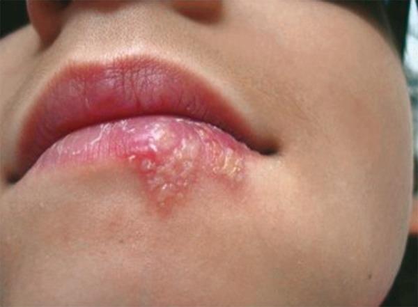
This clinical photograph shows a close-up of a patient's lower lip and perioral region, demonstrating a cluster of herpetic lesions consistent with Herpes Labialis (cold sores). The primary findings include a group of small, fluid-filled vesicles on an erythematous (reddened) base. These vesicles are concentrated on the vermilion border and extend onto the adjacent cutaneous surface of the lower lip. The vesicles exhibit varying degrees of opacity, with some appearing clear and others becoming cloudy as they progress. Mild perilesional inflammation (hyperemia) is visible. In contrast, the upper lip appears normal in color and texture with no active lesions. This image captures the vesicular stage of a recurrent herpes simplex virus (HSV-1) infection, illustrating typical morphology, including clustering, vesicle formation, and location at the mucocutaneous junction. It serves as an educational example for dermatological and oral pathology assessment, highlighting the physical signs used to diagnose and monitor the progression or healing of labial herpes.
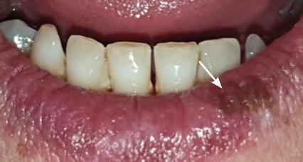
This clinical photograph displays an intraoral view of the lower lip and mandibular anterior teeth, highlighting a localized pathological lesion. The primary finding is a well-defined, crusted lesion located on the lower left lip along the vermillion border, specifically toward the commissure. The lesion exhibits a dark brownish-black coloration with a rough, uneven, and thickened texture, consistent with a dried serosanguinous crust. The surrounding labial mucosa and vermillion show generalized erythema, edematous swelling, and prominent vertical tissue folds, indicating an underlying inflammatory process. This presentation is characteristic of a late-stage Herpes labialis outbreak (cold sore) during the crusting phase. The image serves as a teaching tool for recognizing common viral orofacial infections, focusing on the distribution, morphology, and clinical evolution of herpetic lesions in a dental or dermatological context.
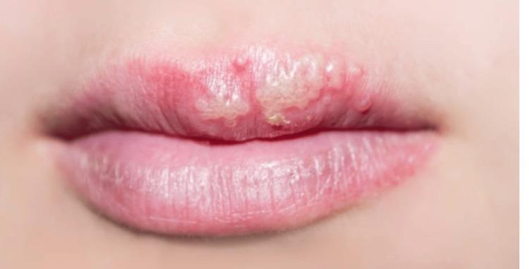
This clinical photograph shows a close-up view of a patient's mouth, specifically focusing on the upper lip. The image demonstrates a cluster of small, translucent to whitish vesicles localized on an erythematous base. These primary skin lesions are characteristic of a herpetic infection. The distribution is clustered and follows the vermillion border of the upper lip, predominantly on the patient's left side. The vesicles appear tense and grouped, causing an irregular disruption of the smooth transition between the lip mucosa and the surrounding cutaneous tissue. There is visible peri-lesional inflammation indicated by localized redness (erythema), while the adjacent skin and the lower lip appear unaffected. This visual is a classic representation of orolabial herpes (Herpes Simplex Virus), often referred to as herpes labialis or cold sores. The clinical significance of this image lies in illustrating the morphology and specific anatomical predilection of HSV-1 manifestations for educational diagnostic purposes in dermatology and primary care.
Herpetic Ulcer of the Lower Lip (Herpes Labialis) - Treatment Protocol
Background
Clinical Stages and Appearance
| Stage | Duration | Features |
|---|---|---|
| Prodrome | Hours | Tingling, burning, itching, pain at the site |
| Erythema | 6-12 hours | Redness, swelling |
| Vesicular | 24-48 hours | Clustered fluid-filled vesicles on erythematous base |
| Ulceration | 2-3 days | Vesicles rupture → shallow, painful ulcers |
| Crusting | 2-3 days | Yellow-brown crust (serosanguinous) |
| Healing | Total: ~7-10 days | Crust falls off, no scarring |
Key fact: Treatment is ONLY effective if started in the prodrome or erythema stage - once frank ulceration occurs, benefit is minimal.
Diagnosis
- Clustered vesicles/ulcers at the vermilion border of the lip
- History of recurrence in the same location
- Prodrome of tingling/burning
- Direct Fluorescent Antibody (DFA) test - fastest, results in hours (preferred)
- Viral culture from vesicle base - gold standard but takes 2-5 days
- Tzanck smear - shows multinucleated giant cells (not HSV-specific, but suggestive)
- PCR - most sensitive, used in atypical or severe cases
- Serology (HSV IgM/IgG) - not useful for acute disease; only establishes serostatus
Treatment Protocol
A. Recurrent Herpes Labialis (Most Common Presentation)
1. Topical Therapy (First-Line for Mild Cases)
| Drug | Dose | Duration | Notes |
|---|---|---|---|
| Penciclovir 1% cream | Every 2 hours while awake | 4 days | FDA-approved; start ASAP in prodrome; reduces healing by ~1 day and pain duration |
| Docosanol 10% cream | 5 times a day | Until healed | FDA-approved OTC; apply at first sign of recurrence |
| Acyclovir 5% cream | 5 times daily | 5 days | Less effective; acyclovir cream/ointment has no consistent clinical benefit for herpes labialis |
"Penciclovir 1% cream every 2 hours while awake for 4 days - when initiated within 1 hour of first symptoms, speeds healing (4.8 vs 5.5 days) and decreases pain duration." - Fitzpatrick's Dermatology
2. Oral Antiviral Therapy (For Moderate-Severe or Faster Resolution)
| Drug | Dose | Duration | Comments |
|---|---|---|---|
| Valacyclovir | 2 g twice daily | 1 day only | FDA-approved for herpes labialis; reduces episode by ~1 day; must start in prodrome |
| Acyclovir | 400 mg 5 times/day | 5 days | Marginal benefit; must be started in earliest 1-2 hours |
| Famciclovir | 1500 mg single dose | Single dose | Reduces healing time by ~2 days vs placebo; highly convenient |
| Famciclovir | 500 mg 3 times/day | 5 days | If started within 48 hours of UV-triggered outbreak |
"For herpes labialis, 1 day of therapy with oral valacyclovir improves time to healing and reduces pain, whereas acyclovir ointment has no consistent clinical benefit." - Goldman-Cecil Medicine
"A single dose of famciclovir (1500 mg) reduced time to healing of herpes labialis lesions by approximately 2 days compared with placebo." - Fitzpatrick's Dermatology
B. Primary (First Episode) Herpetic Gingivostomatitis
| Drug | Adult Dose | Paediatric Dose | Duration |
|---|---|---|---|
| Acyclovir | 400 mg 5 times/day | 15 mg/kg orally 5 times/day (suspension) | 7 days |
| Valacyclovir | 1 g twice daily | Not approved in children | 7-10 days |
| Famciclovir | 250 mg three times/day | Not approved in children | 7-10 days |
"The pediatric dose is 15 mg/kg of acyclovir suspension orally 5 times a day for 7 days. When started within 3 days of onset, this regimen decreases the duration of oral and extraoral lesions, fever, and eating and drinking difficulties." - Fitzpatrick's Dermatology
C. Suppressive Therapy (For Frequent Recurrences ≥6/year)
| Drug | Dose | Frequency |
|---|---|---|
| Acyclovir | 400 mg | Twice daily (long-term) |
| Valacyclovir | 500 mg or 1 g | Once daily |
| Famciclovir | 250 mg | Twice daily |
- Review annually - take a "holiday" each year to reassess whether suppression is still needed
- Sunscreen to lips helps prevent UV-triggered recurrences (benefit shown in some trials)
"The use of suppressive acyclovir for herpes labialis is controversial. Suppressive valacyclovir reduces recurrence frequency and severity in those with frequent episodes." - Fitzpatrick's Dermatology
D. Immunocompromised Patients (HIV, Transplant)
- Lesions are larger, more numerous, more persistent, more frequent
- First-line: oral famciclovir, valacyclovir, or acyclovir for 7 days
- Severe lesions: IV acyclovir (5-10 mg/kg every 8 hours)
- Daily suppressive therapy should be considered for frequent/severe recurrences
- If lesions persist despite adequate treatment: culture for acyclovir resistance
- Acyclovir-resistant HSV: IV foscarnet is the treatment of choice
- Cummings Otolaryngology
Symptomatic / Supportive Care
| Symptom | Management |
|---|---|
| Pain | Oral paracetamol / ibuprofen; topical lidocaine gel |
| Crusting/dryness | Petroleum jelly (Vaseline) to prevent cracking of crust |
| Secondary bacterial infection | Topical fusidic acid or mupirocin if superinfection suspected |
| Nutrition (children) | Cold fluids, ice cream, soft diet; IV fluids if intake severely impaired |
Infection Control / Patient Advice
- Avoid kissing or direct contact during active lesions
- Avoid touching lesions and then touching eyes (risk of herpetic keratitis - sight-threatening)
- Wash hands frequently
- Do not share lip balm, utensils, towels, or cups during active outbreak
- Lesions are most contagious from prodrome through ulceration (before crust forms)
- Sunscreen/lip balm with SPF - apply daily to prevent sun-triggered recurrences
When to Refer / Escalate
| Situation | Action |
|---|---|
| Lesion involving the eye (keratitis risk) | Urgent ophthalmology referral; topical + oral antivirals |
| No healing after 2 weeks | Biopsy to exclude malignancy; consider acyclovir resistance |
| Immunocompromised patient with severe disease | IV acyclovir ± foscarnet (if resistant) |
| Encephalitis symptoms (confusion, fever + headache) | Emergency admission; IV acyclovir |
| Widespread eczema herpeticum | Hospital admission; IV acyclovir 10-15 mg/kg TDS for 14-21 days |
| Neonatal exposure | Urgent neonatology review |
Summary: Quick Treatment Decision Guide
Lower lip herpetic ulcer
       ↓
Prodrome / erythema only (caught early)?
  YES → Penciclovir 1% cream q2h × 4 days
        OR Valacyclovir 2g BD × 1 day
        OR Famciclovir 1500mg single dose
        + Paracetamol for pain
        + Lip balm SPF
  NO (already ulcerated/crusted) →
        Supportive care only (antivirals have minimal benefit at this stage)
        Topical anaesthetic for pain relief
        Keep clean, prevent secondary infection
       ↓
Frequent recurrences (≥6/year)?
  → Add suppressive Acyclovir 400mg BD or Valacyclovir 500mg OD
       ↓
Immunocompromised / severe disease?
  → Oral antivirals × 7-10 days or IV Acyclovir
  → Check for acyclovir resistance if not responding
  → Foscarnet IV if resistant
Sources: Fitzpatrick's Dermatology, Vol 1; Goldman-Cecil Medicine International Edition; Washington Manual of Medical Therapeutics; Textbook of Family Medicine 9th Ed.; Cummings Otolaryngology Head & Neck Surgery