A 50y old male complains of dragging sensation in abdomen. Physical examination revealed pallor and massive spleenomegaly. There is no lymphadenopathy. Total leucocyte count is 1,60,000 cell/mm³ and platelet count is 2 Lakhs/mm³. M a) What is the probable diagnosis? b) Describe cytogenetic change in this case? c) Enumerate peripheral blood smear findings?

Reading File
Finding Sources
Finding Sources
Searching PubMed

chronic myeloid leukemia diagnosis cytogenetics

Reading File
Reading File
Reading File
Finding Sources
Loading Image
Loading Image
I now have comprehensive, authoritative information. Here is the full, structured answer:

Probable Diagnosis: Chronic Myeloid Leukemia (CML)

Clinical reasoning:
  • 50-year-old male (peak incidence is 5th-6th decade)
  • Dragging sensation in abdomen - classic symptom of massive splenomegaly in CML
  • Massive splenomegaly without lymphadenopathy - hallmark distinguishing CML from lymphoproliferative disorders
  • TLC = 1,60,000/mm³ (markedly elevated, typical of CML chronic phase, often 1,00,000-4,00,000/mm³)
  • Platelets = 2 Lakhs/mm³ - normal to elevated (thrombocytosis is characteristic; contrast with acute leukemia where platelets are low)
  • Pallor due to anemia from marrow infiltration and splenic sequestration
"Sometimes the first symptom is a dragging sensation in the abdomen caused by splenomegaly... CML is best differentiated from other myeloproliferative neoplasms by detection of the BCR::ABL fusion gene." - Robbins, Cotran & Kumar Pathologic Basis of Disease, p. 583

a) Probable Diagnosis

Chronic Myeloid Leukemia (CML) - Chronic Phase
This is a myeloproliferative neoplasm arising from a pluripotent hematopoietic stem cell. The combination of:
  • Marked leukocytosis (1,60,000/mm³) without lymphadenopathy
  • Massive splenomegaly
  • Normal/elevated platelets
  • Middle-aged male ...is the classic triad pointing to CML rather than ALL/AML/CLL or lymphoma.

b) Cytogenetic Changes

BCR-ABL pathogenesis diagram showing Philadelphia chromosome formation and downstream signaling
Fig. 13.34 - Robbins Pathology: Pathogenesis of CML showing BCR::ABL fusion gene formation

The Philadelphia (Ph) Chromosome

FeatureDetail
Translocationt(9;22)(q34;q11) - reciprocal translocation
Genes involvedABL gene (chromosome 9q34) + BCR gene (chromosome 22q11)
ResultShortened chromosome 22 = Philadelphia chromosome (Ph)
Fusion geneBCR::ABL chimeric fusion gene
Fusion protein210 kDa BCR-ABL constitutively active tyrosine kinase
FrequencyPresent in >90-95% of CML cases
Mechanism:
  1. DNA breakage and reciprocal translocation between chromosomes 9 and 22
  2. BCR sequences from chr. 22 fuse with ABL tyrosine kinase sequences from chr. 9
  3. This creates the BCR::ABL fusion gene on the derivative (shortened) chromosome 22
  4. The 210 kDa BCR-ABL protein has constitutive, ligand-independent tyrosine kinase activity
  5. It activates downstream pathways - RAS, STAT, AKT - causing growth factor-independent proliferation while preserving normal differentiation (hence mature granulocytes in blood, not blasts)
In the remaining ~5-10% of cases: BCR::ABL fusion is present but the Ph chromosome is not detectable by conventional karyotyping - it requires FISH or PCR for detection. There is no true BCR-ABL-negative CML.
"In more than 90% of cases, BCR::ABL is created by a reciprocal (9;22)(q34;q11) translocation (the so-called Philadelphia chromosome [Ph])." - Robbins, Cotran & Kumar Pathologic Basis of Disease, p. 583
Additional cytogenetic abnormalities (seen in disease progression/accelerated phase):
  • Trisomy 8 (+8)
  • Isochromosome 17q [i(17q)]
  • Duplication of the Ph chromosome (+Ph)
  • These signal transformation toward blast crisis

c) Peripheral Blood Smear Findings

CML peripheral blood smear showing mature neutrophils, metamyelocytes, and myelocytes
Fig. 13.35 - Robbins Pathology: CML peripheral blood smear showing mature neutrophils, metamyelocytes, and myelocytes

Enumerated Peripheral Blood Smear Findings in CML (Chronic Phase):

  1. Leukocytosis - WBC typically 1,00,000-3,00,000/mm³; composed predominantly of granulocytes at various stages of maturation
  2. "Myelocyte bulge" (Myelocyte peak) - The entire myeloid maturation series is represented:
    • Myeloblasts (< 2%)
    • Promyelocytes
    • Myelocytes (predominant immature form - the "bulge") - most conspicuous immature cell
    • Metamyelocytes
    • Band forms (stab cells)
    • Mature segmented neutrophils (predominant cell)
  3. Basophilia - Absolute increase in basophils; a very characteristic and diagnostically important finding. Increasing basophilia signals disease progression/accelerated phase.
  4. Eosinophilia - Mild increase in eosinophils
  5. Thrombocytosis - Platelet count normal to elevated (as in this case, 2 Lakhs/mm³); large/abnormal platelet forms may be seen
  6. Normochromic normocytic anemia - Pallor on smear, mild to moderate reduction in RBCs
  7. Nucleated RBCs (NRBCs) - May be present occasionally
  8. Reduced/absent toxic granulation and Döhle bodies - In contrast to reactive leukocytosis (leukemoid reaction)
  9. Low Leukocyte Alkaline Phosphatase (LAP) score - A key distinguishing feature from leukemoid reaction (in which LAP is high). Note: LAP score is a cytochemical stain result reported with the smear.
  10. No significant lymphocytosis - Differentiates from CLL
"Peripheral blood smear shows many mature neutrophils, some metamyelocytes, and a myelocyte." - Robbins, Cotran & Kumar Pathologic Basis of Disease, Fig. 13.35

Summary Table: Key Differentiating Features

FeatureCMLLeukemoid Reaction
LAP scoreLow/absentHigh
BasophiliaPresentAbsent
SplenomegalyMassiveMild/absent
Philadelphia chromosomePresentAbsent
CauseNeoplasticReactive
Sources: Robbins, Cotran & Kumar Pathologic Basis of Disease (10th ed.), p. 583-584 | Goldman-Cecil Medicine, Chapter on CML | Robbins & Kumar Basic Pathology

A 50y old male complains of dragging sensation in abdomen. Physical examination revealed pallor and massive spleenomegaly. There is no lymphadenopathy. Total leucocyte count is 1,60,000 cell/mm³ and platelet count is 2 Lakhs/mm³. M a) What is the probable diagnosis? b) Describe cytogenetic change in this case? c) Enumerate peripheral blood smear findings?

Reading File
Finding Sources
Finding Sources
Searching PubMed

chronic myeloid leukemia diagnosis peripheral blood

Reading File
Reading File
Reading File
Loading Image
Loading Image
I have everything needed for a thorough, well-illustrated answer. Here is the complete structured response:

a) Probable Diagnosis: Chronic Myeloid Leukemia (CML) - Chronic Phase

Clinical Reasoning:
Feature in this patientSignificance
50-year-old malePeak incidence is 5th-6th decade
Dragging sensation in abdomenClassic symptom of massive splenomegaly in CML
Massive splenomegaly, no lymphadenopathyDistinguishes CML from lymphoproliferative disorders (CLL, lymphoma)
TLC = 1,60,000/mm³Profound leukocytosis - typical of CML chronic phase
Platelets = 2 Lakhs/mm³ (normal-elevated)Thrombocytosis or normal platelets - characteristic of CML; contrast with acute leukemia where platelets fall
PallorMild-to-moderate anemia from marrow infiltration
CML is a myeloproliferative neoplasm (MPN) arising from a pluripotent hematopoietic stem cell (HSC). Because BCR-ABL drives proliferation but does not block differentiation, mature granulocytes accumulate in blood rather than blasts - explaining the clinical picture of extreme leukocytosis with a functioning-appearing smear.
"Sometimes the first symptom is a dragging sensation in the abdomen caused by splenomegaly... The onset is insidious. Mild-to-moderate anemia and hypermetabolism due to increased cell turnover lead to fatigability, weakness, weight loss, and anorexia." - Robbins, Cotran & Kumar Pathologic Basis of Disease, p. 583

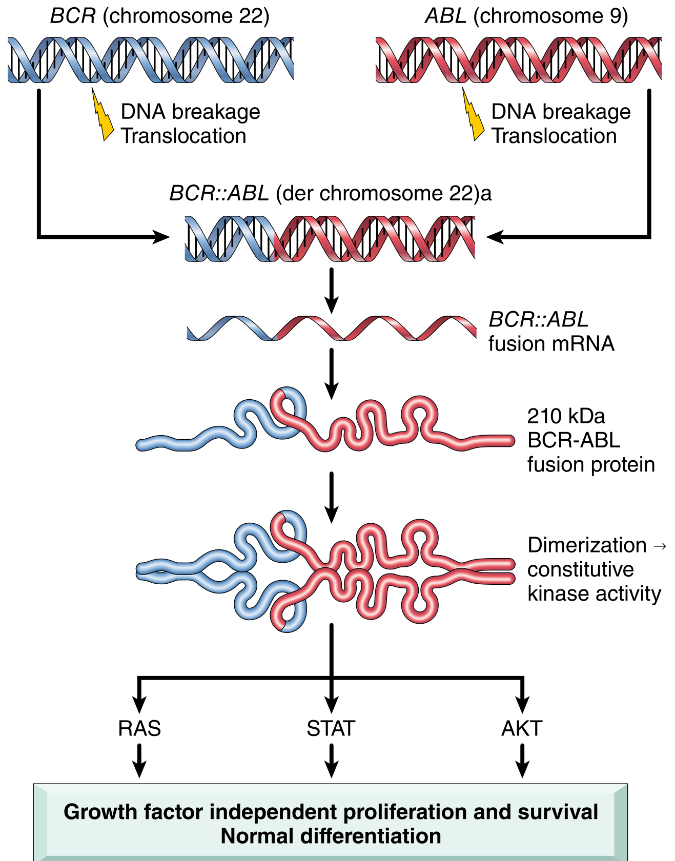
b) Cytogenetic Change: The Philadelphia (Ph) Chromosome

BCR-ABL pathogenesis: t(9;22) translocation forming the Philadelphia chromosome and driving CML
Fig. 13.34 - Robbins Pathology: Pathogenesis of CML - BCR::ABL fusion gene formation and downstream signaling

The Translocation: t(9;22)(q34;q11)

This is a reciprocal translocation between chromosomes 9 and 22:
Chromosome 9 (q34):  ABL proto-oncogene (Abelson tyrosine kinase)
         +
Chromosome 22 (q11): BCR gene (Breakpoint Cluster Region)
         ↓
BCR::ABL fusion gene on the shortened derivative chromosome 22
= Philadelphia (Ph) chromosome

Key Details:

FeatureDetail
Translocation notationt(9;22)(q34;q11)
Genes fusedBCR (chr 22q11) + ABL (chr 9q34)
ResultShortened chromosome 22 = Philadelphia (Ph) chromosome
Elongated chromosome 9Contains ABL sequences lost from chr 9 (der(9))
Fusion protein210 kDa BCR-ABL tyrosine kinase (p210)
MechanismBCR dimerization domain self-associates → constitutive ABL kinase activation
Downstream pathwaysRAS, STAT, AKT → growth factor-independent proliferation + survival, with preserved differentiation
Frequency>90-95% of CML cases by conventional karyotype

Why the Protein is Oncogenic:

Normal ABL kinase is tightly regulated and transiently activated. The BCR moiety provides a dimerization domain causing BCR-ABL to self-associate constitutively, producing a kinase that fires continuously - driving unregulated expansion of myeloid progenitors without arresting maturation.

In the ~5-10% Ph-negative cases:

BCR::ABL fusion still exists but requires FISH or PCR for detection (complex/cryptic rearrangements). There is no true BCR-ABL-negative CML.

Additional cytogenetic abnormalities (disease progression signals):

  • Trisomy 8 (+8)
  • Isochromosome 17q [i(17q)]
  • Duplication of Ph chromosome (+Ph)
  • These emerge in the accelerated/blast phase
"In more than 90% of cases, BCR::ABL is created by a reciprocal (9;22)(q34;q11) translocation (the so-called Philadelphia chromosome). The cell of origin is a pluripotent HSC." - Robbins, Cotran & Kumar Pathologic Basis of Disease, p. 583

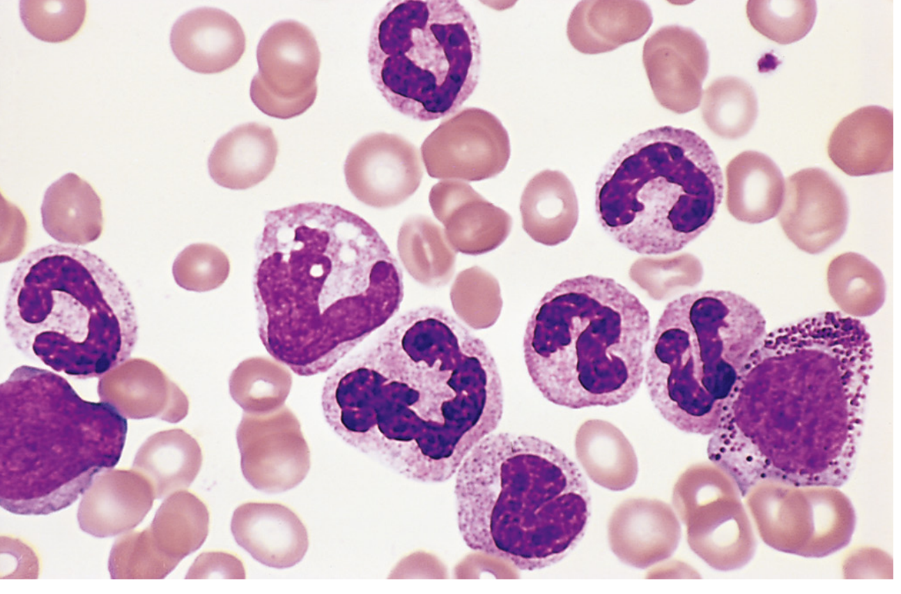
c) Peripheral Blood Smear Findings in CML

CML peripheral blood smear showing mature neutrophils, metamyelocytes, and a myelocyte - the hallmark "myelocyte bulge"
Fig. 13.35 - Robbins Pathology: CML peripheral blood smear - mature neutrophils, metamyelocytes, and myelocytes (Dr. Robert W. McKenna, UT Southwestern)

Enumerated Findings:

1. Marked Leukocytosis
  • WBC typically 1,00,000-4,00,000/mm³ at presentation
  • Composed overwhelmingly of granulocytes at all maturation stages
2. "Myelocyte Bulge" (Myelocyte Peak) - PATHOGNOMONIC The entire granulocytic maturation series is present on the smear in a spectrum:
StageProportion
Myeloblasts<2% (chronic phase)
PromyelocytesFew
MyelocytesProminent - the "bulge"
MetamyelocytesPresent
Band forms (stabs)Present
Segmented neutrophilsPredominant (most numerous)
This "left shift to myelocyte level" with a predominance of mature forms (not blasts) is the hallmark of CML chronic phase.
3. Absolute Basophilia
  • Increased basophils - a very characteristic and diagnostically important feature
  • Rising basophilia signals progression to accelerated phase
4. Absolute Eosinophilia
  • Mild-to-moderate increase in eosinophils
5. Thrombocytosis or Normal Platelets
  • Platelets often elevated (as in this case - 2 Lakhs/mm³)
  • Large, abnormal platelet morphology may be seen
  • Thrombocytopenia in late/accelerated phase signals disease progression
6. Normochromic, Normocytic Anemia
  • Mild-to-moderate; reflects marrow replacement and splenic sequestration
7. Occasional Nucleated Red Blood Cells (NRBCs)
  • Leukoerythroblastic picture in some cases
8. Low/Absent Leukocyte Alkaline Phosphatase (LAP) Score
  • CML neutrophils have markedly reduced LAP (score 0-10; normal 20-100)
  • This is the key cytochemical distinction from a leukemoid reaction (which has very high LAP)
9. No Significant Monocytosis or Lymphocytosis
  • Distinguishes from CMML (high monocytes) and CLL (lymphocytes)
10. Absence of Toxic Granulation and Döhle Bodies
  • These reactive features seen in infective leukocytosis are absent, reflecting a neoplastic rather than reactive process

Summary: CML vs. Leukemoid Reaction

FeatureCMLLeukemoid Reaction
LAP scoreLow/absentVery high
BasophiliaPresentAbsent
EosinophiliaPresentVariable
SplenomegalyMassiveMild
Philadelphia chromosomePresentAbsent
Blasts on smear<2%Absent
Toxic granulationAbsentPresent

Sources: Robbins, Cotran & Kumar - Pathologic Basis of Disease (10th ed.), pp. 582-585 | Goldman-Cecil Medicine, Chapter: Chronic Myeloid Leukemia | Harrison's Principles of Internal Medicine 22E
This is a shared conversation. Sign in to Orris to start your own chat.