
All about kala azhar with native name treatment and how to correct it all
visceral leishmaniasis treatment diagnosis
kala-azar visceral leishmaniasis splenomegaly clinical features Leishmania donovani
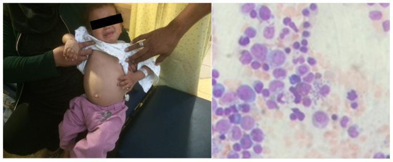
A dual-panel image presenting clinical and diagnostic findings for visceral leishmaniasis (Kala-azar) in a pediatric patient. The left panel is a clinical photograph of a 4-year-old child showing significant abdominal distension and protrusion, characteristic of massive hepatosplenomegaly. The right panel displays a high-magnification light microscopy view of a bone marrow aspirate/biopsy. The microscopic field contains numerous mononuclear cells stained with Giemsa or Wright-Giemsa. Within the cytoplasm of these cells, particularly macrophages, numerous small, ovoid, purple-stained inclusions are visible, identified as Leishmania amastigotes. These intracellular parasites demonstrate the classic morphology of a nucleus and a rod-shaped kinetoplast. This image serves as a teaching tool for recognizing the systemic manifestations and definitive laboratory diagnosis of parasitic infection by Leishmania infantum in endemic regions.
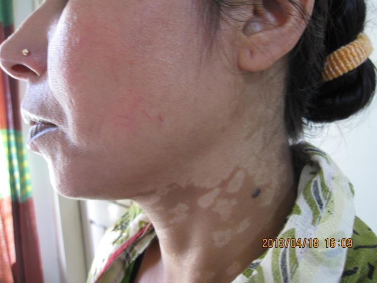
A clinical photograph of the face and neck of a patient from Bangladesh illustrating post-kala-azar dermal leishmaniasis (PKDL). The primary dermatological finding is a widespread, confluent macular rash characterized by hypopigmented to depigmented patches. These pale, irregularly shaped lesions are prominently visible across the cheek and extend inferiorly onto the neck. The lesions vary in size, with some smaller macules coalescing into larger, irregularly bordered areas. Additionally, a single, dark, well-demarcated circular nodule or papule is noted on the lateral aspect of the neck, situated within the hypopigmented region. This visual presentation represents the macular form of PKDL, which is a common cutaneous complication following visceral leishmaniasis (Kala-azar) in Southeast Asian populations. The image serves as a diagnostic reference for identifying characteristic pigmentary changes and distribution patterns of Leishmania donovani-induced skin disease.
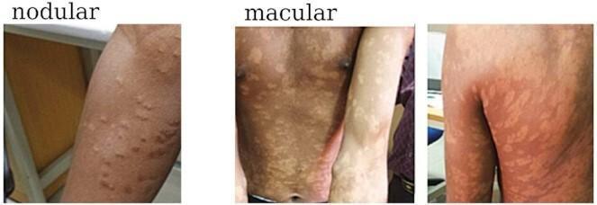
This set of clinical photographs illustrates the diverse dermatological manifestations of Post-Kala-Azar Dermal Leishmaniasis (PKDL). The visual content is divided into two primary morphological categories: nodular and macular. The 'nodular' image depicts multiple small, raised, skin-colored to erythematous bumps on the extensor surface of the forearm. The 'macular' images demonstrate a more extensive and diffuse distribution of pathology, characterized by flat, hypo-pigmented, or discolored patches. These macules are shown across several anatomical regions, including the chest, abdomen, forearms, and the posterior trunk (back and upper buttocks). The comparison highlights the variability in lesion morphology—ranging from discrete, elevated nodules to confluent, flat macules—that can occur following visceral leishmaniasis treatment. This educational material is relevant for tropical medicine and dermatology, specifically for the diagnosis and classification of Leishmania donovani-related skin sequelae.
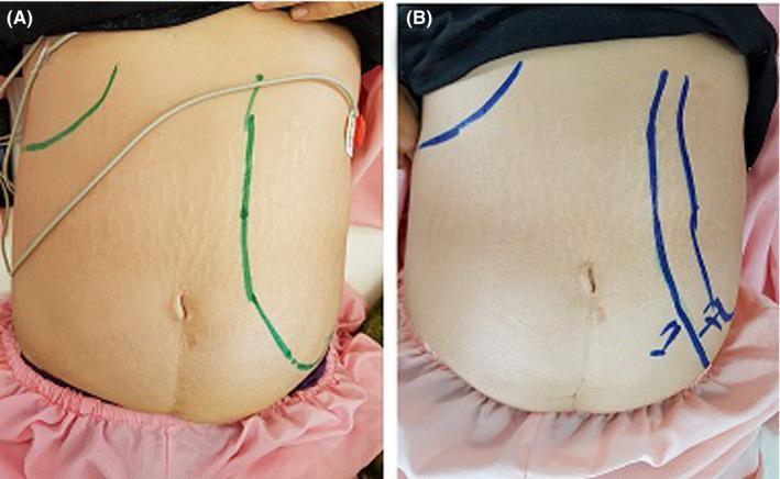
This side-by-side clinical photograph comparison illustrates the regression of massive splenomegaly in a patient with visceral leishmaniasis (kala-azar). Image A shows the patient's abdomen upon admission, displaying significant abdominal distension and striae. Green markings on the skin delineate the palpable borders of the grossly enlarged spleen, which extends from the left costal margin across the midline and deep into the left lower quadrant and pelvic region. Image B, captured one week after the initiation of treatment (Amphotericin B), shows a visible reduction in abdominal girth and more restricted blue markings that indicate the receding splenic margin. The comparison serves as a clinical demonstration of the physical examination findings associated with splenic sequestration and the rapid response to antiprotozoal therapy. The anatomical landmarks include the lower rib cage and iliac crests, used to track the decrease in organomegaly during the recovery phase of the disease.
Kala-Azar (Visceral Leishmaniasis) - Complete Guide
Native Names and Synonyms
| Name | Origin |
|---|---|
| Kala-azar | Hindi/Urdu (South Asia) - "black fever" |
| Dum-dum fever | Named after Dum Dum, Calcutta, India |
| Burdwan fever | Named after Burdwan district, West Bengal |
| Sikari disease | Regional Indian term |
| Sahib disease | Colonial-era South Asian term |
| Death fever | Colloquial, reflecting its lethality |
| Tropical splenomegaly | Descriptive name |
What Is It?
The Parasite
- Causative agents: Leishmania donovani complex
- L. donovani - South Asia and East Africa (anthroponotic: human-to-human)
- L. infantum (syn. L. chagasi) - Mediterranean, Middle East, Americas (zoonotic: dogs as reservoir)
- L. amazonensis, L. colombiensis - rarely cause VL
- South Asia: Phlebotomus argentipes
- East Africa: P. orientalis, P. martini
- Mediterranean: P. perniciosus, P. ariasi
- Promastigote - flagellated form in the sandfly
- Amastigote - non-flagellated, intracellular form in human macrophages (visible in tissue smears)
Epidemiology
- 90% of cases globally occur in Bangladesh, Ethiopia, India, Nepal, Brazil, and Sudan
- India (Bihar state) was historically the worst-hit area; a WHO elimination program achieved a 98.7% decline in India
- WHO declared elimination of VL as a public health problem in Bangladesh and marked decline in Nepal
- East Africa currently has the highest incidence globally
- In Mediterranean Europe, 70% of adult VL cases are associated with HIV co-infection
How It Spreads
- Sandfly bite - infected sandfly injects promastigotes into skin
- Hematogenous dissemination to the reticuloendothelial system (spleen, liver, bone marrow)
- Congenital (transplacental) transmission - rare
- Most infections remain subclinical - only a minority develop active disease
Clinical Features
Cardinal Features (the "FLASK" mnemonic):
- Fever - persistent, prolonged, undulating, high-grade
- Leucopenia (and pancytopenia)
- Anemia - progressive
- Splenomegaly - massive, the most important physical sign
- Kachexia/hypergammaglobulinemia
Additional Features:
- Hepatomegaly (less prominent than splenomegaly)
- Lymphadenopathy
- Thrombocytopenia, epistaxis, mucosal bleeding
- Hyperpigmentation - ashy, dark patches on temple, around mouth, on abdomen, hands, and feet in light-skinned patients - this is why it is called "kala-azar" (the "black" of black fever)
- Burning feet (peripheral neuropathy)
- Hair and skin depigmentation (Kenyan patients)
- Cutaneous nodules and mucosal ulcers (Sudanese patients)
- Trichomegaly (Pitalugo sign) - elongated eyelashes
- Jaundice (less common)
- Emaciation, hepatogastrointestinal disturbances
Fatal Complications (untreated):
- Cachexia
- Secondary bacterial infections
- Enteritis and pneumonia
- Hemolytic anemia
- Acute renal damage
- Severe mucosal hemorrhage
Post-Kala-Azar Dermal Leishmaniasis (PKDL)
- Macular hypopigmented patches (face, neck, trunk)
- Papular/nodular lesions
- Can appear months to years after treatment
Diagnosis
Gold Standard
| Site | Sensitivity |
|---|---|
| Splenic aspirate | >95-97% (most sensitive but invasive and dangerous) |
| Bone marrow aspirate | 60-85% |
| Lymph node aspirate | 50-60% |
Serological Tests
- rK39 rapid diagnostic test (RDT) - immunochromatographic strip; requires only a drop of fingerprick blood; result in 15 minutes
- Sensitivity ~98%, specificity ~90% in immunocompetent patients (outside East Africa)
- Based on recombinant antigen from kinesin region of L. infantum
- Note: Remains positive for years after cure; cannot be used to measure cure or detect relapse
- ELISA and Indirect Immunofluorescent Antibody Test (IFAT) - used in specialized labs
- rK28 - newer synthetic polyprotein antigen, better performance in Sudan but not commercially available
Molecular Tests
- PCR (qualitative) and Real-time PCR (quantitative) - highly sensitive, allows species identification; requires specialized lab
- LAMP (Loop-mediated Isothermal Amplification) - better than microscopy, simpler equipment, suitable for point-of-care
Montenegro (Leishmanin) Skin Test
- Similar to tuberculin test; killed promastigotes injected intradermally
- Detects exposure but cannot distinguish past from active disease
- Up to 50% of people in endemic areas test positive without disease
- Negative in active VL (patient is anergic during active disease)
Differential Diagnosis
| Mimicking Condition | Distinguishing Point |
|---|---|
| Malaria | Most common mimic; blood smear, RDT |
| Typhoid fever | Widal test, blood culture |
| Tuberculosis | AFB, CXR, Mantoux |
| Brucellosis | Brucella serology, exposure history |
| Schistosomiasis | Eosinophilia, serology, travel history |
| Histoplasmosis | Fungal cultures, antigen test |
| Portal hypertension | Imaging, LFTs |
| Chronic myeloid leukemia | BCR-ABL, peripheral blood film |
| Tropical splenomegaly syndrome | High malarial antibody titers |
Treatment
Principles
1. Liposomal Amphotericin B (L-AmB) - Drug of Choice in Most Settings
- First-line in South Asian subcontinent and Mediterranean countries
- Preferentially taken up by reticuloendothelial tissues (where Leishmania hides)
- Terminal half-life ~150 hours; detectable in liver/spleen for weeks after a single dose
- Dose (India): Single IV infusion of 10 mg/kg (WHO-recommended for Indian subcontinent); OR 3-5 mg/kg/day for several days
- Less toxic than conventional AmB because very little free drug circulates
2. Pentavalent Antimonial Compounds (SbV) - Still First-Line in Most of the World
- Sodium stibogluconate (SSG / Pentostam) - 100 mg SbV/mL
- Meglumine antimoniate (Glucantime) - 85 mg SbV/mL
- <50% in Bihar, India due to widespread resistance - antimonials are NOT used there
- Arthralgia, myalgia
- Elevated aminotransferases (hepatotoxicity)
- ECG changes: Concave ST elevation (not significant) vs. QTc prolongation >0.5 s (dangerous - may herald ventricular arrhythmia and sudden death - discontinue immediately)
- Chemical pancreatitis (common, usually mild; severe in immunosuppressed patients)
3. Conventional Amphotericin B Deoxycholate
- Dose: 0.75-1.0 mg/kg on alternate days for 15 infusions (total 15 doses)
- Used where liposomal formulation is unavailable
- Relapses uncommon in immunocompetent patients
- Fever with chills (almost universal with each infusion - premedicate with chlorpheniramine + acetaminophen)
- Nausea and vomiting
- Thrombophlebitis at infusion site
- Renal dysfunction and hypokalemia (monitor closely)
- Rare: Hypersensitivity reactions, bone marrow suppression, myocarditis (fatal)
4. Miltefosine - Oral Drug
- The only oral antileishmanial drug available
- Preferred in Bihar, India (where antimonials have failed)
- Dose: 2.5 mg/kg/day orally for 28 days (max 150 mg/day)
- Cure rates: ~95% in India
- Gastrointestinal (nausea, vomiting, diarrhea) - most common
- Teratogenic - absolutely contraindicated in pregnancy; requires effective contraception for 3 months after treatment
5. Paromomycin (Aminosidine)
- An aminoglycoside antibiotic with anti-leishmanial activity
- Dose: 11 mg/kg/day IM for 21 days
- Used in India and East Africa
- Can be combined with other drugs (combination therapy)
Regional Treatment Recommendations Summary
| Region | Preferred Treatment |
|---|---|
| Indian subcontinent (India, Nepal, Bangladesh) | Liposomal AmB (single dose 10 mg/kg) OR miltefosine 28 days |
| Bihar, India (antimonial-resistant) | Liposomal AmB or miltefosine (NOT antimonials) |
| Mediterranean/Europe | Liposomal AmB (cost not an issue) |
| East Africa (Sudan, Ethiopia) | Sodium stibogluconate 30 days ± paromomycin; liposomal AmB |
| Americas (Brazil) | Meglumine antimoniate OR conventional AmB OR miltefosine |
Combination Therapy
- AmB + miltefosine
- AmB + paromomycin
- Miltefosine + paromomycin
VL/HIV Co-infection
- More difficult to treat; higher relapse rates
- Immune reconstitution with antiretrovirals (ART) is essential alongside antileishmanial treatment
- Liposomal AmB is preferred
- Secondary prophylaxis may be required to prevent relapse in severely immunosuppressed patients
Monitoring Cure
- Clinical: Resolution of fever, regression of splenomegaly, weight gain, normalization of blood counts
- Serological tests (rK39, ELISA) cannot be used for cure assessment - remain positive for years
- Splenic aspirate at 6 months may be done in research settings
- PCR is the best objective marker for parasitological cure but not routinely available
Follow-Up
Recent Evidence (2024-2025)
- A 2025 network meta-analysis in Frontiers in Cellular and Infectious Microbiology confirms liposomal AmB and miltefosine as the most efficacious and well-tolerated regimens across leishmaniasis forms [PMID: 41473767]
- A 2024 systematic review of VL therapeutic efficacy studies (2000-2021) highlights major methodological heterogeneity in published trials, underscoring the need for standardized outcome reporting [PMID: 38955166]
Key Points Summary
| Feature | Detail |
|---|---|
| Native name | Kala-azar (Hindi: "black fever"); also Dum-dum fever, Burdwan fever |
| Cause | Leishmania donovani complex |
| Vector | Phlebotomus sandfly |
| Hallmark sign | Massive splenomegaly |
| Name-giving sign | Dark hyperpigmentation of skin |
| Gold standard Dx | Amastigotes on splenic/bone marrow smear |
| Best field test | rK39 RDT (fingerprick, 15 min) |
| If untreated | ~100% fatal within 2 years |
| Best drug globally | Liposomal amphotericin B |
| Only oral drug | Miltefosine |
| Resistant region | Bihar, India (antimonials fail) |
| Post-treatment complication | PKDL (months-years later) |
Sources: Harrison's Principles of Internal Medicine 22E (2025), Fitzpatrick's Dermatology, Andrews' Diseases of the Skin, with supplementary evidence from PubMed systematic reviews (2024-2025).