


A itchy scaly lesion with silvery plucks at upper neck and back of ear involving some part of hairs what is probable diagnosis and treatment in detail
scalp psoriasis silvery scales hairline posterior neck
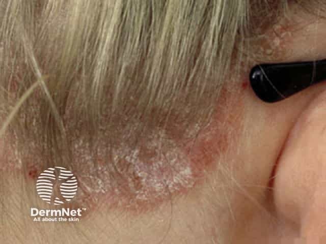
Clinical photography of dermatologic disease. This is a close-up, lateral view of the posterior neck and occipital scalp near the hairline, showing erythematous, well-demarcated plaques with thick, silvery scales consistent with active plaque psoriasis. Lesions extend along the nuchal region and behind the ear, with mild induration and irregular borders in places. The skin surface exhibits adherent scale and subtle fissuring; surrounding skin is relatively normal. The color is red-erythematous with a metallic sheen from the scales, and there is minimal lichenification. This plaque morphology—sharply demarcated erythematous plaques with overlying silvery scale—reflects classic psoriasis pattern, commonly seen on scalp, neck, and extensors. No pustules or erosions are evident. The image demonstrates typical disease distribution in the head and neck area and highlights the keratotic scale with underlying epidermal hyperplasia. Clinically, this appearance supports the diagnosis of plaque psoriasis; differential considerations include seborrheic dermatitis, atopic dermatitis, tinea capitis/corporis, and pityriasis rosea in variant presentations. Diagnostic significance lies in recognizing plaque psoriasis morphology for treatment planning, including topical corticosteroids or vitamin D analogues, calcineurin inhibitors in selected cases, and consideration of phototherapy for extensive disease. Educationally, this image aids lesion recognition, distribution mapping, and patient counseling. It is suitable for teaching reference.
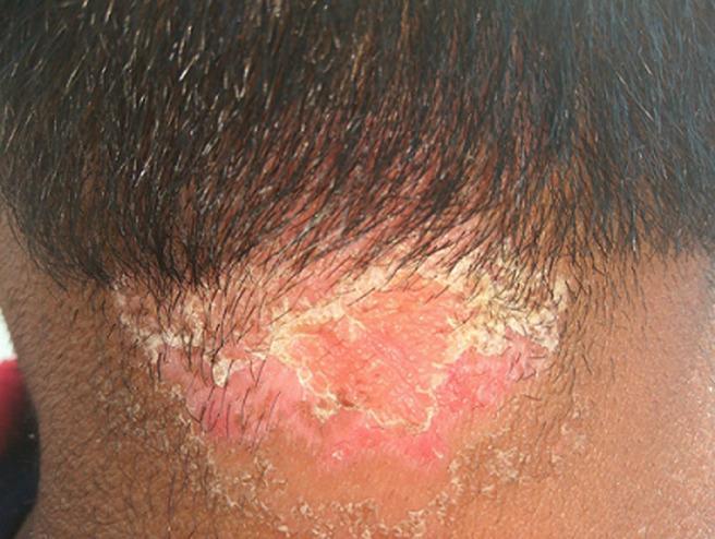
This clinical photograph displays a well-demarcated skin lesion characteristic of scalp psoriasis, located at the posterior hairline and extending onto the neck. The affected area is a prominent erythematous plaque with a vivid reddish-pink hue, indicating underlying inflammation. The lesion is characterized by thick, micaceous (silvery-white to yellowish) scales that are most densely concentrated along the irregular borders, partially adhering to the hair shafts and the scalp surface. The central portion of the plaque shows visible skin thickening and hyperkeratotic texture with less dense scaling. The surrounding skin appears relatively normal, though the plaque exhibits sharp demarcation from the healthy tissue. This image serves as a classic educational example of plaque psoriasis affecting the scalp and hairline, demonstrating key diagnostic features such as well-defined erythema and characteristic scaling patterns relevant for dermatology and primary care education.
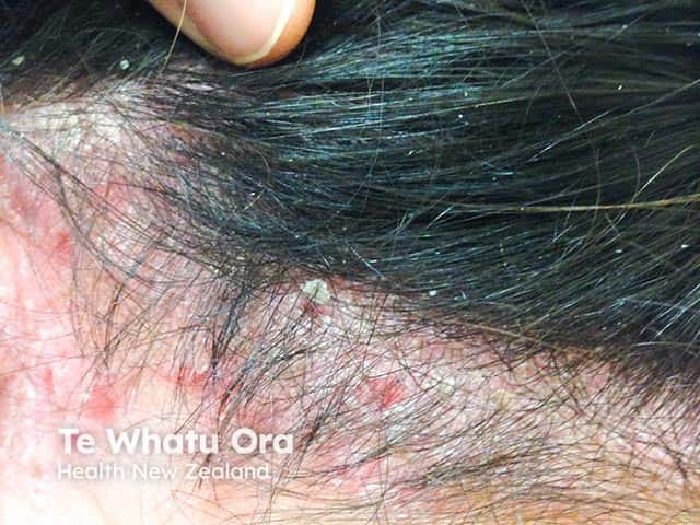
Clinical photograph of the scalp and posterior neck demonstrating active psoriasis vulgaris with scalp involvement extending to the nuchal (occipital) region. Imaging modality: noninvasive dermatologic photography; technique: wide-field, non-magnified view under standard lighting. The primary subject is human skin within the scalp dermatome area and neck, featuring erythematous plaques with finely adherent silvery-white scales, variable plaque thickness, and marginal scaling at the hairline. Notable features include well-demarcated, raised erythema, diffuse desquamation, and minimal excoriation in exposed areas. The lesions appear on the vertex and temporal regions as well as the posterior scalp transitioning to the occipital neck, consistent with psoriatic plaque morphology. From a diagnostic standpoint, these findings are characteristic of psoriasis vulgaris involving the scalp, with potential involvement of the nuchal region; differential diagnoses include seborrheic dermatitis, tinea capitis, eczema, and lichen planus. Clinically, the image underscores the importance of recognizing psoriasis in hair-bearing areas due to treatment implications (topical corticosteroids, vitamin D analogs, coal tar preparations, or phototherapy) and possible assessment for psoriatic arthritis in long-standing disease. This image is relevant for clinical education, dermatology research, and narrative case assist in medical literature reviews and teaching modules. It supports diagnosis, therapy planning, and patient education resources for clinicians.
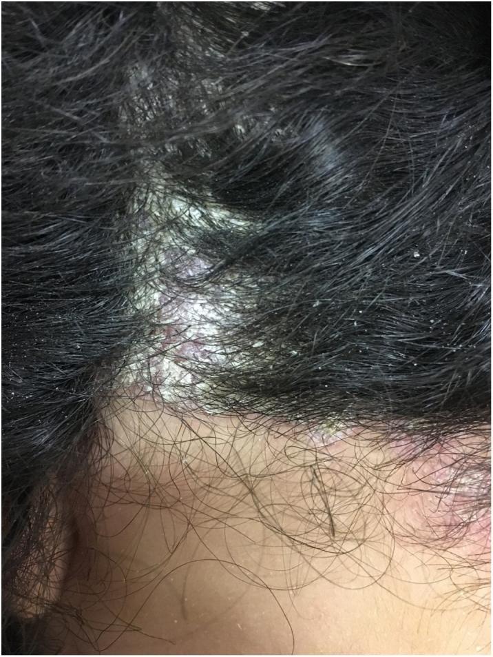
This clinical photograph shows a close-up view of the human scalp and posterior hairline, demonstrating well-demarcated erythematous plaques characteristic of scalp psoriasis. The lesions are covered with thick, silvery-white micaceous scales that adhere to both the underlying inflamed skin and the proximal hair shafts. The distribution is patchy, with prominent plaques extending beyond the frontal and temporal hair-bearing areas onto the adjacent non-terminal hair skin. The surrounding dark hair remains intact but contains scattered fine, flaky white debris (secondary scaling). The visible borders of the primary plaque are distinct and irregular. This image is an educational example of inflammatory dermatoses of the scalp, specifically illustrating the classic morphology and scaling pattern of psoriasis and pseudotinea amiantacea.
"scalp psoriasis" treatment
Probable Diagnosis: Scalp Psoriasis (Psoriasis Capitis)
Diagnostic Features Supporting Scalp Psoriasis
| Feature | Significance |
|---|---|
| Well-demarcated silvery scaly plaques | Classic psoriatic plaque morphology |
| Location: posterior neck + retroauricular | Named "special location" in psoriasis literature |
| Scales adhering to hair shafts | Called pityriasis amiantacea (asbestos-like) - most common cause is psoriasis |
| Intense itching | Common in scalp psoriasis |
| Extends beyond hairline onto neck | Characteristic - unlike seborrheic dermatitis |
Differential Diagnoses to Consider
- Seborrheic dermatitis - yellow/greasy scales, less well-demarcated; can coexist with psoriasis ("sebopsoriasis")
- Tinea capitis - fungal infection, usually in children; can cause pityriasis amiantacea pattern
- Atopic dermatitis - more diffuse, poorly demarcated, family history of atopy
- Contact dermatitis - consider earring metal allergy given the retroauricular location
Treatment in Detail
Step 1 - Descaling (First, Always)
- Salicylic acid 5-10% in oil or ointment base applied to scalp before shampooing - keratolytic, promotes penetration of other agents
- Coconut oil / olive oil soaks overnight under a shower cap can also soften thick scale
Step 2 - First-Line Topical Treatments
A. Topical Corticosteroids (Most Widely Used)
- High-potency corticosteroids are preferred on scalp (e.g., betamethasone valerate 0.1% lotion/foam, clobetasol propionate 0.05% scalp solution/foam)
- Use lotion, gel, foam, or spray bases - preferred over creams/ointments on hair-bearing scalp
- Apply once or twice daily; 2-week course then weekend pulse therapy to reduce atrophy risk
- Maximum: 50 g/week of superpotent corticosteroid; 100 g/week of potent corticosteroid
- Intralesional triamcinolone acetonide (2.5-5 mg/mL) for resistant individual plaques
B. Vitamin D3 Analogues (First-Line)
- Calcipotriol/Calcipotriene (Dovonex) - ointment, cream, or solution
- Achieves ~60% PASI reduction as monotherapy at 8 weeks
- Combination: Calcipotriol + Betamethasone dipropionate (Daivobet gel) - most effective option: ~70% PASI reduction and clearing in ~70% of scalp psoriasis patients; approved first-line for scalp
C. Coal Tar Preparations
- Coal tar shampoo (2-5%) - used 2-3 times/week; leave on 5-10 minutes before rinsing
- Tar bath oils, LCD (liquor carbonis detergens) in shampoo form
- Anti-inflammatory, anti-proliferative; odor can be a concern; useful adjunct
Step 3 - Second-Line Topical Treatments
- Tazarotene (topical retinoid) - reduces keratinocyte hyperproliferation; combine with topical steroid to reduce irritation; once daily
- Tacrolimus / Pimecrolimus (calcineurin inhibitors) - especially useful for thin lesions, retroauricular area, and face-adjacent regions where steroid atrophy risk is higher; can cause initial burning sensation
- Anthralin (Dithranol) - effective anti-proliferative; use Short Contact Anthralin Treatment (SCAT): apply for 15-30 minutes then wash off; stains skin/clothing brown
Step 4 - Phototherapy (Moderate-Extensive Disease)
- Narrowband UVB (NB-UVB) - mainstay for moderate-to-severe scalp disease; part the hair for better penetration; 3 sessions per week
- Excimer laser (308 nm) - targeted phototherapy for localized refractory scalp plaques
Step 5 - Systemic Therapy (Severe/Refractory Cases)
- Methotrexate 7.5-25 mg/week (oral or SC) - traditional mainstay
- Cyclosporine 2.5-5 mg/kg/day - rapid response, for short-term control
- Acitretin (oral retinoid) - especially if nail/hair involvement
- Biologics (for moderate-to-severe): IL-17 inhibitors (secukinumab, ixekizumab), IL-23 inhibitors (guselkumab, risankizumab), anti-TNF agents (adalimumab, etanercept) - a recent network meta-analysis (2026) shows IL-17/IL-23 class biologics have highest relative efficacy for scalp psoriasis
Step 6 - Medicated Shampoos (Ongoing Maintenance)
| Shampoo Type | Active Ingredient | Use |
|---|---|---|
| Ketaconazole 2% | Antifungal | If sebopsoriasis component |
| Coal tar | Coal tar 2-5% | Anti-inflammatory |
| Salicylic acid | Keratolytic | Scale removal |
| Zinc pyrithione | Antimicrobial | Mild cases / maintenance |
| Selenium sulfide 2.5% | Antifungal/keratolytic | Adjunct |
Practical Regimen for This Patient
- Start with overnight coconut or salicylic acid oil application to soften scales
- Shampoo with coal tar or zinc pyrithione shampoo (leave-on 5-10 min)
- Apply Calcipotriol + Betamethasone gel (Daivobet) to affected areas once daily for 4 weeks
- After clearing, maintenance with twice-weekly coal tar shampoo and intermittent weekend steroid pulse
- Refer to dermatologist if no response in 4-6 weeks for consideration of phototherapy or systemic agents
Important note: This assessment is based on the clinical description and images provided. A proper dermatological examination is needed for definitive diagnosis. If Wood's lamp examination or fungal scraping is needed to exclude tinea capitis, please consult a dermatologist. Do not use high-potency topical steroids for extended periods without medical supervision.