
Dysplasia medicine
dysplasia treatment management
dysplasia histology cells epithelium
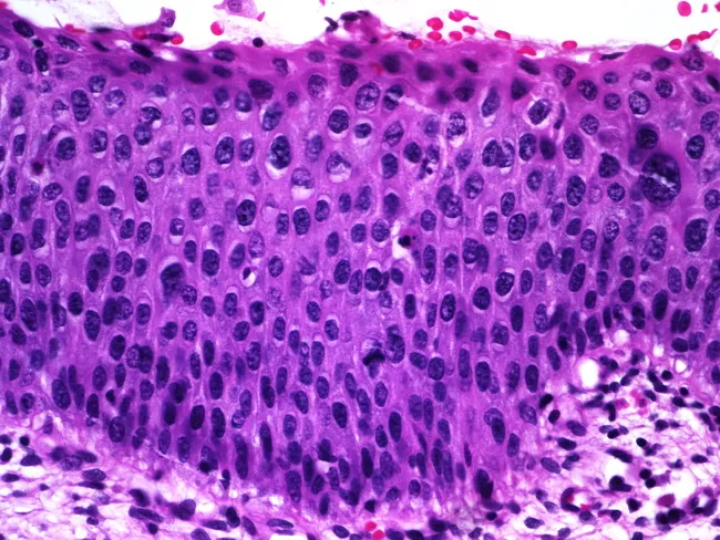
Histology image of mucosal epithelium showing features of squamous epithelial dysplasia on light microscopy. Prepared as a paraffin-embedded hematoxylin and eosin (H&E) stained section and viewed under brightfield microscopy at high magnification (~400x). The epithelium is stratified, with orderly basal cell layer but marked nuclear atypia throughout the lower and mid portions. Nuclear enlargement and hyperchromasia are evident, along with variation in nuclear size (anisokaryosis) and coarse, clumped chromatin. There is partial maturation at the superficial aspect, with surface layer showing more differentiated cells, suggesting some preserved maturation toward the lumen. Occasional mitotic figures are observed in the lower half of the epithelium, indicative of increased proliferative activity. Overall architecture reveals thickened epithelium (acanthosis) with preserved polarity in some cells but dysplastic changes in others. No invasion is evident in this field, though the mitotic activity and cytologic atypia raise concern for preinvasive squamous intraepithelial lesion. The imaging is relevant for diagnosis of epithelial dysplasia or squamous intraepithelial lesion and has clinical significance for risk stratification, biopsy correlation, and surveillance. Potential clinical applications include screening for precancerous lesions in mucosal surfaces, monitoring response to therapy, and educational demonstration of dysplastic cytology for medical students and residents.
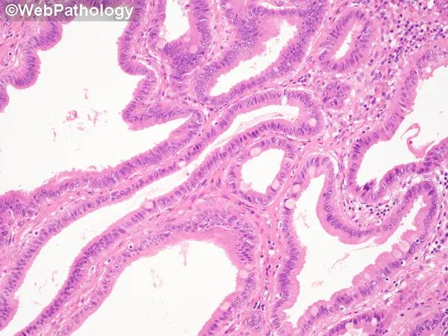
Bright-field hematoxylin and eosin stained histology slide of gallbladder mucosa demonstrating an intracholecystic papillary-tubular neoplasm (ICPN) with low‑grade dysplasia. The lesion exhibits papillary and tubular architectural complexity with mucin-producing epithelium. Epithelial cells show hyperchromatic nuclei that are mildly enlarged but largely uniform in size and shape. Nuclear polarity is maintained and the dysplastic epithelium is confined predominantly to the lower portion of the lining (basal third). Nucleoli are inconspicuous or small. Goblet cells are present, indicating intestinal-type differentiation within the ICPN. Mitotic activity is not markedly elevated. Clinical note: while this image focuses on low-grade changes, high-grade dysplasia is characterized by marked nuclear enlargement and hyperchromasia, nuclear pleomorphism, loss of polarity, prominent nucleoli, mucin depletion, and increased mitotic figures, often seen in larger lesions or extension into flat adjacent mucosa. This slide illustrates premalignant biliary neoplasia; accurate histologic grading guides surgical management and prognosis. Relevance to clinical practice includes differentiation from reactive/benign biliary mucosa and from higher‑risk lesions such as high‑grade dysplasia or invasion. Potential uses: pathology education, atlas reference, surgical pathology reporting, and research on ICPN progression. This image is suitable for teaching histologic criteria, grading challenges, and illustrating the transition from dysplasia to invasive carcinoma in gallbladder neoplasms.
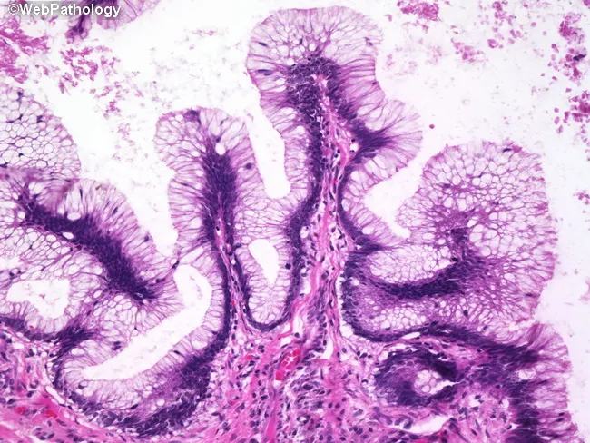
Histology image demonstrating mucosal villous projections lined by intestinal-type columnar epithelium with goblet cells. The architecture consists of elongated, finger‑like villi with uniform mucinous epithelium and goblet cell abundance. Nuclei are hyperchromatic and basally located with preserved polarity, consistent with low- to intermediate-grade dysplasia. Mitotic activity is not prominently increased, and the glandular crowding typical of high-grade dysplasia is absent. The superficial mucosa shows a columnar epithelial lining with a crypt-heavy layout and surface mucin production, surrounded by a fibrous lamina propria and mild accompanying inflammatory infiltrate. Overall, the mucosal lesion exhibits a villous adenomatous pattern rather than a serrated or hyperplastic configuration. Notably, goblet cells appear scattered within the epithelium, and the nuclei remain aligned basally without marked pseudostratification or mitotic atypia. This histomorphology supports a premalignant colorectal polyp with low-grade dysplasia and a risk of progression if left untreated. Clinically, such lesions are associated with colorectal polyps detected on colonoscopy and warrant endoscopic excision and histopathologic surveillance. Immunohistochemical staining for mismatch repair proteins or p53 is not necessary for routine diagnosis but may be employed in equivocal cases. The image underscores the importance of correlating endoscopic appearance with microscopic villous architecture to guide management and cancer prevention strategies today.
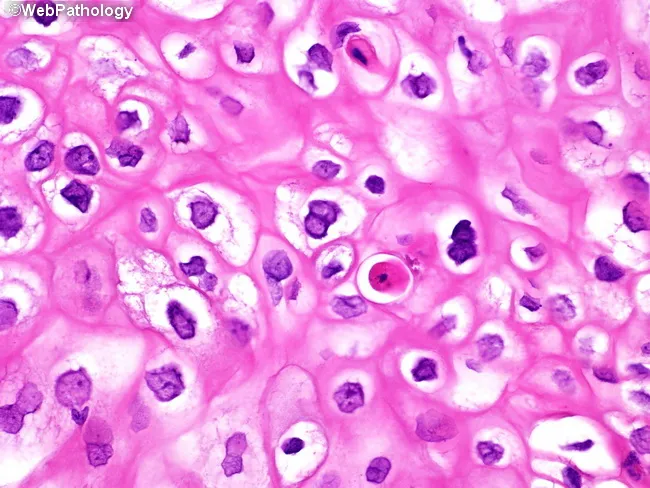
This histopathology micrograph depicts HPV-associated condyloma acuminatum involving urinary bladder mucosa, captured at high magnification with Hematoxylin and Eosin staining. The epithelium shows acanthotic, papillary squamous hyperplasia with koilocytotic cells featuring perinuclear halos, wrinkled raisinoid nuclei, and occasional binucleation. Dyskeratotic (dyskeratosis) cells are present in the upper spinous layer. Overall maturation is preserved, and cytologic atypia is limited, arguing against invasive squamous carcinoma. The lesion exhibits characteristic koilocytic change and surface/epithelial thickening consistent with a condylomatous pattern and verruciform architecture. In the bladder, such condylomas are recognized risk factors for subsequent development of squamous cell carcinoma or verrucous carcinoma; therefore, clinical management emphasizes surveillance and follow-up. The image supports an HPV-driven lesion and correlates with infectious etiologies rather than urothelial carcinoma. Differential diagnoses include squamous metaplasia, flat condylomatous dysplasia, and verrucous carcinoma when invasion is suspected; immunohistochemistry or HPV testing can aid confirmation. This depiction is valuable for educational purposes in pathology, urology, and gynecologic oncology, illustrating classic cytopathic changes (koilocytosis, nuclear wrinkling, binucleation, dyskeratosis) and the importance of correlating histology with clinical risk factors and follow-up imaging. Correlation with HPV typing and patient history may guide prevention and counseling.
Dysplasia in Medicine
Definition
Histological Features (Shared Across Types)
- Nuclear enlargement and pleomorphism
- Hyperchromasia (dark-staining nuclei)
- Loss of normal cell polarity and stratification
- Atypical mitoses - mitotic figures in abnormal locations
- Crowding of glands/tubules
- Loss of cytoplasmic maturation toward the surface
| Grade | Features |
|---|---|
| Low-grade (LGD) | Mild atypia, partial maturation preserved, lower half involvement |
| High-grade (HGD) | Marked atypia, full-thickness involvement, near-carcinoma |
Major Types of Dysplasia
1. Barrett's Esophagus Dysplasia
- Nondysplastic Barrett's → cancer: ~0.25%/year
- LGD → cancer progression: ~0.4%/year (but contested; some studies show 85% cumulative risk over 9 years in confirmed LGD)
- HGD → cancer: ~6%/year
- All Barrett's patients: once-daily PPI therapy (chemoprevention per ACG guidelines)
- LGD: endoscopic therapy preferred; surveillance every 6-12 months acceptable
- HGD: endoscopic eradication therapy (EET) - radiofrequency ablation (RFA) is first-line; historically esophagectomy was standard but carries 3-10% mortality
- Biopsies: Seattle protocol (4-quadrant every 1-2 cm); dysplasia concentrated in the proximal half of the Barrett segment
2. Cervical Dysplasia (CIN - Cervical Intraepithelial Neoplasia)
- CIN 1: mild dysplasia, lower third of epithelium - usually regresses spontaneously
- CIN 2: moderate - treatment often recommended
- CIN 3/CIS: severe dysplasia/carcinoma in situ - requires treatment
- Treatment: LEEP (Loop Electrosurgical Excision Procedure), cryotherapy, cold knife conization
- HIV-positive women have higher rates of persistent/recurrent dysplasia after treatment and should also receive anal Pap tests (Goldman-Cecil Medicine)
3. Colorectal Adenomatous Dysplasia
- Villous adenomas: higher dysplasia risk than tubular
- Low-grade dysplasia: nuclei basally located, goblet cells preserved
- High-grade dysplasia: marked nuclear atypia, pseudostratification, glandular crowding
- Treatment: endoscopic polypectomy; surveillance colonoscopy
4. Laryngeal Dysplasia (Premalignant Laryngeal Lesions)
- Seen in leukoplakia/erythroplakia of the larynx
- ~18% of lesions undergo malignant transformation
- Severe dysplasia and CIS behave similarly clinically
- Diagnosis: incisional/excisional biopsy (gold standard)
- Treatment of early lesions: endoscopic excision; radiotherapy not generally recommended for high-grade dysplasia (K.J. Lee's Essential Otolaryngology)
5. Gastric Dysplasia
- Occurs in background of chronic gastritis, atrophy, and intestinal metaplasia
- Gastric adenomas exhibit dysplasia (low or high grade)
- Risk of adenocarcinoma is related to adenoma size - particularly elevated for lesions >2 cm
- Carcinoma may be found in up to 30% of gastric adenomas at time of excision (Robbins Basic Pathology)
- H. pylori eradication can cause regression of hyperplastic polyps
6. Hip Dysplasia (Developmental Dysplasia of the Hip - DDH)
- Screening: ultrasound in neonates
- Early treatment: Pavlik harness (infants)
- Adult treatment: periacetabular osteotomy (PAO) for symptomatic hip dysplasia; total hip replacement for advanced OA (Miller's Orthopaedics, Campbell's Operative Orthopaedics)
7. Fibrous Dysplasia
- Associated with McCune-Albright syndrome (polyostotic + cafe-au-lait spots + endocrinopathy)
- Treatment: bisphosphonates for pain; surgical stabilization for fracture risk
8. Renal Dysplasia
9. Ectodermal Dysplasia
10. Fibromuscular Dysplasia (FMD)
Key Concepts
| Feature | Dysplasia | Hyperplasia | Metaplasia | Carcinoma in situ |
|---|---|---|---|---|
| Cell number | Increased | Increased | Normal/increased | Increased |
| Cell type | Abnormal | Normal | Changed | Markedly abnormal |
| Architecture | Disordered | Maintained | Maintained | Severely disordered |
| Reversible? | Often yes (low-grade) | Yes | Usually yes | No |
| Basement membrane | Intact | Intact | Intact | Intact (by definition) |
| Invasive? | No | No | No | No |
Summary
Vasculitis medicine
vasculitis classification vessel size histology
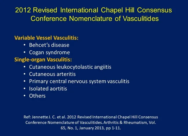
This image is an educational slide detailing the 2012 Revised International Chapel Hill Consensus Conference (CHCC) nomenclature for vasculitis. The primary subject is the modern framework used to classify inflammatory vessel disease by vessel involvement rather than presumed etiology. The slide contrasts Variable Vessel Vasculitis, with examples such as Behçet’s disease and Cogan syndrome, against Single-organs Vasculitis, which includes conditions like cutaneous leukocytoclastic angiitis, cutaneous arteritis, primary central nervous system vasculitis, and isolated aortitis. The visual emphasizes taxonomy, terminology, and the practical implications for diagnostic coding, case reporting, and research design. While not depicting histology or imaging, the slide anchors clinical reasoning about systemic versus organ-restricted vasculitis and guides clinicians toward appropriate laboratory workup, imaging strategies, and treatment planning. The CHCC framework aims to harmonize nomenclature across specialties, reduce diagnostic ambiguity, and improve comparability of epidemiologic data and therapeutic trials. Users include clinicians, residents, fellows, and scientists seeking consistent vocabulary for case documentation, literature searches, and educational materials. Overall, the image serves as a concise reference for vasculitis classification, differential diagnoses, and clinical correlates within rheumatology, dermatology, neurology, and internal medicine. This slide therefore supports evidence synthesis, guideline interpretation, and cross-disciplinary teaching by providing a stable reference point for enrolling vasculitis patients in clinical studies.
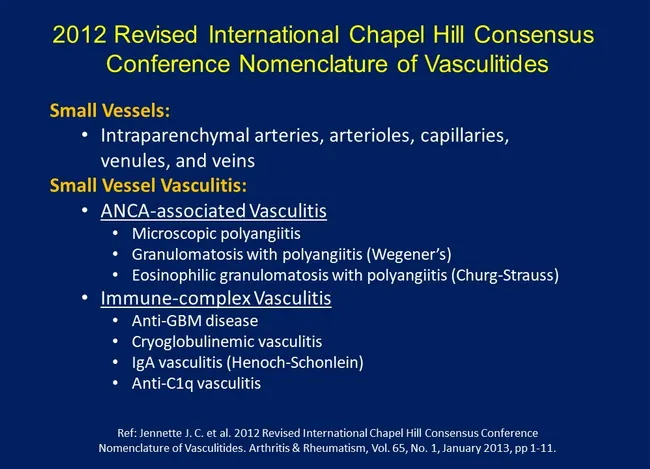
Educational slide illustrating the 2012 Revised International Chapel Hill Consensus Conference nomenclature for vasculitis, with emphasis on small-vessel disease. Core subject: classification of small vessel vasculitis into ANCA-associated vasculitis and immune-complex vasculitis, plus related disorders. Primary focus areas include Small Vessels: intraparenchymal arteries, arterioles, capillaries, venules, and veins within the microvasculature; Small Vessel Vasculitis as a clinical-radiologic-pathologic framework. The ANCA-associated vasculitis subgroup comprises Microscopic polyangiitis; Granulomatosis with polyangiitis (Wegener’s); Eosinophilic granulomatosis with polyangiitis (Churg–Strauss). The immune-complex vasculitis subgroup includes Anti-GBM disease, Cryoglobulinemic vasculitis, IgA vasculitis (Henoch–Schönlein). Anti-C1q vasculitis is also listed. The slide conveys nomenclature variants and synonymous terms to aid searchability and education. This image is not a patient study but a teaching aid for clinicians, residents, and students in rheumatology, nephrology, dermatology, pulmonology, and internal medicine. Clinical relevance includes understanding diagnostic categories, guiding serologic testing (ANCA panels, anti-GBM, cryoglobulins, IgA), directing biopsy strategy, and informing treatment decisions and prognosis. It supports case discussions, exam preparation, and scholarly review of vasculitis taxonomy and disease spectrum. Keywords for indexing include vasculitis taxonomy, Chapel Hill criteria, systemic vasculitis, pediatric and adult references, differential diagnoses, serology interpretation, biopsy targets. This facilitates rapid retrieval in teaching files, clinical decision support, and research datasets.
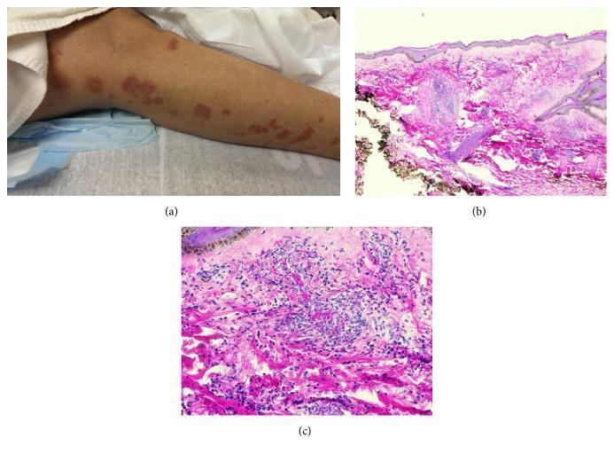
This composite image illustrates the clinical and histopathological findings of drug-induced small vessel vasculitis. Panel (a) is a clinical photograph of a lower extremity showing a multi-focal skin rash characterized by erythematous papules and plaques, some appearing purpuric or urticarial in nature. Panels (b) and (c) present hematoxylin and eosin (H&E) stained sections of a skin punch biopsy at low (20x) and high (200x) magnification, respectively. The histology reveals classic features of leukocytoclastic vasculitis, including a dense perivascular inflammatory infiltrate predominantly composed of neutrophils and eosinophils. Visible vascular damage is evidenced by fibrinoid change and the extravasation of red blood cells into the surrounding dermis. These findings collectively demonstrate a hypersensitivity reaction leading to small vessel inflammation, typical of drug-induced cutaneous vasculitis syndromes such as those triggered by antibiotics.
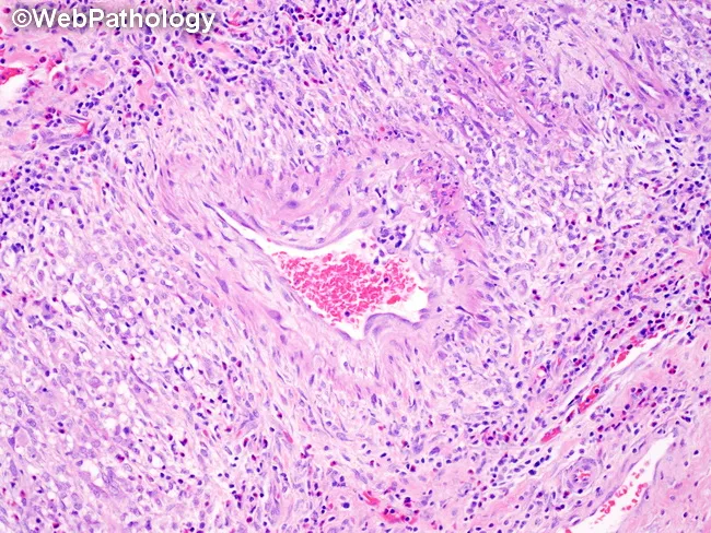
Histopathology of gallbladder tissue reveals necrotizing transmural vasculitis of medium-sized muscular arteries within the gallbladder wall. The vessel wall exhibits fibrinoid necrosis and inflammatory infiltrates that are initially neutrophilic and eosinophilic, with histiocytes; the surrounding tissue typically shows minimal inflammatory reaction. Lesions are discontinuous, with skip areas, and multiple vessels commonly display varying stages of evolution within a single specimen. In active lesions, transmural inflammation encroaches on all vascular layers, culminating in destruction of the tunica media and internal elastic lamina; elastic stains most clearly demonstrate lamina destruction. In healed or healing stages, fibrointimal proliferation produces luminal narrowing or occlusion. The inflammatory infiltrate may transition to a lymphocytic predominance during resolution. Importantly, bystander inflammation related to adjacent cholecystitis must be distinguished from true vasculitis. The grossly preserved surrounding gallbladder parenchyma contrasts with the targeted vascular injury. These features are characteristic of polyarteritis nodosa and may reflect systemic vasculitis when observed in the gallbladder, prompting clinical correlation for systemic symptoms and laboratory markers (ANCA status is often negative in classic PAN). This histology underlines the need for immunosuppressive therapy and vigilance for renal, gastrointestinal, and vascular complications. Correlation with clinical data guides targeted treatment and prognosis assessment in systemic vasculitis workup.
vasculitis classification treatment
giant cell arteritis temporal arteritis granuloma histology
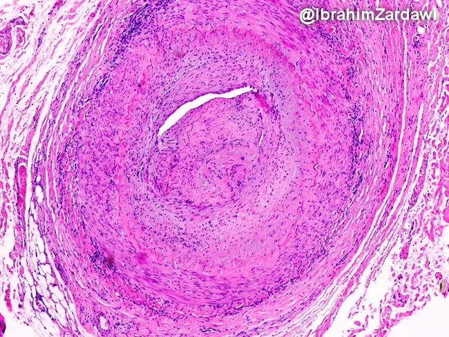
Histology image obtained from a temporal artery biopsy, prepared with Hematoxylin and Eosin stain and viewed under light microscopy at low magnification. The specimen demonstrates classic giant cell arteritis with transmural, granulomatous inflammation encompassing the full thickness of the arterial wall (intima, media, and adventitia) in a segmental distribution. Multinucleated giant cells are interspersed with a dense lymphohistiocytic infiltrate, and fragmentation or disruption of the internal elastic lamina is evident. The inflammatory process extends to the vasa vasorum and small venules within the adventitia, indicating widespread perivascular involvement. Accompanying features include marked intimal hyperplasia producing marked luminal narrowing and, in this field of view, near-occlusive lumen occlusion. The arterial media may show loss of elastic fibers and necrosis is not conspicuous here. These findings are diagnostic of GCA and help differentiate from other large-vessel vasculitides and nonspecific atherosclerotic changes. Clinically, such histology supports a diagnosis of temporal arteritis in patients with new headaches, visual symptoms, jaw claudication, and elevated inflammatory markers, guiding urgent corticosteroid therapy to prevent ischemic complications. This image exemplifies the radiologic-pathologic correlation typical of vasculitic arteritis. The slide highlights diagnostic relevance, research utility, and educational value for trainees and clinicians in practice. For medical education and research.
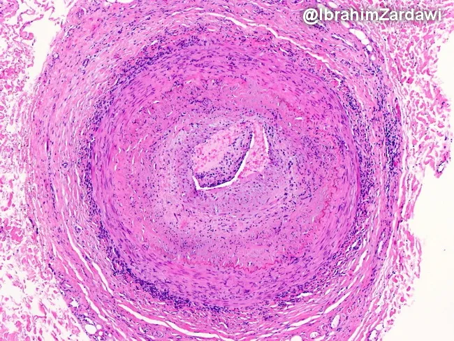
This histology image captures a cross-sectional temporal artery biopsy illustrating the hallmarks of giant cell arteritis. Prepared as a paraffin-embedded tissue section and stained with Hematoxylin and Eosin, the slide reveals a transmural inflammatory infiltrate that traverses all arterial wall layers. At the center, a narrowed lumen is surrounded by concentric intimal hyperplasia with marked medial disruption. Multinucleated giant cells and mononuclear macrophage clusters are present within the media, accompanied by lymphocytic infiltrate along the vessel wall. The elastic lamina appears disrupted, and fragmentation of the media is evident, consistent with robust granulomatous vasculitis. These features produce near-occlusive narrowing of the artery, explaining the ischemic risk associated with temporal arteritis. Clinically, such histopathology confirms a diagnosis of GCA and helps differentiate from atherosclerotic disease, infectious vasculitis, or other large-vessel vasculitides. The image underscores the systemic nature of the disease, as inflammation can extend from cranial vessels to the aorta. The diagnostic significance lies in correlating histology with patient presentation, including age-related cranial symptoms and risk of vision loss; timely immunosuppressive therapy can prevent irreversible ischemic complications. This specimen exemplifies classic angiitis with granulomatous inflammation and intimal hyperplasia driving luminal compromise.
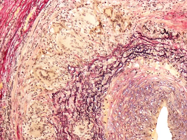
This histopathology image shows a temporal artery biopsy stained with an elastic stain (e.g., Verhoeff-Van Gieson) to highlight elastic laminae within the arterial wall. The specimen demonstrates classic giant cell arteritis features: fragmentation and partial duplication of the internal elastic lamina with loss of elastic fibers, and prominent intimal hyperplasia producing marked luminal narrowing. Inflammatory infiltrates, including mononuclear cells and multinucleated giant cells, surround the media in a granulomatous vasculitis pattern. Elastic lamina disruptions create irregular vascular borders and eccentric thickening of the intima. Some sections reveal medial edema and preserved outer adventitia, without definite necrosis. The overall morphology supports inflammatory arteritis affecting cranial arteries, particularly the superficial temporal artery. Clinically this histology aligns with temporal headache, jaw claudication, decreased vision risk, and elevated ESR/CRP in patients over 50. The diagnostic significance is high when elastic lamina fragmentation is present with compatible clinical symptoms. Differential considerations include atherosclerotic changes with intimal thickening and luminal narrowing, other large-vessel vasculitides such as Takayasu arteritis in younger patients, and other granulomatous diseases. Correlation with serology, imaging, and clinical presentation is essential for accurate diagnosis and timely glucocorticoid therapy. This image supports education, research, and diagnostic case reviews in rheumatology and pathology practice today.
Vasculitis in Medicine
Definition and Overview
- Immune-mediated inflammation (the vast majority)
- Direct vascular invasion by infectious pathogens
The 2012 Chapel Hill Consensus Conference (CHCC) Classification
Classification Table (Robbins & Kumar / CHCC)
Large-Vessel Vasculitis
| Disease | Key Features |
|---|---|
| Giant Cell Arteritis (GCA) | Granulomatous arteritis; temporal/carotid/vertebral arteries; age >50; risk of blindness |
| Takayasu Arteritis | Granulomatous; aorta + branches; age <50; "pulseless disease" |
Medium-Vessel Vasculitis
| Disease | Key Features |
|---|---|
| Polyarteritis Nodosa (PAN) | Necrotizing arteritis; NOT in pulmonary vessels or glomeruli; ANCA-negative; up to 30% associated with HBsAg immune complexes |
| Kawasaki Disease | Mucocutaneous lymph node syndrome; coronary artery aneurysms; mainly infants/children |
Small-Vessel Vasculitis
| Disease | ANCA Type | Key Features |
|---|---|---|
| Granulomatosis with Polyangiitis (GPA) - formerly Wegener's | PR3-ANCA (c-ANCA) in 95% | Necrotizing granulomatous inflammation of upper + lower respiratory tract; necrotizing glomerulonephritis |
| Microscopic Polyangiitis (MPA) | MPO-ANCA (p-ANCA) | Necrotizing vasculitis with few immune deposits; glomerulonephritis + pulmonary capillaritis |
| Eosinophilic Granulomatosis with Polyangiitis (EGPA) - formerly Churg-Strauss | MPO-ANCA in >50% | Eosinophilic necrotizing granulomas; asthma + eosinophilia |
| Disease | Immune Deposits | Key Features |
|---|---|---|
| IgA Vasculitis (Henoch-Schönlein Purpura) | IgA + C3 | Palpable purpura, arthritis, abdominal pain, renal vasculitis; often post-strep/staph |
| Cryoglobulinemic Vasculitis | Cryoglobulins | Often HCV-related |
| Anti-GBM Disease (Goodpasture) | Anti-GBM IgG | Pulmonary-renal syndrome |
Variable-Vessel Vasculitis
- Behcet Disease: oral/genital ulcers, uveitis, skin lesions, venous thrombosis
- Cogan Syndrome
Single-Organ Vasculitis
- Primary CNS vasculitis
- Cutaneous leukocytoclastic angiitis
- Cutaneous arteritis
Pathogenic Mechanisms
1. Immune Complex Deposition
- Complexes deposit in vessel walls → complement activation → neutrophil recruitment → tissue damage
- Seen in: SLE, IgA vasculitis, mixed cryoglobulinemia, drug hypersensitivity, hepatitis B-associated PAN
- Drug hypersensitivity vasculitis resolves with removal of the offending agent
2. ANCA-Mediated
- ANCAs (autoantibodies against neutrophil cytoplasmic antigens) are key diagnostic and pathogenic markers
- PR3-ANCA (c-ANCA): mainly GPA - shares homology with microbial peptides
- MPO-ANCA (p-ANCA): mainly MPA and EGPA
- ANCA titers mirror clinical severity; rising titers predict relapse
3. Anti-Endothelial Cell Antibodies
- Cause endothelial activation and complement-dependent cytotoxicity
4. Autoreactive T Cells
- Drive granuloma formation in GCA and GPA
Individual Disease Profiles
Giant Cell Arteritis (Temporal Arteritis)
- New temporal headache, scalp tenderness
- Jaw claudication (pathognomonic)
- Sudden visual loss (ophthalmic artery involvement - medical emergency)
- Elevated ESR (>50 mm/hr), CRP
- Often coexists with polymyalgia rheumatica (PMR)
- High-dose prednisone (40-60 mg/day) immediately - do not wait for biopsy if vision threatened
- Tocilizumab (IL-6 inhibitor) - approved as steroid-sparing agent
- Methotrexate (MTX) + corticosteroids - used in tocilizumab-unavailable/contraindicated cases (Firestein & Kelley's Rheumatology)
GPA (Granulomatosis with Polyangiitis / Wegener's)
- PR3-ANCA positive in 95%
- Treatment: Rituximab or cyclophosphamide + high-dose glucocorticoids for induction; rituximab or azathioprine for maintenance
- MTX + corticosteroids for non-severe/early disease
MPA (Microscopic Polyangiitis)
- Most common cause of pulmonary-renal syndrome (diffuse alveolar hemorrhage + rapidly progressive GN)
- MPO-ANCA positive
- Pauci-immune on renal biopsy (few immune deposits)
- Treatment: same as GPA
EGPA (Eosinophilic Granulomatosis with Polyangiitis / Churg-Strauss)
- Prodrome: asthma, allergic rhinitis
- Eosinophilic phase: peripheral blood and tissue eosinophilia
- Vasculitic phase: systemic necrotizing vasculitis
- MPO-ANCA in >50%
- Treatment: glucocorticoids ± cyclophosphamide for severe disease; mepolizumab (anti-IL-5) for remission maintenance
Kawasaki Disease
- Acute febrile illness in children <5 years
- CRASH criteria: Conjunctivitis, Rash, Adenopathy, Strawberry tongue, Hand/foot changes (erythema + desquamation) + fever >5 days
- Feared complication: coronary artery aneurysms (20-25% if untreated)
- Treatment: High-dose aspirin + IVIG (reduces aneurysm risk to <5%)
IgA Vasculitis (Henoch-Schönlein Purpura)
- Most common vasculitis in children
- Classic tetrad: palpable purpura (lower limbs) + arthritis + abdominal pain + renal involvement (hematuria/proteinuria)
- Triggered by upper respiratory infections (strep/staph)
- Perivascular IgA + C3 deposits on skin biopsy
- Treatment: supportive; steroids for severe abdominal/renal involvement
Leukocytoclastic Vasculitis (LCV)
- Colchicine 0.6 mg twice daily
- Prednisone 1 mg/kg/day
- Dapsone up to 200 mg/day
- Remove offending drug if drug-induced
- Immunosuppressives for severe/refractory cases
Vasculitis of Specific Organs
CNS Vasculitis (Grainger & Allison's Diagnostic Radiology)
- Heterogeneous group affecting small parenchymal and leptomeningeal vessels
- Causes: primary CNS angiitis, infection, cocaine, SLE, PAN, sarcoidosis, Wegener's
- MRI: non-specific white matter hyperintensities, microhemorrhage, leptomeningeal enhancement
- DSA catheter angiography is superior to MRA/CTA for small vessel disease; shows stenosis, occlusion, aneurysms - but may be negative
- Brain/meningeal biopsy often required for definitive diagnosis
Pulmonary Vasculitis (Murray & Nadel's Respiratory Medicine)
- Systemic vasculitis is the most common cause of diffuse alveolar hemorrhage (DAH)
- Major causes of DAH: GPA (most common), anti-GBM disease (Goodpasture), MPA, SLE
- Also: isolated pulmonary capillaritis, mixed cryoglobulinemia, Behcet syndrome
Diagnostic Approach
| Test | Utility |
|---|---|
| ESR, CRP | Elevated in active disease |
| ANCA panel (PR3 + MPO) | Essential for small-vessel ANCA vasculitis |
| Urinalysis + creatinine | Screen for renal involvement (hematuria, RBC casts) |
| CBC | Eosinophilia in EGPA |
| Complement (C3, C4, CH50) | Low in immune-complex vasculitis |
| HBsAg, HCV serology | Screen for infection-associated vasculitis |
| Cryoglobulins | Cryoglobulinemic vasculitis |
| Temporal artery biopsy | Gold standard for GCA |
| Skin/renal biopsy | Direct IF for immune deposits |
| Angiography (DSA/CTA) | Large/medium vessel disease |
Treatment Principles
| Category | First-line Induction | Maintenance / Steroid-sparing |
|---|---|---|
| GCA | Prednisone 40-60 mg/day | Tocilizumab; MTX |
| GPA/MPA | Rituximab or cyclophosphamide + steroids | Rituximab or azathioprine |
| EGPA | Steroids ± cyclophosphamide | Mepolizumab (anti-IL-5) |
| PAN | Steroids; cyclophosphamide for severe | MTX + steroids for nonsevere (Firestein & Kelley) |
| Kawasaki | IVIG + aspirin | - |
| Cutaneous LCV | Colchicine / dapsone / prednisone | Immunosuppressives if refractory |
| Takayasu | Steroids | MTX ± biologics |
Key Differentiating Points
| Feature | GCA | Takayasu | PAN | GPA | MPA |
|---|---|---|---|---|---|
| Age | >50 | <50 | Any | Any | Any |
| Vessel size | Large | Large | Medium | Small-medium | Small |
| ANCA | Negative | Negative | Negative | PR3-ANCA | MPO-ANCA |
| Granulomas | Yes | Yes | No | Yes | No |
| Renal | Rare | Rare (renovascular HT) | Yes (renal arteries) | GN common | GN common |
| Lung | Aortitis | No | No (spares lung) | Cavities, hemorrhage | Hemorrhage |
In general medicine
What would you like to know about vasculitis in general medicine?
vasculitis skin palpable purpura clinical presentation
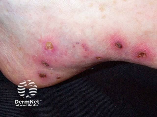
Clinical photograph of the lower extremity skin showing palpable purpura typical of cutaneous small-vessel vasculitis. Modality: digital close‑up photography using visible light; magnification approximately macro scale; the image captures multiple erythematous–violaceous papules and patchy plaques with central crusting on the anterior-lateral aspect of the ankle and distal leg. Edema is mild; lesions appear non-blanching with surrounding erythema; some lesions demonstrate necrotic crusts and pigmented halos. The distribution is predominantly on extensor surfaces of the lower leg, consistent with common presentation of leukocytoclastic vasculitis. Skin biopsy, if performed, would be expected to show leukocytoclasia with perivascular neutrophilic infiltration and immune complex deposition; correlate with immune-mediated processes. The image illustrates a dermatologic finding that may be isolated to the skin but can accompany systemic vasculitis or secondary etiologies such as infection, drugs, autoimmune diseases, or IgA vasculopathy (Henoch-Schönlein purpura). Clinically relevant significance includes differentiation from purpura due to thrombocytopenia or coagulopathy; the presence of palpable purpura warrants laboratory evaluation (CBC, inflammatory markers, complements, IgA, serum cryoglobulins) and careful history for recent medications or infections. Use cases include dermatology education, vasculitis teaching files, and assisting in triage for systemic workup. This image supports clinical correlation, patient counseling, and multidisciplinary consultation as needed promptly.
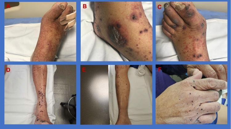
This clinical photograph series illustrates the progression and treatment response of palpable purpura in a patient with IgA vasculitis (Henoch-Schönlein purpura). Panels A-C display the clinical presentation at admission: Panel A shows a dorsal view of the foot with dense, confluent, reddish-purple purpuric macules and papules; Panel B highlights the lateral ankle featuring larger ecchymotic areas and bullous lesions indicating skin breakdown; and Panel C focuses on the toes, demonstrating dark, necrotic-appearing patches and digital ulceration. Panels D-F illustrate the clinical state post-steroid therapy: Panels D and E show a significant reduction in the density and inflammatory appearance of the lesions on the lower legs, which have transitioned to darker, resolving crusts and post-inflammatory pigmentary changes. Panel F displays the hand, showing resolving, sparse, small purpuric spots. This comparison highlights the hallmark dermatologic manifestation of leukocytoclastic vasculitis, characterized by symmetric, gravity-dependent palpable purpura, and demonstrates the efficacy of systemic corticosteroids in managing cutaneous inflammation and associated systemic symptoms like edema and arthralgia.
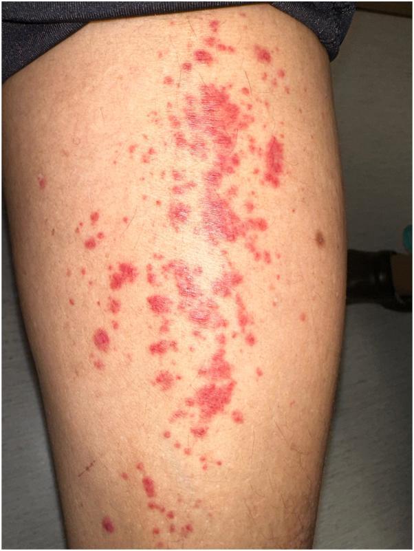
This clinical photograph displays a dermatological presentation of palpable purpura on the lower extremity. The lesions are characterized by numerous erythematous to violaceous macules and small papules that range from pinpoint petechiae to larger confluent patches. These non-blanching lesions exhibit irregular borders and a scattered distribution, primarily concentrated along the pretibial area. The morphology and coloring are consistent with blood extravasation into the dermis. This visual presentation is a hallmark clinical sign of small-vessel vasculitis, specifically leukocytoclastic vasculitis. The image serves as a diagnostic educational resource for identifying inflammatory vascular skin disorders and drug-induced cutaneous eruptions in a clinical setting.
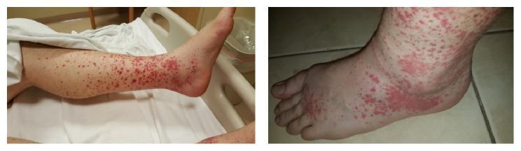
Clinical photograph of the lower extremities demonstrating classic palpable purpura, a hallmark of small-vessel vasculitis such as IgA vasculitis (Henoch-Schönlein purpura). The images show multiple erythematous to violaceous, non-blanching macules and papules. The lesions exhibit a dependent distribution, being most densely concentrated around the ankles and dorsal aspect of the feet, with progressive thinning toward the knees and mid-calf. In areas of high density, particularly the distal leg and ankle, individual purpuric spots coalesce into larger, confluent ecchymotic patches. Significant soft tissue edema is visible around the ankle and foot, evidenced by a loss of anatomical landmarks and a shiny appearance of the skin surface. This clinical presentation illustrates the typical cutaneous manifestation of leukocytoclastic vasculitis, characterized by inflammatory cell infiltration and hemorrhage within the superficial dermis. The presence of concurrent edema suggests systemic involvement or localized inflammation associated with the underlying vasculitic process.
How Vasculitis Presents in General Medicine
The Core Clinical Challenge
- Is the lesion/symptom actually due to vasculitis?
- Are other organ systems involved?
- Can history, exam, and tests narrow the specific type?
- Can a specific vasculitis diagnosis be made?
- Does the patient need immediate treatment and/or hospitalization?
How Vasculitis Enters General Medicine
The Most Common "Entry Points"
| Presenting Complaint | What it Signals |
|---|---|
| Palpable purpura (non-blanching, raised, lower limbs) | Small-vessel vasculitis - the classic calling card |
| Unexplained fever + weight loss + fatigue | Systemic inflammation from almost any vasculitis |
| New headache in a patient >50 | Giant cell arteritis - a medical emergency |
| Haematuria/proteinuria on routine dipstick | Renal vasculitis (often silent until significant damage done) |
| Sinusitis not responding to antibiotics | Possible GPA (Wegener's) |
| Adult-onset asthma + eosinophilia | Think EGPA (Churg-Strauss) |
| Young woman with absent pulses or BP asymmetry | Takayasu arteritis |
| Mononeuritis multiplex (foot drop + wrist drop) | Vasculitic neuropathy - PAN, EGPA, GPA |
Organ-System Map of Vasculitis Presentation
Constitutional (Almost Universal)
- Fever - seen in most systemic vasculitides
- Fatigue/malaise - most systemic vasculitides
- Weight loss - most systemic vasculitides
Eyes
| Symptom | Disease Process | Vasculitis Type |
|---|---|---|
| Red eye | Episcleritis, scleritis, uveitis | GPA, MPA, Behcet's |
| Acute visual loss / amaurosis fugax | Arterial insufficiency | GCA, Takayasu - URGENT |
| Proptosis | Orbital granuloma | GPA |
| Tearing | Dacryocystitis / lacrimal duct occlusion | GPA, EGPA |
Ears
| Symptom | Vasculitis Type |
|---|---|
| Sensorineural hearing loss | GPA, MPA, EGPA, GCA |
| Ear pain + fullness (mastoiditis) | GPA, EGPA |
| External ear redness/tenderness (chondritis) | Relapsing polychondritis, GPA, EGPA |
Nose and Sinuses
| Symptom | Vasculitis Type |
|---|---|
| Epistaxis, nasal crusting, bloody discharge | GPA, EGPA |
| Saddle nose deformity (nasal bridge collapse) | GPA (pathognomonic), relapsing polychondritis |
| Nasal polyps | EGPA |
| Sinusitis not responding to antibiotics | GPA, EGPA |
| Anosmia | GPA, EGPA |
Respiratory
| Symptom | Disease Process | Vasculitis Type |
|---|---|---|
| Haemoptysis | Pulmonary haemorrhage / capillaritis | GPA, MPA |
| Dry cough / dyspnoea | Interstitial lung disease | GPA, MPA, EGPA |
| Adult-onset asthma | Eosinophilic airway inflammation | EGPA |
Cardiovascular
| Symptom | Disease Process | Vasculitis Type |
|---|---|---|
| Jaw claudication | Masseteric artery ischaemia | GCA (highly specific) |
| Limb claudication | Large/medium vessel stenosis | Takayasu, GCA |
| Absent pulses, BP asymmetry | Aortic arch branch stenosis | Takayasu |
| Hypertension (new, young patient) | Renal artery stenosis | Takayasu, PAN |
| Coronary aneurysms / MI in a child | Coronary arteritis | Kawasaki |
Gastrointestinal
| Symptom | Disease Process | Vasculitis Type |
|---|---|---|
| Abdominal pain post-eating | Mesenteric ischaemia | PAN, GPA, MPA |
| Abdominal pain in a child | Gut ischaemia / intussusception | IgA vasculitis (HSP) |
| Rectal bleeding | Gut vasculitis | PAN, IgA vasculitis |
Renal (Often Clinically Silent)
| Finding | Disease Process | Vasculitis Type |
|---|---|---|
| Microscopic haematuria (RBC casts) | Glomerulonephritis | GPA, MPA, EGPA, IgA vasculitis, cryoglobulinaemia |
| Gross haematuria | Renal infarction | PAN |
| Rapidly rising creatinine | Rapidly progressive GN (RPGN) | GPA, MPA - EMERGENCY |
Critical point: Renal vasculitis is silent in many patients. A routine urinalysis (dipstick + microscopy) must be performed in every patient with suspected vasculitis, even if asymptomatic - Fishman's Pulmonary Diseases
Nervous System
| Symptom | Disease Process | Vasculitis Type |
|---|---|---|
| Mononeuritis multiplex | Vasculitic nerve ischaemia | EGPA, GPA, MPA, PAN, cryoglobulinaemia |
| Headache + scalp tenderness | Cranial arteritis | GCA, Takayasu |
| Cranial nerve palsy | Nerve inflammation | GCA, GPA, MPA |
| Stroke in young adult | CNS vasculitis | Primary CNS angiitis, Behcet's |
Musculoskeletal
| Symptom | Disease Process | Vasculitis Type |
|---|---|---|
| Polyarthralgia / polyarthritis | Joint inflammation | GPA, GCA, Takayasu, cryoglobulinaemia, IgA vasculitis |
| Shoulder and hip girdle pain + morning stiffness | Polymyalgia rheumatica | GCA (50% co-exist) |
| Muscle weakness | Myositis | EGPA |
Skin - The Most Visible Clue
| Lesion | Disease Process | Vasculitis Type |
|---|---|---|
| Palpable purpura (raised, non-blanching, gravity-dependent) | Small-vessel vasculitis | GPA, MPA, EGPA, PAN, IgA vasculitis, cryoglobulinaemia |
| Painful nodules, deep ulcers | Medium-vessel vasculitis | PAN, GPA, MPA, cryoglobulinaemia |
| Digital ischaemia / gangrene | Medium/large artery stenosis | GCA, Takayasu, PAN, GPA, MPA |
| Superficial nodules | Granulomas | GPA, EGPA |
| Papulopustular lesions | Papulopustular lesions | Behcet's |
| Livedo reticularis | Vessel wall ischaemia | PAN, cryoglobulinaemia |
Why Vasculitis Is Missed in General Medicine
Common Masquerades
| What It Looks Like | What It Actually Is |
|---|---|
| Sinusitis (antibiotic-resistant) | GPA |
| Asthma + allergies | EGPA |
| UTI / haematuria | Renal vasculitis |
| Tension headache in elderly | GCA |
| Peripheral neuropathy (diabetes?) | Vasculitic neuropathy |
| Skin rash + joint pain (lupus?) | IgA vasculitis / cryoglobulinaemia |
| Malignancy work-up (weight loss, fatigue) | Systemic vasculitis |
Initial Approach in General Medicine
Step 1: Recognise the Pattern
Step 2: History
- Full review of systems - all organ systems
- Duration, sequence of symptoms
- Drug history (all medications, last 6-12 months, recreational drugs)
- Recent infections, travel, sick contacts
- Family history of autoimmune disease
Step 3: Examination Red Flags
| Finding on Exam | Urgency |
|---|---|
| Visual loss / temporal artery tenderness | EMERGENCY - GCA |
| Absent pulses / BP asymmetry | Urgent - Takayasu |
| Haemoptysis + haematuria | EMERGENCY - pulmonary-renal syndrome (GPA/MPA) |
| Mononeuritis multiplex | Urgent referral |
| Purpura + systemic signs | Urgent workup |
Step 4: First-Line Investigations
| Test | Rationale |
|---|---|
| Urinalysis + microscopy | Screen for haematuria, RBC casts (silent renal vasculitis) |
| ESR + CRP | Elevated in active disease; ESR >50 in GCA |
| Full blood count | Eosinophilia (EGPA), anaemia of chronic disease |
| U&E + creatinine | Renal function - rapidly rising creatinine = RPGN |
| ANCA (PR3 + MPO) | Small-vessel ANCA vasculitis (GPA, MPA, EGPA) |
| ANA, anti-dsDNA, complement | Screen for SLE-related vasculitis |
| Hepatitis B & C serology | HBV-associated PAN; HCV-associated cryoglobulinaemia |
| Cryoglobulins | Cryoglobulinaemic vasculitis |
| Serum IgA | IgA vasculitis (HSP) |
| Skin biopsy | Histology + direct immunofluorescence for immune deposits |
| Temporal artery biopsy | Gold standard for GCA (do urgently, before steroids where possible) |
The Washington Manual of Medical Therapeutics summarises GPA labs: anaemia of chronic disease, elevated ESR/CRP, elevated creatinine, haematuria, proteinuria, and positive PR3-ANCA; imaging may show lung nodules, infiltrates, or cavities.
When to Refer vs. When to Act Immediately
| Situation | Action |
|---|---|
| GCA suspected (headache + visual symptoms, age >50) | Start high-dose prednisone NOW before biopsy; refer urgently to rheumatology/ophthalmology |
| Pulmonary haemorrhage + renal failure | Emergency admission - RPGN/DAH is rapidly fatal |
| Kawasaki (child, fever >5 days + CRASH features) | Admit, IVIG + aspirin urgently |
| Confirmed ANCA-positive with renal/lung involvement | Urgent rheumatology referral |
| Isolated cutaneous vasculitis, no systemic features | Manage/investigate in general medicine; refer if persistent/severe |
| IgA vasculitis (HSP) with mild disease | Supportive care; monitor urine/renal function closely |
Summary: The General Medicine Mindset for Vasculitis
- Think multisystem - never accept a single-organ explanation for a patient with multiple seemingly unrelated symptoms
- Always dip the urine - renal vasculitis is clinically silent and devastating if missed
- Age matters - GCA only above 50; Kawasaki in young children; Takayasu in young women
- Drugs first - always exclude drug-induced vasculitis before labelling primary vasculitis
- Don't wait for pathology in GCA - visual loss is irreversible; treat first
- ANCA testing is high yield in unexplained multisystem disease with renal or pulmonary involvement
- Vasculitis is not rare - it is underdiagnosed because it mimics common conditions