
Write a note. All. Nefron. Eee? Enumerate. It's... Function.
nephron anatomy diagram parts
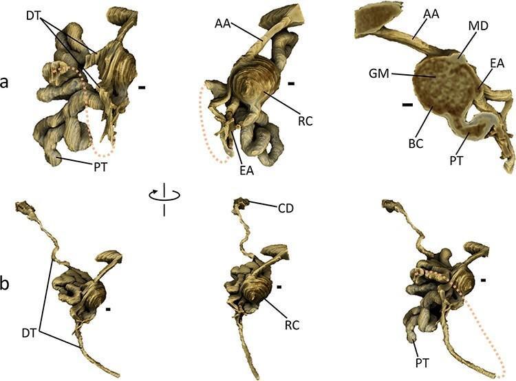
Anatomical diagram featuring high-resolution 3D renderings of a single mouse nephron, highlighting renal microstructure and spatial arrangement. Panel (a) provides detailed views of the renal corpuscle (RC), demonstrating the spherical Bowman's capsule (BC) and internal glomerulus (GM). It clearly illustrates the vascular pole with the afferent arteriole (AA), efferent arteriole (EA), and the macula densa (MD) of the distal tubule (DT) forming a v-shaped contact point. The tubular pole shows the exit of the highly convoluted proximal tubule (PT). Panel (b) illustrates the longitudinal progression from the renal corpuscle through the PT and DT to the collecting duct (CD). The rendering distinguishes between the intensely convoluted segments near the corpuscle and the straighter distal segments of the tubules. Key educational concepts include the juxtaglomerular apparatus anatomy, the morphology of glomerular filtration components, and the physical relationship between various segments of the nephron and their associated vasculature, essential for understanding renal physiology and pathology.
Educational panel illustrating the surgical anatomy and complications of an ileal conduit urinary diversion. Part A is an anatomical diagram showing a cross-section of the abdomen with a focal stoma. Key structures labeled include the ileal conduit (1), its associated mesentery containing vascular supply (2), and the right (3) and left (4) ureters. Asterisks mark the uretero-ileal anastomoses. The posterior abdominal wall is noted as deperitonealized (5) following radical cystectomy. Parts B and C are clinical laparoscopic photographs showing intraoperative views of the same anatomy. Photograph B demonstrates complex fibrous adhesions involving the ileal conduit, a common challenge in parastomal hernia repair. Photograph C shows the pinkish, smooth surface of the deperitonealized posterior abdominal wall (5) adjacent to the textured, tubular ileal conduit (1). Small orientation icons indicate the stoma position on the lower abdominal wall. This content is designed to teach the anatomical changes following cystectomy and the specific challenges of treating parastomal hernias in patients with urinary diversions.
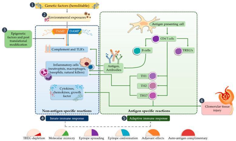
This pathophysiology flow diagram illustrates the multi-step immune signaling pathway leading to glomerular tissue injury in the nephron. The process is initiated by three primary drivers: genetic factors (heritable), environmental exposures, and epigenetic factors coupled with post-translational modifications. These drivers trigger the 'Innate Immune Response' (Non-antigen specific reactions), characterized by the activation of Pathogen-Associated Molecular Patterns (PAMPs) and Damage-Associated Molecular Patterns (DAMPs). This activates the complement system, Toll-Like Receptors (TLRs), and inflammatory cells (neutrophils, macrophages, basophils, NK cells), leading to the release of cytokines, chemokines, and growth factors. The pathway then transitions to the 'Adaptive Immune Response' (Antigen-specific reactions), where antigen-presenting cells activate CD4+ T cells, B cells, and TREGs. Further differentiation into TH1, TH2, and TH17 cells, along with antigen-antibody complex formation, culminates in glomerular tissue injury. The diagram also identifies key mechanisms of immune dysregulation below the main flow, including TREG depletion, molecular mimicry, epitope spreading, epitope conformation, adjuvant effects, and auto-antigen complementarity.
The Nephron - Structure and Functions
Definition and Overview
- Brenner and Rector's The Kidney, p. 83
- NKF Primer on Kidney Diseases, 8e, p. 24
Anatomy: Parts of the Nephron
| Segment | Location |
|---|---|
| Glomerulus + Bowman's capsule (renal corpuscle) | Cortex |
| Proximal convoluted tubule (PCT) | Cortex |
| Proximal straight tubule | Cortex/outer medulla |
| Thin descending limb of Henle | Medulla |
| Thin ascending limb of Henle | Inner medulla |
| Thick ascending limb (TAL) | Outer medulla/cortex |
| Macula densa | Cortex (at DCT-TAL junction) |
| Distal convoluted tubule (DCT) | Cortex |
| Connecting tubule | Cortex |
| Cortical collecting duct (CCD) | Cortex |
| Outer medullary collecting duct (OMCD) | Outer medulla |
| Inner medullary collecting duct (IMCD) | Inner medulla |
Enumerated Functions of Each Nephron Segment
1. Glomerulus (Renal Corpuscle)
- Ultrafiltration of blood plasma across the glomerular filtration barrier (endothelium, glomerular basement membrane, podocytes with slit diaphragms)
- Produces approximately 180 L/day of ultrafiltrate (GFR ~125 mL/min)
- Filters water, electrolytes, small solutes; retains large proteins and blood cells
- The glomerular filtration rate (GFR) is the key measure of renal function
2. Proximal Tubule (PCT + Straight Segment)
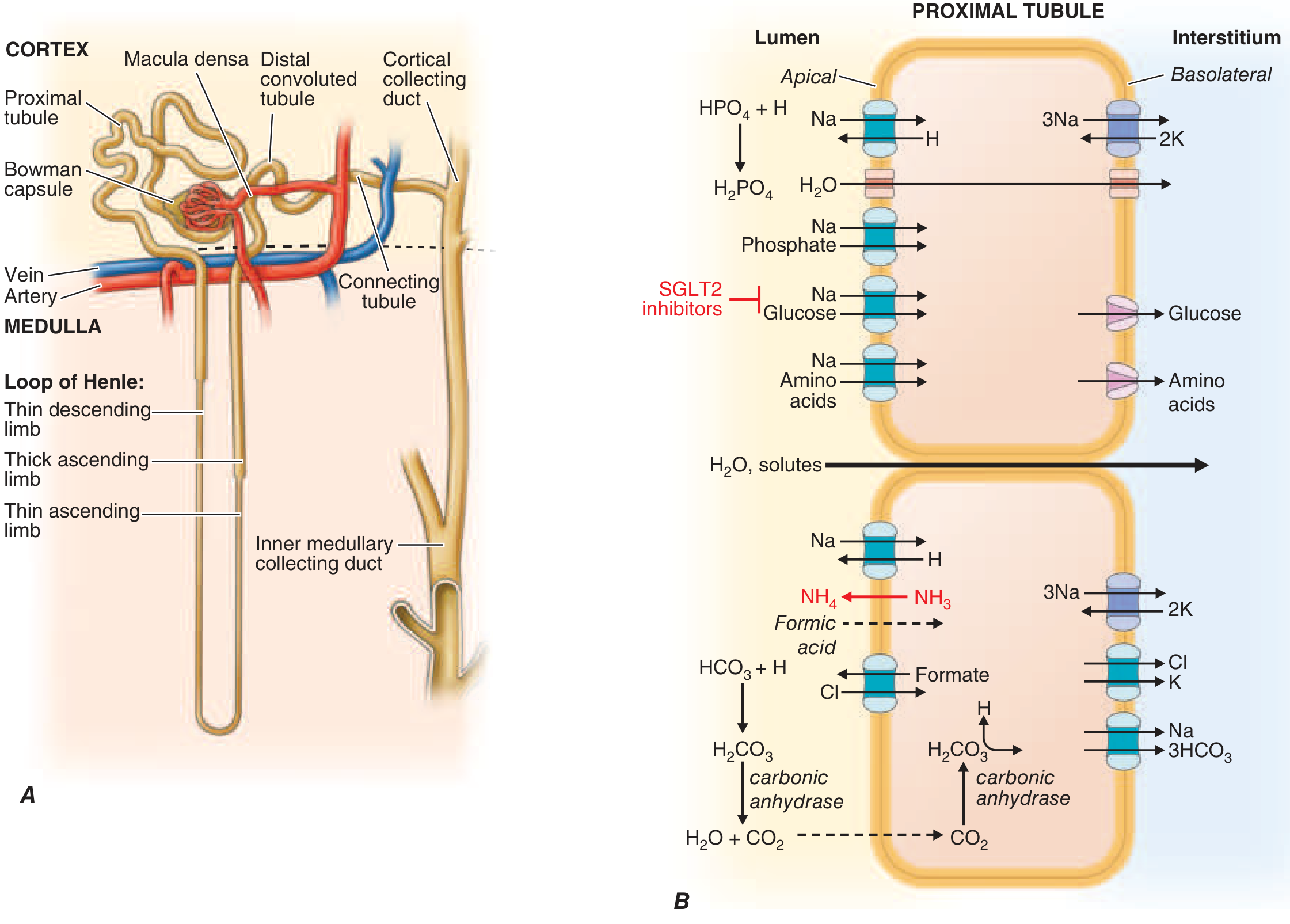
- NaCl and water reabsorption - ~60% of filtered NaCl and water is reabsorbed isosmotically. Primary apical transporter is Na+/H+ exchanger 3 (NHE3). Water follows through aquaporin-1 channels (constitutively active - no hormonal regulation needed).
- Glucose reabsorption (~100%) - via SGLT2 (Na+-glucose cotransporter) in early PCT; SGLT1 in late PCT. (SGLT2 inhibitors block this to lower blood glucose in diabetes.)
- Amino acid reabsorption (~100%) - Na+-coupled amino acid transporters on apical membrane.
- Bicarbonate reclamation (~90%) - filtered HCO3- is titrated by H+ (from NHE3) → H2CO3 → CO2 + H2O (carbonic anhydrase IV on brush border), CO2 diffuses into cell → reconverted to HCO3- (carbonic anhydrase II) → exits basolaterally via NBC1.
- Phosphate reabsorption - via Na+-phosphate cotransporters; regulated by PTH (which inhibits reabsorption, causing phosphaturia).
- Uric acid, organic acids, and drug secretion - proximal tubule is the main site of organic anion/cation secretion (e.g., penicillin, furosemide, methotrexate, creatinine).
- Ammonia generation - from glutamine metabolism; NH4+ is excreted via NHE3 (K+ position); critical for acid-base homeostasis.
- Protein catabolism - small filtered proteins are endocytosed and degraded by lysosomes.
- Harrison's Principles of Internal Medicine 22E, p. 2410
- NKF Primer on Kidney Diseases, 8e, p. 24
3. Loop of Henle
- Permeable to water (but NOT solutes) - water exits into the hypertonic medullary interstitium, concentrating tubular fluid
- Fluid becomes progressively hyperosmotic as it descends
- Impermeable to water - solutes (NaCl) passively diffuse out into the interstitium
- Active NaCl reabsorption (~25-30% of filtered NaCl) - via apical NKCC2 (Na+/K+/2Cl- cotransporter); inhibited by loop diuretics (furosemide, bumetanide)
- Impermeable to water - dilutes tubular fluid ("diluting segment")
- K+ recycling - K+ secreted back into lumen via ROMK channel to sustain NKCC2 (luminal K+ is limiting)
- Ca2+ and Mg2+ reabsorption - paracellular, driven by lumen-positive voltage generated by K+ recycling; accounts for ~50-60% of Mg2+ reabsorption
- NH4+ reabsorption - absorbed via NKCC2 in the K+ position; important for acid-base handling
- The net result: medullary interstitium becomes highly hyperosmotic (up to ~1200 mOsm/kg), the only compartment in the body with higher osmolality than serum
- NKF Primer on Kidney Diseases, 8e, p. 24
4. Macula Densa
- Tubuloglomerular feedback (TGF) - senses luminal NaCl concentration via NKCC2; if NaCl is high → signals afferent arteriole to constrict → reduces GFR (autoregulation)
- Renin secretion control - low luminal NaCl → stimulates juxtaglomerular cells to release renin → activates RAAS
5. Distal Convoluted Tubule (DCT)
- NaCl reabsorption (~5-8%) - via apical NCC (Na+/Cl- cotransporter); inhibited by thiazide diuretics
- Ca2+ reabsorption (regulated) - transcellular, stimulated by PTH and 1,25(OH)2 vitamin D; via apical TRPV5 channels and basolateral NCX1
- Mg2+ reabsorption - via apical TRPM6 channels; this is the main regulated segment for Mg2+
- Fine-tuning of K+ - some K+ reabsorption and secretion
6. Collecting Duct (Cortical + Medullary) - Principal and Intercalated Cells
- Na+ reabsorption - via apical ENaC (epithelial Na+ channel); tightly regulated by aldosterone (which increases ENaC expression and activity) and proteolytic enzymes. Blocked by amiloride/triamterene.
- K+ secretion - via apical K+ channels (ROMK); driven by high intracellular K+ (from Na+/K+-ATPase) and lumen electronegativity created by Na+ reabsorption. Promoted by aldosterone and fast flow.
- Water reabsorption - via aquaporin-2 (AQP2) on the apical membrane, inserted in response to ADH/vasopressin (V2 receptor → cAMP → AQP2 trafficking). AQP3/AQP4 on basolateral membrane. Absence of ADH → dilute urine; presence of ADH → concentrated urine.
Summary Table: Functions by Segment
| Segment | % NaCl reabsorbed | Key Functions | Key Transporter/Channel |
|---|---|---|---|
| Glomerulus | - | Filtration | Slit diaphragm/GBM |
| Proximal tubule | ~60% | NaCl, H2O, glucose, AA, HCO3-, PO4, organic secretion | NHE3, SGLT1/2, AQP1, NBC1 |
| Thin descending limb | - | H2O reabsorption (passive) | AQP1 |
| TAL | ~25-30% | NaCl reabsorption, dilution, Ca2+/Mg2+ reabs. | NKCC2, ROMK |
| DCT | ~5-8% | NaCl, Ca2+, Mg2+ reabsorption | NCC, TRPV5, TRPM6 |
| Collecting duct | ~2-3% | Na+/K+/H2O/acid-base final regulation | ENaC, AQP2, H+-ATPase |
Types of Nephrons
- Cortical nephrons (~80%): short loops of Henle, remain in cortex/outer medulla; primarily involved in reabsorption
- Juxtamedullary nephrons (~20%): long loops of Henle extending deep into inner medulla; critical for generating the medullary concentration gradient and producing concentrated urine
Overall Renal Functions Performed by the Nephron
- Excretion of metabolic waste products (urea, creatinine, uric acid)
- Regulation of body fluid volume (Na+ and H2O balance)
- Regulation of electrolyte composition (Na+, K+, Ca2+, Mg2+, PO4)
- Acid-base homeostasis (H+ secretion, HCO3- reabsorption/generation)
- Blood pressure regulation (RAAS activation via renin)
- Erythropoiesis (erythropoietin secretion by peritubular fibroblasts)
- Vitamin D activation (1-alpha hydroxylase converts 25-OH-D to active 1,25(OH)2-D)
- Gluconeogenesis (proximal tubule during fasting)