
White fat in fetus
white adipose tissue lipoblast fetal development histology
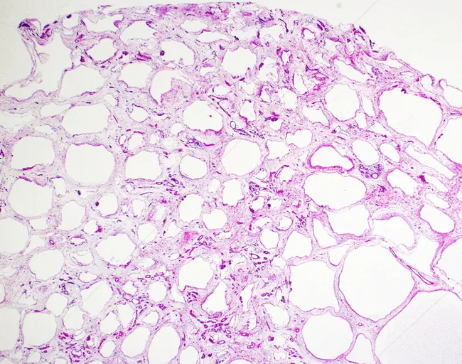
This histology image depicts white adipose tissue from subcutaneous hypodermis, prepared as a paraffin-embedded section and evaluated by bright-field light microscopy. The tissue demonstrates classic adipocyte lobules separated by delicate fibrovascular septa. Individual adipocytes appear as large, round to polygonal cells with abundant clear cytoplasm that contains a single, large lipid droplet, producing the characteristic empty-appearing space on hematoxylin–eosin staining. The nuclei are flattened and displaced to the periphery, creating a rim-like chromatin boundary. The extracellular matrix shows sparse collagenous septa and sparse vascular channels, with minimal inflammatory infiltrate. No cellular atypia or malignant features are evident. Overall, the architecture corresponds to normal white adipose tissue with relatively uniform adipocyte size, though physiologic variation in cell size can occur. This image is suitable as a morphologic reference for adipose tissue histology, comparisons in obesity-related studies, or teaching demonstrations of adipocyte biology and lipid storage. Clinically, white fat functions in energy storage, insulation, and endocrine signaling through adipokines; histology aids in evaluating conditions such as lipodystrophy, panniculitis, or lipomatosis when accompanied by clinical data. Keywords: adipocytes, unilocular, lipid droplet, peripherally displaced nucleus, H&E, hypodermis, subcutaneous fat. This description supports retrieval for educational, diagnostic, and research queries related to normal adipose histology and adipokine biology.
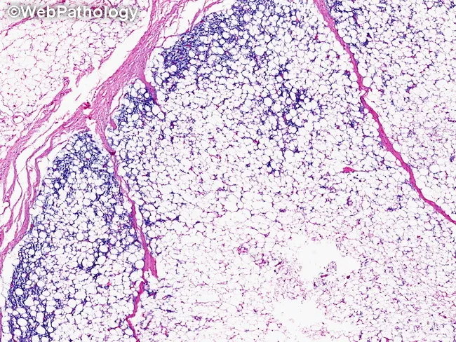
This is a hematoxylin and eosin (H&E) stained histology section of soft tissue lipoblastoma. The tissue is organized in lobules separated by delicate fibrous septa. Peripheral zones contain primitive mesenchymal cells with small oval to spindle nuclei, scant cytoplasm, and a high nuclear-to-cytoplasmic ratio, whereas central cores consist of mature adipocytes with clear lipid vacuoles. Lipoblasts with multivacuolated cytoplasm indent the nucleus, producing a scalloped silhouette. The observed zonal maturation pattern—immature cells at the periphery transitioning toward mature adipocytes centrally—supports a benign adipocytic neoplasm. The stroma may be variably myxoid or fibrous, and mitotic activity is not prominent. No overt cytologic atypia or necrosis is evident at this magnification. Overall architecture favors lipoblastoma, a pediatric-origin tumor of adipose tissue, frequently arising in subcutaneous soft tissue of limbs or trunk. Clinically, this histology helps differentiate from liposarcoma, where cellular atypia, mitotic activity, and dedifferentiation may be present, particularly in older patients. The image therefore has diagnostic relevance for surgical planning, prognosis, and longitudinal surveillance in pediatric patients, and it illustrates classic lipoblast maturation with peripheral immature cells and central mature adipocytes, a hallmark of this entity. Awareness of this pattern aids pathologists in reporting and guiding targeted excision with clear margins to guide therapy.
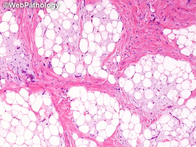
This histopathology image depicts a lipoblastoma arising in soft tissue, analyzed by light microscopy after Hematoxylin and Eosin staining. The specimen shows a lobulated adipocytic proliferation with irregular lobules separated by dense fibrous septa. Within each lobule, adipocytes are present in multiple maturation stages, ranging from primitive, stellate or spindle-shaped mesenchymal cells (preadipocytes) to multivacuolated lipoblasts and mature univacuolated adipocytes. Some lipoblasts exhibit signet-ring morphology with cytoplasmic lipid vacuoles pushing the nucleus to the periphery. The proportion of cells at each maturation stage varies both between lobules and within a given lobule, producing a heterogeneous histologic appearance. The fibrous septa may contain sparse spindle cells and delicate capillaries but show no overt cytologic atypia, mitotic figures, necrosis, or infiltrative invasion. This combination of lobulated architecture, lipoblast-rich adipose tissue, and absence of malignant features is characteristic of lipoblastoma, a benign pediatric adipocytic tumor. Clinically, these histologic features support diagnosis and guide management toward conservative excision with careful margin assessment to minimize recurrence. Differential considerations include myxoid liposarcoma in older patients or atypical lesions; molecular testing (e.g., PLAG1 rearrangement) can aid confirmation. Overall, the image illustrates classic histology with diagnostic significance for pediatric soft tissue tumors.
White Fat (White Adipose Tissue) in the Fetus
Origin and Cellular Lineage
- Histology: A Text and Atlas (Eroschenko/Ross), 17e
- Junqueira's Basic Histology, 17e
Timing of Appearance
| Gestational age | White fat as % body weight |
|---|---|
| First trimester | ~1% |
| 26-29 weeks | ~3.5% |
| 30-34 weeks | ~8% |
| Full term (38 weeks) | ~16% |
- The Developing Human: Clinically Oriented Embryology (Moore et al.), 11e
Stages of Lipoblast (Preadipocyte) Differentiation
-
Early lipoblasts - Look like fibroblasts; elongated with cytoplasmic processes, abundant RER and Golgi. Small lipid inclusions appear at one pole. A thin external lamina forms (distinguishing them from fibroblasts). Collections of these are called "primitive fat organs" - characterized by proliferating early lipoblasts and proliferating capillaries.
-
Midstage lipoblasts - Assume an oval shape. Vesicles and small lipid droplets accumulate around the nucleus. Glycogen particles appear at the periphery of droplets.
-
Late lipoblasts - Cells enlarge and become spherical. Small lipid droplets coalesce into one large lipid droplet that occupies the central cytoplasm. sER becomes prominent while rER decreases.
-
Mature adipocyte - The single large lipid droplet fills the entire cell, compressing the nucleus and cytoplasm into a thin peripheral rim - giving the characteristic "signet ring" appearance on H&E sections. Diameter can exceed 100 µm.
- Histology: A Text and Atlas (Eroschenko/Ross), 17e
Histological Structure of Mature White Adipocytes
- Large unilocular (single-droplet) cells, up to 100 µm in diameter
- Nucleus flattened and eccentrically displaced to one side
- In paraffin sections, the lipid is dissolved by solvents (xylene), leaving a delicate meshwork of polygonal profiles
- Capillaries lie at the angles where adjacent adipocytes meet
- Surrounded by reticular fibers (type III collagen) secreted by the adipocytes
- Also contain unmyelinated nerve fibers and numerous mast cells
Lipid Accumulation Mechanisms
- ~50% of increased body fat comes from increased lipid transport across the placenta (facilitated by rising fetal albumin)
- ~50% from de novo fatty acid synthesis in the fetal liver (stimulated by insulin acting on fetal hepatocytes)
- Insulin promotes lipogenesis and substrate availability (glucose, lactate)
- Medical Physiology (Boron & Boulpaep), 3e
Locations of White Fat Depots
- Subcutaneous fat (insulation, energy reserve)
- Visceral fat (around organs)
Functional Significance
- Energy reserve - stored triacylglycerols ensure adequate fuel for postnatal survival
- Thermal insulation - protects the newborn against heat loss
- Endocrine function - adipokine secretion (leptin, adiponectin, etc.) begins postnatally
Clinical Significance: Low Birth Weight / IUGR
- The Developing Human, 11e