
Glagine
insulin glargine mechanism of action pharmacology 2025
Insulin Glargine (Gla-rgine)
What It Is
Structural Modifications (Why It Works Differently)
- A21 substitution: Asparagine at position A21 on the A-chain is replaced with glycine
- B-chain addition: Two arginine residues are added to the C-terminus of the B-chain (positions B31 and B32)
- Katzung's Basic and Clinical Pharmacology, 16e, p. 1180
- Goodman & Gilman's, p. 1051
Pharmacokinetics
| Parameter | Glargine (U-100) |
|---|---|
| Onset | ~0.5-1 hour |
| Peak | No significant peak ("peakless") |
| Duration | ~24 hours |
| Injection | Subcutaneous only, once daily |
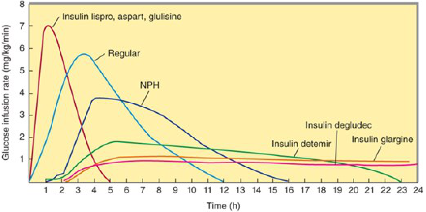
Mechanism of Action
- Promotion of glucose uptake into muscle and adipose tissue
- Inhibition of hepatic gluconeogenesis
- Promotion of glycogen synthesis and lipogenesis
- Inhibition of lipolysis and ketogenesis
Clinical Indications
- Type 1 Diabetes Mellitus (adults and children) - as basal insulin in combination with prandial insulin
- Type 2 Diabetes Mellitus (adults) - typically initiated when 2-3 oral antidiabetics fail to achieve glycemic control, or when HbA1c > 9% with symptoms
- Can be given at any time of day (consistent daily timing preferred); efficacy is equivalent regardless of time of administration
Formulations
| Formulation | Concentration | Brand |
|---|---|---|
| Glargine U-100 | 100 units/mL | Lantus, Basaglar |
| Glargine U-300 | 300 units/mL | Toujeo |
| Biosimilars | Available | Multiple |
Important Practical Points
- Cannot be mixed with other insulins - the acidic pH (4.0) of glargine will alter the pharmacodynamic profile of short-acting insulins formulated at neutral pH
- Injection site - absorption is independent of anatomic site (abdomen, thigh, upper arm are all equivalent)
- Splitting doses: Most patients use once daily; very insulin-sensitive patients (< ~20 units/day) or very insulin-resistant patients may benefit from twice-daily dosing
- Pregnancy: Systematic reviews support safety in pregnancy; provides stable basal insulin release throughout the day
Adverse Effects
| Effect | Notes |
|---|---|
| Hypoglycemia | Lower risk than NPH, especially overnight |
| Injection site reactions | Pain, lipodystrophy |
| Weight gain | Class effect of insulin therapy |
| Hypokalemia | Insulin shifts K+ intracellularly |
| Hypersensitivity | Rare |
Comparison with Other Basal Insulins
| Insulin | Mechanism of prolongation | Duration | Peak |
|---|---|---|---|
| NPH | Protamine-zinc complex | 10-20 h | Yes (6-7 h) |
| Glargine | pH precipitation / microcrystals | ~24 h | None |
| Detemir | Albumin binding (fatty acid) | 12-24 h | Minimal |
| Degludec | Multihexamer depot | 42+ h | None |
- Katzung's Basic and Clinical Pharmacology, 16th Ed., p. 1180
- Goodman & Gilman's Pharmacological Basis of Therapeutics, p. 1051
- Lippincott Illustrated Reviews: Pharmacology, p. 806
- Textbook of Family Medicine, 9th Ed.
Clinical Analysis: Severe Plantar Dermatosis
What the Image Shows
- Severe diffuse hyperkeratosis - markedly thickened, yellowish-brown, plaque-like stratum corneum covering the entire plantar surface
- Deep linear fissures cutting through thick plaques, especially over the metatarsal heads and lateral arch
- Coarse, adherent scaling with loss of normal skin lines
- Dark grey-black discoloration near the heel
- The distribution covers the entire plantar surface, heel, and lateral borders - the classic "moccasin" pattern
Most Likely Diagnosis: Moccasin-Type Tinea Pedis (Chronic Hyperkeratotic Tinea Pedis)
"Chronic hyperkeratotic (moccasin) type tinea pedis is characterized by patchy or diffuse scaling on the soles and the lateral and medial aspects of the feet, in a distribution similar to a moccasin on a foot."
- Diffuse hyperkeratosis, erythema, scaling, and fissures on the plantar surfaces
- Frequently chronic and difficult to cure
- Most common causative organism: Trichophyton rubrum, followed by Epidermophyton floccosum
- Can be completely asymptomatic or mildly pruritic
- Occasionally associated with immune deficiency (important to consider in this patient)
Differential Diagnoses
| Condition | Supporting Features | Against |
|---|---|---|
| Moccasin Tinea Pedis | Entire sole, hyperkeratosis, fissures, chronic | Need KOH/culture to confirm |
| Plantar Psoriasis | Thick yellowish plaques, fissures | Usually with nail changes, other sites involved |
| Severe Xerosis/Asteatotic Eczema | Dry skin, cracking, elderly patient | Usually less thick, diffuse |
| Palmoplantar Keratoderma | Diffuse hyperkeratosis | Usually inherited/genetic, bilateral symmetry |
| Contact Dermatitis (chronic) | Lichenification, scaling | History of exposure needed |
The Dark Spot on the Heel
- Talon noir (black heel) - harmless hemosiderin deposits from friction
- Deep bruising/old hematoma from the fissures
- Melanoma (acral lentiginous type) - rare but must be excluded if the lesion has irregular borders, is growing, or has unusual features. A dermatologist must assess this.
Treatment Plan
"Topical antifungal plus product with urea or lactic acid; may require oral antifungal therapy"
-
Keratolytic therapy first - to thin the thick plaque so antifungals can penetrate:
- Urea cream 20-40% or salicylic acid 5-10% applied daily to soften and remove hyperkeratotic scale
- Soaking feet in warm water before application helps
-
Topical antifungal (for milder/early cases):
- Terbinafine 1% cream twice daily for 2-4 weeks
- Alternatives: clotrimazole, miconazole, ciclopirox
-
Oral antifungal (strongly recommended for moccasin type due to poor topical penetration through thick scale):
- Terbinafine 250 mg once daily for 2-6 weeks (first-line)
- Itraconazole 200 mg/day for 2-4 weeks (alternative)
- Oral therapy is particularly indicated in diabetic or immunocompromised patients
-
Wound care for fissures:
- Keep clean and covered to prevent bacterial superinfection (cellulitis, Staphylococcus, Pseudomonas)
- Dilute acetic acid soaks if bacterial co-infection suspected
-
Prevent recurrence:
- Keep feet dry, avoid occlusive footwear
- Treat any concurrent onychomycosis (nail infection), as untreated nails are a reservoir for reinfection
- Antifungal powder in shoes
When to See a Doctor - Urgently
- The fissures are deep enough to risk bacterial superinfection / cellulitis - especially dangerous in diabetics or elderly patients
- The dark lesion on the heel requires dermoscopy or biopsy to exclude acral melanoma
- KOH preparation and fungal culture are needed to confirm the diagnosis
- The severity suggests this has been present for a long time and may need prescription-strength oral antifungals
⚠️ Important: This is a visual assessment only and not a formal medical diagnosis. Please see a dermatologist or podiatrist promptly, particularly given the fissuring (infection risk) and the dark discoloration on the heel.
Also diabetic
⚠️ HIGH RISK: Diabetic Foot - Urgent Attention Required
Why Diabetes Makes This Foot So Dangerous
- Bacterial infections (streptococcal, necrotizing fasciitis)
- Fungal infections (onychomycosis, candidal)
- Foot ulcers
- All driven by neuropathy, peripheral vascular disease, and immune dysfunction
The Fissures = Open Wounds in a Diabetic
"Diabetic foot infections occur contiguous to open skin wounds - ulceration, skin fissure, cut"
Risk Assessment
| Risk Factor | Present in This Patient? |
|---|---|
| Deep skin fissures (portal of entry) | ✅ YES - visible |
| Likely chronic fungal infection | ✅ YES - probable |
| Diabetes (impaired immunity) | ✅ YES - confirmed |
| Dark heel lesion (unknown) | ✅ YES - needs evaluation |
| Possible peripheral neuropathy | ⚠️ Unknown - must assess |
| Possible peripheral vascular disease | ⚠️ Unknown - must assess |
What Must Happen NOW - Urgency Level
🔴 Seek medical care within 24-48 hours (or sooner if any of the warning signs below appear)
- Redness spreading up the foot or leg (cellulitis)
- Swelling, warmth, or heat around the cracks
- Pus or discharge from the fissures
- Fever, chills, or feeling generally unwell
- Any new pain (or strangely, absence of expected pain)
- Black or discolored skin spreading (could indicate ischemia/gangrene)
Treatment Framework for This Diabetic Patient
Step 1 - Investigations (at the clinic)
- KOH scraping + fungal culture to confirm tinea pedis
- Blood glucose and HbA1c - assess glycemic control
- Neurological exam - monofilament test (10g Semmes-Weinstein) for peripheral neuropathy
- Vascular assessment - ankle-brachial index (ABI) for peripheral arterial disease
- Examine the dark heel lesion - dermoscopy to exclude melanoma or ischemic change
Step 2 - Treat the Fungal Infection Aggressively
"Oral antifungal therapy should be considered in diabetics, immunocompromised patients, and those with moccasin-type tinea pedis"
- Dermatology 2-Volume Set 5e
- Oral terbinafine 250 mg once daily for 2-6 weeks (first-line for tinea pedis in diabetics)
- Combined with topical antifungal (terbinafine 1% or clotrimazole cream) applied twice daily
- High-concentration urea cream (20-40%) as a keratolytic - softens thick scale and allows antifungal penetration; also acts as a moisturizer
Step 3 - Wound Care for Fissures
- Cleanse gently with mild soap and water daily
- Hydrocolloid dressings or liquid bandage over deep fissures to protect them and promote closure
- Watch for signs of infection at every dressing change
- Do NOT apply anything between the toes that keeps them moist (increases maceration risk)
Step 4 - Glycemic Control
Step 5 - Footwear and Prevention
- Wear closed, cushioned footwear - no flip-flops or bare feet
- Daily foot inspection - use a mirror for the sole; check between every toe
- Moisturize every day with urea or emollient cream (but never between the toes)
- See a podiatrist regularly for callus management (do not attempt to cut or file thick skin at home)
"Education regarding daily foot care that includes inspection for any scratch, blister or erythema, daily washing in warm water, appropriate footwear selection, skin moisturizing, and treatment of nail and foot fungal infection should be provided."
Referral Pathway
| Specialist | Why |
|---|---|
| GP/Endocrinologist | Urgent - HbA1c review, systemic antifungal prescription |
| Podiatrist | Debridement of hyperkeratosis, fissure management, orthotics |
| Dermatologist | Confirm diagnosis, assess dark heel lesion |
| Vascular surgeon | If ABI suggests peripheral arterial disease |
⚠️ Bottom line: In a diabetic patient, the deep fissures on this foot are open wounds. Left untreated, they can lead to cellulitis, deep tissue infection, and osteomyelitis - a chain of events that sometimes ends in amputation. This foot needs professional medical attention this week, not next month.
Pain present
Pain Present - Updated Clinical Assessment
The Good News About Pain
- Sensory neuropathy has not fully destroyed protective sensation yet
- The patient can feel that something is wrong
- This makes it more likely they will seek help - which is exactly the right response
Why This Pain Needs Assessment Now
| Type of Pain | Character | What It Means |
|---|---|---|
| Fissure/wound pain | Sharp, on pressure/walking, localised to crack | Open wound - portal for infection |
| Inflammatory pain | Throbbing, warmth, spreading redness | Early infection / cellulitis |
| Ischemic rest pain | Burning, constant even at night, worse lying down | Peripheral arterial disease - urgent vascular review |
| Neuropathic pain | Burning, tingling, "electric," worse at night | Diabetic neuropathy |
Where This Foot Sits on the IWGDF/IDSA Diabetic Foot Infection Scale
| Grade | Criteria | This Foot |
|---|---|---|
| 1 - Uninfected | No signs of infection | Possibly here... |
| 2 - Mild | Pain/tenderness + erythema ≤2cm + no systemic illness | Likely at minimum here |
| 3 - Moderate | Cellulitis >2cm, lymphangitis, deep tissue spread | Must rule out |
| 4 - Severe | Fever, chills, metabolic instability | Must rule out |
🔴 Action Plan - Revised for Painful Diabetic Foot
TODAY - Do These at Home (Temporary Measures Only)
- Keep weight off the foot as much as possible - every step drives bacteria deeper through fissures
- Gently clean the fissures with clean water and mild soap - pat dry (don't rub)
- Cover the cracks with a clean bandage or dressing - this reduces pain on walking and prevents contamination
- Do NOT apply any strong chemical (hydrogen peroxide, iodine, alcohol) directly to the fissures - these damage fragile diabetic tissue
- Check blood sugar - high glucose impairs healing and increases infection risk
- No tight footwear over the fissures
WITHIN 24 HOURS - See a Doctor
- Examine for signs of early cellulitis (redness, warmth, swelling spreading beyond the fissures)
- Check pedal pulses (dorsalis pedis, posterior tibial) - absence suggests arterial disease
- Monofilament test for neuropathy
- Blood tests: full blood count (WBC), CRP, blood glucose, HbA1c
- Prescribe oral antibiotics if any sign of infection - typically targeting Staphylococcus and Streptococcus (e.g., co-amoxiclav or cefalexin for mild infection)
- Prescribe oral terbinafine for the underlying fungal infection
- Consider X-ray of the foot if pain is bone-deep (to screen for osteomyelitis)
🚨 Go to Emergency Now If Any of These Are Present
- Spreading red line up the leg (lymphangitis)
- Fever or chills
- Pus coming from the fissures
- Black or bluish discoloration of any part of the foot
- Foot feels cold and numb compared to the other foot
- Severe pain that is getting rapidly worse
- You feel generally unwell, confused, or have a very high blood sugar
Bottom Line
Create a diabetic foot care checklist and warning signs poster
A professional medical patient education poster titled "DIABETIC FOOT CARE" with two main sections. TOP SECTION - "DAILY FOOT CARE CHECKLIST" with a green header and checkboxes listing: Inspect both feet top and bottom (use a mirror for the sole), Check between all toes for cracks, blisters, or redness, Wash feet in lukewarm water (test with elbow, NOT bare hand), Dry thoroughly especially between toes, Apply moisturizing cream to heels and soles (NOT between toes), Wear clean dry socks - no tight elastic, Wear closed protective footwear - never walk barefoot, Check inside shoes for foreign objects before wearing, Keep blood sugar controlled daily. BOTTOM SECTION - "WARNING SIGNS - SEE A DOCTOR TODAY" with a bold red header and warning symbols listing in red text: Deep cracks or fissures in skin, Any wound that is not healing, Spreading redness or warmth around a wound, Swelling or pus from any area, Black blue or dark discoloration, Burning tingling or electric pain at night, Cold foot or absent pulse, Fever or chills with a foot wound, Pain that is getting worse rapidly. A small footer in orange: "IN DIABETES - A SMALL WOUND CAN BECOME SERIOUS QUICKLY. NEVER IGNORE FOOT PAIN." Clean clinical layout, white background, easy to read large font, medical illustration style, suitable for a clinic waiting room.
404 Not Found. {'message': '{\n "error": {\n "code": 404,\n "message": "Publisher model `projects/engaged-beaker-474810-u2/locations/global/publishers/google/models/gemini-3-pro-image-preview` was not found or your project does not have access to it. Ensure you are using a valid model name and that the model is available in the specified region. For more information, see: https://docs.cloud.google.com/gemini-enterprise-agent-platform/resources/locations.",\n "status": "NOT_FOUND"\n }\n}\n', 'status': 'Not Found'}
We encountered an error while processing your request. Please try again or reach out to us for help.
Retty
A professional medical patient education poster for diabetic foot care. Clean white background with two clear sections. TOP HALF - green banner header "DIABETIC FOOT CARE - DAILY CHECKLIST" with a foot icon. Below in a clean list with green checkboxes: ✓ Inspect both feet daily - top, bottom, between toes ✓ Use a mirror to see the sole ✓ Wash in lukewarm water - test with elbow ✓ Dry thoroughly, especially between toes ✓ Moisturize heels and soles - never between toes ✓ Wear clean dry socks, no tight elastics ✓ Always wear closed protective footwear ✓ Check inside shoes before wearing ✓ Control blood sugar every day BOTTOM HALF - bold red banner "WARNING SIGNS - SEE A DOCTOR TODAY" with a red alert icon. Below in red text with warning symbols: ⚠ Deep skin cracks or fissures ⚠ Any wound not healing after 2 days ⚠ Spreading redness or warmth ⚠ Pus or discharge from any area ⚠ Black, blue or dark discoloration ⚠ Burning or electric pain at night ⚠ Cold foot or weak pulse ⚠ Fever or chills with a foot problem ⚠ Pain that is rapidly getting worse Orange footer box: "IN DIABETES - SMALL WOUNDS CAN BECOME SERIOUS FAST. NEVER IGNORE FOOT PAIN." Professional clinic poster style, large readable fonts, medical infographic design.
404 Not Found. {'message': '{\n "error": {\n "code": 404,\n "message": "Publisher model `projects/engaged-beaker-474810-u2/locations/global/publishers/google/models/gemini-3-pro-image-preview` was not found or your project does not have access to it. Ensure you are using a valid model name and that the model is available in the specified region. For more information, see: https://docs.cloud.google.com/gemini-enterprise-agent-platform/resources/locations.",\n "status": "NOT_FOUND"\n }\n}\n', 'status': 'Not Found'}
We encountered an error while processing your request. Please try again or reach out to us for help.