
Prepare a content for making a presentation about neck masses for ENT anfrom a standard ENT book
neck mass classification anatomy cervical lymph nodes levels
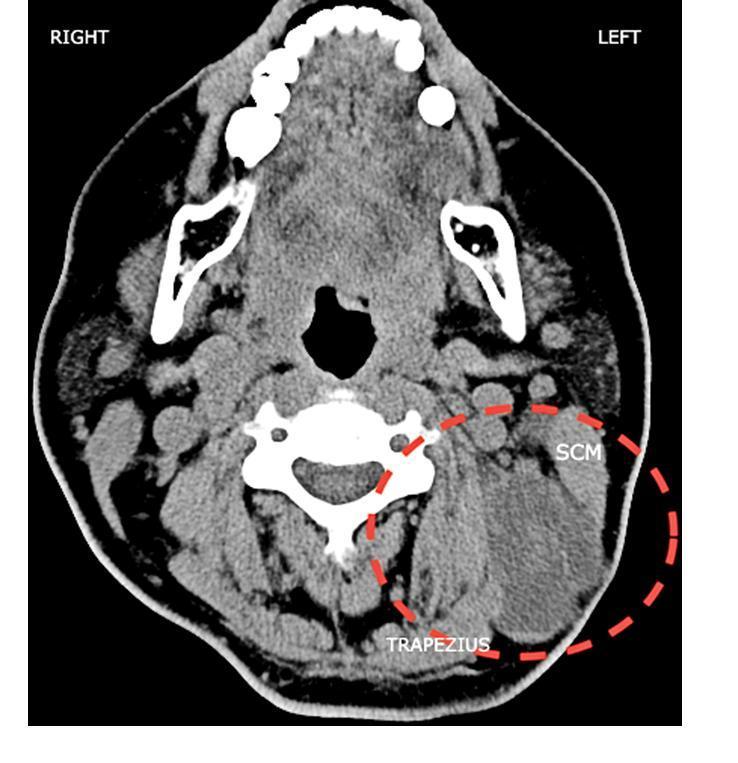
This diagnostic image is an axial computed tomography (CT) scan of the neck at the level of the upper cervical spine, illustrating significant cervical lymphadenopathy. The primary finding is a large, heterogeneous, soft-tissue mass in the left lateral neck, delineated by a red dashed circle. The mass measures approximately 6.8 x 5.1 cm and is situated within levels II, III, and V of the cervical lymph node stations. It is seen abutting the deep surface of the sternocleidomastoid (SCM) muscle and extending posteriorly toward the trapezius muscle, with poorly defined margins that suggest extracapsular extension or infiltration. In contrast, the right side of the neck demonstrates normal anatomical symmetry with smaller, discrete lymph nodes of uniform density. Clinical context indicates this mass represents metastatic squamous cell carcinoma originating from a scalp ulcer. The image highlights the use of cross-sectional imaging in staging head and neck malignancies and evaluating the relationship between metastatic nodes and vital musculoskeletal structures.
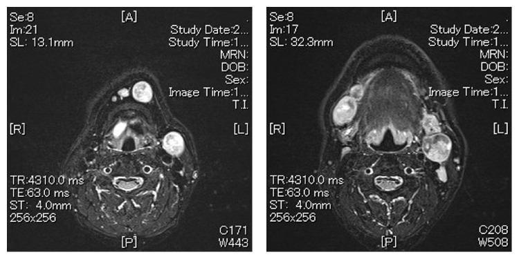
Two axial T2-weighted magnetic resonance imaging (MRI) slices of the neck at different anatomical levels demonstrate extensive bilateral cervical lymphadenopathy. The images show multiple enlarged, hyperintense (high signal intensity) rounded masses consistent with metastatic lymph nodes. In the superior slice, level Ia and right level Ib lymph nodes are prominently swollen, appearing as well-defined oval structures. In the inferior slice, a large heterogeneous hyperintense mass is visible at left level IIa, measuring approximately 37 mm, along with bilateral enlargement of level Ib nodes. These findings are consistent with metastatic spread from an oral cavity malignancy, specifically involving the submental (Level I) and upper jugular (Level II) chains. The images provide clinical evidence of nodal staging for head and neck squamous cell carcinoma (HNSCC), highlighting the use of MRI in assessing nodal size, signal characteristics, and anatomical distribution for treatment planning and surgical consideration.
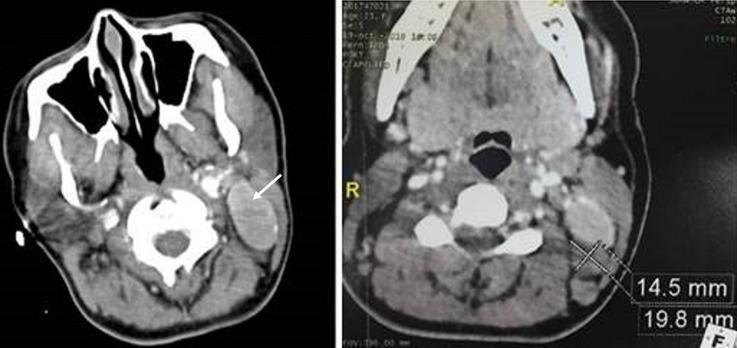
This diagnostic image consists of two axial cervical CT scan slices with intravenous contrast enhancement. The images demonstrate significant lymphadenopathy in the left lateral neck, specifically involving anatomical levels IIB and V. The first image (left) shows a large, well-circumscribed, oblong lymph node at level IIB, indicated by a white arrow, characterized by strong and relatively homogeneous enhancement compared to surrounding musculature. The second image (right) provides a quantitative assessment of a level V lymph node, measuring approximately 14.5 mm by 19.8 mm. Notable surrounding anatomy includes the cervical vertebrae, paraspinal muscles, and portions of the nasopharyngeal airway. The high degree of contrast enhancement in these enlarged lymph nodes is a key diagnostic feature, often associated with hypervascular conditions such as Castleman's disease or certain metastatic processes. This visual is used in medical education to illustrate radiographic neck level classification and the appearance of hypervascular lymphadenopathy.
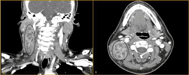
This diagnostic image is a contrast-enhanced computerized tomography (CT) scan of the neck presented in two planes: a coronal section (A) and a transverse (axial) section (B). Both views highlight a large, glomerate lymph node mass in the right cervical region, measuring approximately 4.2 x 7.4 cm, indicated by black ovals. The mass is characterized by its hypoattenuating appearance relative to surrounding musculature, containing multiple internal low-density areas consistent with central necrosis or cystic degeneration. Anatomically, the coronal view shows the mass extending vertically along the right lateral neck, superficial to the cervical spine. The transverse view demonstrates the mass situated lateral to the vertebral column and posterior to the oropharynx, maintaining relatively well-defined borders with adjacent soft tissues. This finding is characteristic of significant cervical lymphadenopathy, frequently seen in infectious processes like tuberculosis (scrofula) or metastatic malignancy. The image is intended for medical students and clinicians to learn the radiological presentation of necrotic lymph nodes and the relevant anatomy of the neck compartments.
thyroglossal duct cyst branchial cyst neck swelling
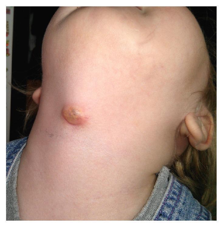
This clinical photograph displays a 2-year-old female patient with a prominent anterior neck swelling, characteristic of an infected thyroglossal duct cyst (TGDC). The lesion is located near the midline in the thyrohyoid region, slightly below the mandibular level. The swelling is approximately 1-2 cm in diameter, presenting as a raised, nodular mass with a circular to oval morphology. The surface exhibits a yellowish-brown crusting and erythema of the surrounding skin, suggesting an inflammatory or infectious process, such as an abscess or a draining fistula. This image serves as a clinical example of a common congenital midline neck mass in pediatric otolaryngology, illustrating the typical presentation of a thyroglossal duct cyst when secondary infection occurs. It is an essential visual reference for differential diagnosis of pediatric neck masses, distinguishing TGDC from other entities like dermoid cysts or lymphadenopathy based on its characteristic anatomical position.
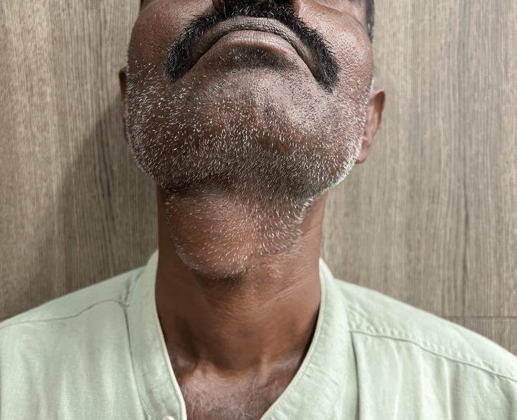
A clinical photograph of an adult male depicting a prominent midline neck mass, consistent with a thyroglossal duct cyst. The patient's neck is slightly extended, revealing a well-circumscribed, ovoid, and globular swelling located in the anterior midline of the neck, specifically at the level of the hyoid bone and inferior to the submental region. The overlying skin is intact and appears unremarkable, without signs of erythema, inflammation, ulceration, or fistulous tracts. This visual presentation is characteristic of a congenital thyroglossal duct anomaly, which typically manifests as a painless, mobile midline mass that often moves superiorly with tongue protrusion or swallowing. The image serves as a clinical example for Otolaryngology and General Surgery education, emphasizing the anatomical localization and physical characteristics necessary for the differential diagnosis of midline neck swellings, such as dermoid cysts or ectopic thyroid tissue.
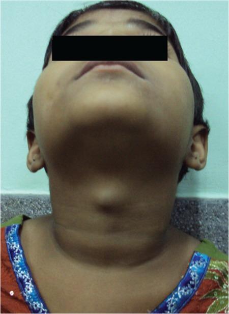
Clinical photograph of a pediatric patient's neck, viewed from an inferior-anterior perspective (looking up toward the chin). The image demonstrates a prominent, well-circumscribed, midline cystic swelling in the anterior neck, located superior to the thyroid cartilage. The overlying skin appears normal with no visible erythema, inflammation, or ulceration. The morphology is characteristic of a thyroglossal duct cyst, a common congenital abnormality occurring along the descent pathway of the thyroid gland. In this clinical context, such a mass often contains ectopic functional thyroid tissue and typically moves upward upon protrusion of the tongue or swallowing. The visual findings illustrate a classic presentation of a midline neck mass in childhood, serving as an educational example for differential diagnosis in pediatric otolaryngology and endocrinology, specifically regarding ectopic thyroid tissue and thyroglossal duct remnants.
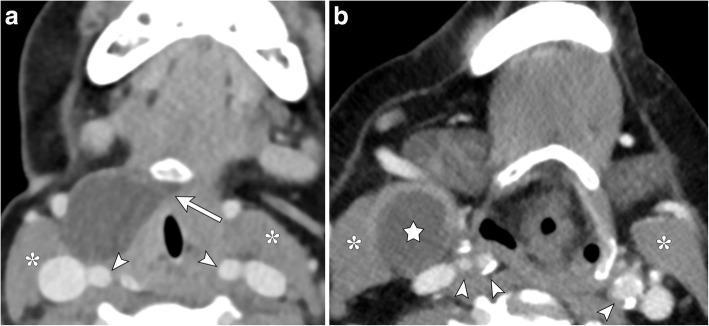
This dual-panel axial contrast-enhanced CT of the neck provides a side-by-side comparison of two distinct congenital cystic neck masses. Image A illustrates a thyroglossal duct cyst presenting as a well-circumscribed, hypoattenuating mass. It is located anterior to the carotid sheath vessels (indicated by arrowheads) and anteromedial to the sternocleidomastoid muscle (indicated by asterisks). A diagnostic 'tail sign' or medial extension (arrow) toward the hyoid bone is visible, characteristic of thyroglossal duct remnants. Image B demonstrates a second branchial cleft cyst (indicated by a star) in a similar lateral neck position. This lesion is rounded, thin-walled, and fluid-filled, located adjacent to the right sternocleidomastoid muscle (asterisks) and lateral to the carotid space. Unlike the thyroglossal duct cyst, the branchial cleft cyst lack a medial extension to the hyoid bone. Both images emphasize the importance of anatomical landmarks, such as the relationship to the carotid vessels and the presence of a medial tract, in differentiating these common pediatric and young-adult neck pathologies.
NECK MASSES
Presentation Content for ENT
SLIDE 1 - TITLE SLIDE
- Department of Otorhinolaryngology - Head & Neck Surgery
- Source: Scott-Brown's & Cummings Otolaryngology
SLIDE 2 - OVERVIEW / TABLE OF CONTENTS
- Introduction and Classification
- Anatomical Framework - Neck Levels
- Clinical Assessment
- Congenital Neck Masses
- Inflammatory/Infective Neck Masses
- Neoplastic Neck Masses
- Investigation Approach
- Management Principles
SLIDE 3 - INTRODUCTION
- Neck masses are among the most common presenting complaints in ENT
- Aetiology is strongly age-dependent: congenital in neonates/children, inflammatory in young adults, neoplastic in older adults
- The "Rule of 80s" (adults >40 years): 80% of non-thyroid neck masses are neoplastic; 80% of those are malignant; 80% of those malignancies are metastatic
| Category | Examples |
|---|---|
| Congenital | Thyroglossal cyst, branchial cyst, dermoid cyst, vascular malformations |
| Inflammatory | Reactive lymphadenopathy, TB, NTM, cat scratch disease |
| Neoplastic | Metastatic SCC, lymphoma, salivary gland tumors, thyroid cancer |
SLIDE 4 - ANATOMICAL FRAMEWORK: NECK LEVELS
| Level | Group | Anatomical Boundaries |
|---|---|---|
| IA | Submental | Between anterior bellies of digastric, above hyoid |
| IB | Submandibular | Anterior belly of digastric to posterior border of submandibular gland |
| II (A & B) | Upper Jugular | Skull base to hyoid; divided by accessory nerve |
| III | Mid Jugular | Hyoid to inferior border of cricoid |
| IV | Lower Jugular | Inferior cricoid to clavicle |
| V (A & B) | Posterior Triangle | Posterior SCM to anterior trapezius; divided at inferior cricoid |
| VI | Anterior Compartment | Between common carotid arteries; hyoid to sternum |
| VII | Superior Mediastinum | Below sternal notch |
SLIDE 5 - ANATOMICAL LOCATIONS AND DIFFERENTIAL DIAGNOSIS
By Location:
- Lymph nodes (most common) - along superficial and deep cervical chains
- Branchial cleft cysts
- Vascular malformations, haemangiomas
- Neural/connective tissue tumors
- Metastatic lymph node deposits
- Thyroglossal duct cysts (most common)
- Lymph nodes
- Dermoid/epidermoid cysts
- Thyroid disease (inflammatory or neoplastic)
- Salivary gland disease, sialadenitis
- Lymphadenopathy (reactive or neoplastic)
- Vascular malformations, plunging ranula
- Lymph nodes (most common)
- Branchial anomalies
- Vascular malformations
- Neoplasia
SLIDE 6 - CLINICAL HISTORY
- Age: Most acute lymphadenitis > 6 months. Neonatal masses are more likely pathological/neoplastic
- Duration: Short history (days) = acute inflammation. >6 weeks = chronic, warrants investigation
- Size: Very large or progressively enlarging swellings despite antibiotics = investigate
- Associated Symptoms:
- Acute: Preceding URTI, fever, rhinorrhoea, sore throat
- Chronic: Weight loss, night sweats, chronic cough, distant lymphadenopathy
- Exposure History: TB contacts, cats (cat scratch disease), farm animals/ticks, travel history
- Medications: Carbamazepine, phenytoin, isoniazid can cause lymphadenopathy
- Family History: Congenital anomalies, familial disease
SLIDE 7 - CLINICAL EXAMINATION
- Site (level, midline vs. lateral, depth)
- Size, shape, surface
- Consistency: soft, firm, hard, fluctuant
- Fixity: mobile vs. tethered vs. fixed
- Tenderness, warmth, overlying skin changes
- Pulsatile or compressible (vascular lesions)
- Moves on swallowing + tongue protrusion = Thyroglossal cyst
- Moves on swallowing only = Thyroid swelling
- Classical "cold abscess" (no signs of acute inflammation) = Tuberculous lymphadenopathy
- Fluctuance = Abscess formation
- Head and neck: pharynx, nose, ears, scalp - search for primary infection/tumor
- General: fever, rash, hepatosplenomegaly, distant lymphadenopathy
SLIDE 8 - RED FLAGS (Cummings, Box 204.1)
- Age < 12 months
- Lymph node is non-tender and hard
- Diameter > 3 cm
- Supraclavicular location (Virchow's node - left supraclavicular = Troisier's sign)
- Persistent generalized lymphadenopathy
- Associated mediastinal or abdominal mass
- Persistent unexplained: pruritus, fever, weight loss, pallor, fatigue, petechiae, haemorrhagic lesions, hepatosplenomegaly
KEY POINT (Scott-Brown's): In patients over the age of 35, cystic neck lumps should be considered metastatic unless proven otherwise - usually HPV-related head and neck squamous cell carcinoma.
SLIDE 9 - CONGENITAL NECK MASSES: THYROGLOSSAL DUCT CYST
- Thyroid primordia migrates from foramen caecum (base of tongue) down along thyroglossal duct to lower anterior neck
- Duct normally involutes at 8th-10th week of gestation
- If persistence occurs → cyst formation from epithelial lining secretions
- Tract is intimately related to the hyoid bone (passes through/around it)
- ~95% present as asymptomatic cystic midline mass at/near hyoid bone
- Moves on swallowing AND tongue protrusion (pathognomonic)
- No sex preponderance; mean age ~5 years (range: 4 months to old age)
- Location: 75% midline, 10% off-midline (95% of these on the left)
- 65% below hyoid; 15% at hyoid level; 5% above
- Up to 5% present as acute inflammatory episode
- 15% may have associated discharging fistula
SLIDE 10 - TDC: IMAGING & TREATMENT
- Homogenous anechoic mass with posterior wall enhancement
- Pseudosolid appearance (proteinaceous secretions)
- Heterogeneous echo (haemorrhage/infection)
- Key imaging points: relationship to hyoid, presence of normal thyroid tissue, any solid component
- Invariably high T2 signal; variable T1 depending on proteinaceous content
- Always confirm presence of normal thyroid before surgery!
- Sistrunk's procedure = surgery of choice
- Excision of cyst + central portion of hyoid body + tissue tract to foramen caecum
- Recurrence rate with adequate Sistrunk's: ~8%
- Failure to remove hyoid body = main cause of recurrence
SLIDE 11 - CONGENITAL NECK MASSES: BRANCHIAL CYSTS
- Derived from branchial apparatus (arches form 3rd-8th week of gestation)
- Second branchial cleft cysts account for 95% of all branchial cysts
- Account for up to 19% of all pediatric cervical masses
- Peak incidence: 2nd-3rd decade (young adults)
- Lined by stratified squamous epithelium (90%); rarely respiratory epithelium (10%)
- 80% have lymphoid tissue in outer wall
- Contain straw-coloured fluid with cholesterol crystals
- Branchial apparatus theory (remnants of pouches/clefts)
- Cervical sinus theory (remnants of cervical sinus of His)
- Thymopharyngeal duct theory
- Inclusion theory (epithelial inclusions in lymph nodes - most widely accepted)
- Presents as mass at angle of mandible, superficial to carotid sheath
- Posterior to submandibular gland, along anteromedial border of SCM
- 60% on the left side
- Soft, compressible, non-tender unless infected
- May present with acute infection and abscess
SLIDE 12 - BRANCHIAL FISTULAE AND SINUSES
- Complete tract from pharynx to neck skin
- 2nd arch fistula: internal opening at anterior tonsillar pillar; external opening at anterior border of SCM at junction of middle and lower thirds; passes between internal and external carotid arteries
- External opening only, along anterior border of SCM from tragus to sternoclavicular joint
- Complete surgical excision
- Identify and preserve: marginal branch of facial nerve, vagus, accessory, and hypoglossal nerves
- No need to routinely look for internal tract during branchial cyst excision
SLIDE 13 - OTHER CONGENITAL MASSES
| Mass | Key Features |
|---|---|
| Dermoid cyst | Submental/suprasternal, midline; contains skin appendages (sebaceous glands, hair); fatty content pathognomonic on imaging |
| Epidermoid cyst | Similar location; entirely ectodermal; often present in infancy |
| Ranula | Retention cyst of sublingual gland; simple = floor of mouth; plunging ranula = extends through mylohyoid into submandibular space ("diving ranula") |
| Lymphatic malformation (cystic hygroma) | Multilocular cystic mass; often posterior triangle; high T2 on MRI; mostly present at birth; 90% by age 2 |
| Lipoma | Feathery/striped on US; low attenuation on CT; high T1/T2 on MRI |
| Nerve sheath tumors | Schwannoma, neurofibroma; mimic lymph nodes on US; displace carotid vessels rather than distort them |
SLIDE 14 - INFLAMMATORY/INFECTIVE NECK MASSES
- EBV (Infectious Mononucleosis): Fever, exudative tonsillitis, massive cervical lymphadenopathy, hepatosplenomegaly; atypical lymphocytes on blood film; Monospot/Paul-Bunnell positive; Ampicillin/Amoxicillin contraindicated (causes rash)
- Adenovirus, Rhinovirus, Enterovirus: Reactive lymphadenopathy; self-limiting
- HIV: Persistent generalized lymphadenopathy; small, symmetrical, non-tender nodes; follicular hyperplasia on biopsy
- Acute cervical lymphadenitis: Most commonly Staph. aureus, Strep. pyogenes; tender, warm, unilateral; may progress to abscess
- Tuberculosis: Scrofula; "cold abscess" (no acute inflammatory signs); calcification on X-ray; Mantoux/IGRA testing
- Non-tuberculous mycobacteria (NTM): Common in toddlers; especially parotid region; NTM, unlike TB, often causes skin color change (violaceous)
- Cat scratch disease (Bartonella henselae): Tender regional lymphadenopathy following cat scratch; serology confirmatory
- Toxoplasmosis: Posterior triangle nodes; serological diagnosis
SLIDE 15 - NEOPLASTIC NECK MASSES
Metastatic Disease (Most common in adults):
- Primary sites by level:
- Level I: Lip, floor of mouth, anterior tongue, submandibular gland
- Level II: Oropharynx, nasopharynx, oral cavity, parotid
- Level III: Oral cavity, oropharynx, larynx, hypopharynx
- Level IV: Larynx, hypopharynx, thyroid, cervical esophagus
- Level V: Nasopharynx, oropharynx, scalp (posterior)
- Level VI: Thyroid, larynx, cervical esophagus
Lymphoma:
- Most common pediatric head and neck malignancy
- Hodgkin lymphoma: Bimodal distribution; cervical nodes most common
- Non-Hodgkin lymphoma: More common in adults; systemic features prominent
- Both present as painless, rubbery, non-tender cervical lymphadenopathy
Primary Thyroid Carcinoma:
- Midline/paratracheal mass
- Often associated with a primary thyroid nodule
- Fine needle aspiration cytology (FNAC) is first-line investigation
SLIDE 16 - INVESTIGATIONS
Step-by-Step Investigation Algorithm:
- FBC: Neutrophilia (bacterial), lymphocytosis (viral/lymphoma), pancytopenia (haematological malignancy)
- CRP/ESR: Raised in bacterial infection, lymphoma
- Monospot/Paul-Bunnell: EBV
- Serology: Toxoplasma, Bartonella, CMV, HIV
- Mantoux/IGRA: Tuberculosis
- LDH: Lymphoma activity marker
- TFTs: Thyroid masses
| Modality | Role |
|---|---|
| Ultrasound (US) | First-line; differentiates cystic vs. solid; assesses internal architecture; guides FNAC; benign criteria: echogenic hilum, oval shape, L/T ratio >2 |
| CT neck (contrast) | Nodal staging; abscess mapping; surgical planning; identifies calcification |
| MRI | Vascular malformations, salivary gland masses, soft tissue tumors; not ideal for acute inflammation |
| PET-CT | Unknown primary search; staging lymphoma/metastatic disease |
| Chest X-ray | Mediastinal involvement; tuberculosis |
- FNAC: First-line; accuracy >90%; can be performed under US guidance; HPV/EBV testing on aspirate for unknown primary
- Core biopsy: For lymphoma subtyping; when anaplastic carcinoma suspected
- Open biopsy / Excision biopsy: For lymphoma when core biopsy non-diagnostic; last resort (risk of field contamination)
SLIDE 17 - INVESTIGATION: IMAGING EXAMPLES
SLIDE 18 - MANAGEMENT PRINCIPLES
Congenital Masses:
- Thyroglossal Duct Cyst: Sistrunk's procedure (excision + central hyoid + suprahyoid tract to foramen caecum)
- Branchial Cyst: Complete surgical excision; protect cranial nerves (VII marginal, hypoglossal, accessory, vagus)
- Lymphatic Malformation: Sclerotherapy (OK-432, bleomycin), surgical excision for macro-cystic forms
- Ranula (plunging): Excision including sublingual gland to prevent recurrence
Inflammatory Masses:
- Acute bacterial lymphadenitis: IV antibiotics (Staph./Strep. cover); surgical drainage if abscess forms
- EBV: Supportive; steroids for airway obstruction; avoid ampicillin/amoxicillin
- TB: RNTCP/standard anti-TB therapy (4-drug regimen); surgical excision rarely needed
- NTM: Surgical excision (curettage or complete excision) preferred over antibiotics alone
Neoplastic Masses:
- Metastatic SCC: Neck dissection (selective or modified radical); concurrent chemoradiation
- Lymphoma: Chemotherapy ± radiotherapy; MDT essential for subtype-directed therapy
- Thyroid carcinoma: Thyroidectomy + level VI dissection; selective lateral neck dissection for metastatic nodes
SLIDE 19 - NECK DISSECTION OVERVIEW (Cummings, Chapter 118)
| Type | Structures Removed | Indication |
|---|---|---|
| Radical Neck Dissection (RND) | Levels I-V + SCM + IJV + CN XI | Extensive disease; largely historical |
| Modified Radical Neck Dissection (MRND) | Levels I-V; preserves ≥1 of: SCM, IJV, CN XI | N+ neck; standard in most cases |
| Selective Neck Dissection (SND) | Selected levels based on primary site | N0 neck; limited nodal metastasis |
| Extended Neck Dissection | Adds non-lymphatic structures (carotid, skin, paraspinal muscles) | Advanced disease with ENE |
SLIDE 20 - SPECIAL SITUATIONS
Unknown Primary with Neck Metastasis:
- 3-5% of all head and neck malignancies
- Work-up: PET-CT + panendoscopy + directed biopsies + tonsillectomy
- HPV/EBV testing on nodal FNAC guides investigation toward oropharynx/nasopharynx
- Treatment: Definitive neck treatment ± bilateral mucosal irradiation
Paediatric Neck Mass - Special Considerations (Cummings, Table 204.2):
- Syndromes with increased neck malignancy risk: Down syndrome (leukemia), NF-1/2, Li-Fraumeni syndrome, MEN-2a/2b (medullary thyroid carcinoma + pheochromocytoma), Gorlin syndrome, Beckwith-Wiedemann syndrome
- Lymphoma = 3rd most common childhood malignancy overall; most common head and neck pediatric malignancy
HPV-related Cystic SCC:
- Rising incidence in younger patients (30s-40s)
- Presents as cystic lateral neck mass at Level II
- Often without obvious primary on routine examination
- p16 immunostaining on FNAC; PET-CT + transoral robotic surgery (TORS) biopsy of base of tongue/tonsillar tissue
SLIDE 21 - SUMMARY: CLINICAL APPROACH FLOWCHART
NECK MASS
    |
    ├── Midline?
    |       ├── Moves with swallowing + tongue protrusion → THYROGLOSSAL CYST
    |       ├── Moves with swallowing only → THYROID
    |       └── Fixed, fluctuant → DERMOID / ABSCESS
    |
    └── Lateral?
            ├── Age < 15 → Likely CONGENITAL or INFLAMMATORY
            |       ├── Tender, warm → REACTIVE LYMPHADENOPATHY / ABSCESS
            |       └── Soft, cystic, angle of jaw → BRANCHIAL CYST
            |
            ├── Age 15-40 → LYMPHOMA / INFLAMMATORY / CONGENITAL
            |
            └── Age > 40 → METASTATIC until proven otherwise
                    ├── US + FNAC → FNAC HPV/EBV testing
                    ├── CT/PET-CT for staging
                    └── Panendoscopy for unknown primary
SLIDE 22 - KEY LEARNING POINTS
- Location is diagnosis - midline = congenital or thyroid; lateral = lymph node (most common); posterior triangle = lymph node or branchial anomaly
- Age determines probability - inflammatory in children, metastatic in adults > 40
- >35 years + cystic lateral mass = HPV-related cystic SCC until proven otherwise
- Cold abscess = tuberculosis (no acute inflammatory signs)
- Sistrunk's procedure = treatment for TDC (must remove central hyoid bone)
- FNAC > open biopsy - open biopsy before full workup risks compromising oncological surgery
- Physical examination sensitivity 81%, specificity 77% - always supplement with US + FNAC
- Red flags (hard, fixed, supraclavicular, >3 cm, constitutional symptoms) = urgent malignancy workup
- Ampicillin/Amoxicillin contraindicated in EBV mononucleosis
- Lymphoma = most common head and neck malignancy in children
SLIDE 23 - REFERENCES
- Scott-Brown's Otorhinolaryngology Head & Neck Surgery, 9th Ed. - Chapter 36 (Clinical Examination of the Neck), Chapter 37 (Imaging of the Neck), Chapter 18 (Assessment of Cervical Lymphadenopathy)
- Cummings Otolaryngology Head and Neck Surgery, 7th Ed. - Chapter 118 (Neck Dissection), Chapter 204 (Neck Masses in Children)
- Scott-Brown's Vol 2 - Chapter 37 (Paediatric Neck Masses: Clinical Assessment)
- Scott-Brown's Vol 1 - Cervical Lymphadenopathy in HIV
Total Slides: 23 | Ready for PowerPoint conversion | All content sourced from Scott-Brown's Otorhinolaryngology and Cummings Otolaryngology (standard ENT references)