Duct ectasia

Reading File
Finding Sources
Finding Sources
Searching Images

mammary duct ectasia breast histology

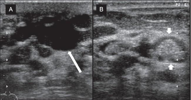
This diagnostic ultrasound image demonstrates chronic duct ectasia in the breast parenchyma, presented in two planes. Image A (longitudinal view) shows a significantly dilated, tubular, and elongated duct. The lumen is predominantly filled with anechoic fluid, but also contains focal areas of increased echogenicity (white arrow) representing inspissated debris. Image B (cross-sectional view) displays the same pathology where the intraductal debris appears as a rounded, focal lesion with heterogeneous echogenicity (white arrowheads). The lesion is contained within the margins of the ductal structure and is surrounded by typical breast parenchymal echotexture. These visual features are characteristic of mammary duct ectasia, where accumulated secretions and cellular debris can sometimes mimic solid masses. The modality is high-resolution gray-scale ultrasonography, used here to distinguish between simple cysts, solid masses, and benign ductal dilation with internal echoes.
This diagnostic ultrasound image demonstrates chronic duct ectasia in the breast parenchyma, presented in two planes. Image A (longitudinal view) shows a significantly dilated, tubular, and elongated duct. The lumen is predominantly filled with anechoic fluid, but also contains focal areas of increased echogenicity (white arrow) representing inspissated debris. Image B (cross-sectional view) displays the same pathology where the intraductal debris appears as a rounded, focal lesion with heterogeneous echogenicity (white arrowheads). The lesion is contained within the margins of the ductal structure and is surrounded by typical breast parenchymal echotexture. These visual features are characteristic of mammary duct ectasia, where accumulated secretions and cellular debris can sometimes mimic solid masses. The modality is high-resolution gray-scale ultrasonography, used here to distinguish between simple cysts, solid masses, and benign ductal dilation with internal echoes.

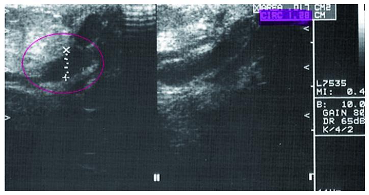
This diagnostic image consists of two side-by-side breast ultrasonography panels demonstrating mammary ductal ectasia. On the left panel, a pink circle highlights a dilated, elongated, and hypoechoic (dark) fluid-filled structure, characteristic of an enlarged lactiferous duct. Calipers are visible within the circle, measuring the transverse diameter of the duct, which appears to contain minor internal echogenic foci or debris. The right panel provides a longitudinal view of the same or a continuous dilated duct, showing its tubular morphology and well-defined but irregular borders against the surrounding heterogeneous, more echogenic fibroglandular stroma. Annotations on the right margin include ultrasound settings (gain, mechanical index) and measurement data. The image serves as a clinical example of benign ductal changes often evaluated in the context of nipple aspirate fluid (NAF) collection or BI-RADS assessment for inconclusive mammographic findings.
This diagnostic image consists of two side-by-side breast ultrasonography panels demonstrating mammary ductal ectasia. On the left panel, a pink circle highlights a dilated, elongated, and hypoechoic (dark) fluid-filled structure, characteristic of an enlarged lactiferous duct. Calipers are visible within the circle, measuring the transverse diameter of the duct, which appears to contain minor internal echogenic foci or debris. The right panel provides a longitudinal view of the same or a continuous dilated duct, showing its tubular morphology and well-defined but irregular borders against the surrounding heterogeneous, more echogenic fibroglandular stroma. Annotations on the right margin include ultrasound settings (gain, mechanical index) and measurement data. The image serves as a clinical example of benign ductal changes often evaluated in the context of nipple aspirate fluid (NAF) collection or BI-RADS assessment for inconclusive mammographic findings.

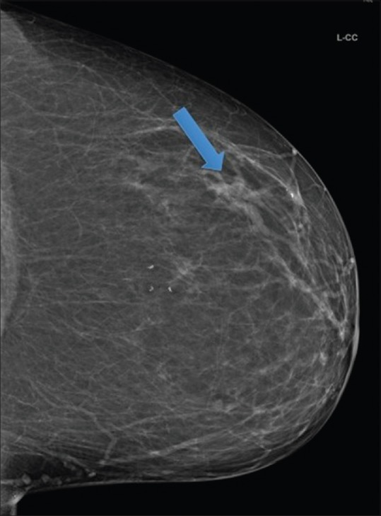
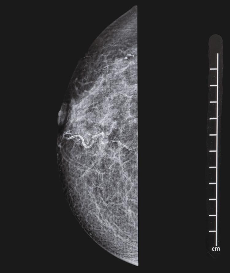
**Imaging Modality:** Diagnostic mammography.

**Anatomical Region:** Left breast, Craniocaudal (L-CC) view.

**Observed Pathology:** Duct ectasia/dilated mammary ducts.

**Characteristic Visual Features:** The image displays a focal area of prominent, branching tubular radiodensities located within the outer hemisphere of the breast parenchyma. These structures originate from the retroareolar region and extend peripherally, consistent with dilated lactiferous ducts. A blue arrow highlights the primary site of these tubular densities. The surrounding breast tissue appears predominantly fatty-replaced with scattered fibroglandular densities (BI-RADS Breast Composition Category B). No suspicious microcalcifications, speculated masses, or significant architectural distortion are visualized in the immediate vicinity of the dilated ducts.

**Key Diagnostic Features:** Branching tubular patterns in a ductal distribution, absence of associated solid mass or suspicious pleomorphic calcifications, and localized manifestation in the outer quadrant. These findings are characteristic of benign ductal dilatation or ectasia.
**Imaging Modality:** Diagnostic mammography. **Anatomical Region:** Left breast, Craniocaudal (L-CC) view. **Observed Pathology:** Duct ectasia/dilated mammary ducts. **Characteristic Visual Features:** The image displays a focal area of prominent, branching tubular radiodensities located within the outer hemisphere of the breast parenchyma. These structures originate from the retroareolar region and extend peripherally, consistent with dilated lactiferous ducts. A blue arrow highlights the primary site of these tubular densities. The surrounding breast tissue appears predominantly fatty-replaced with scattered fibroglandular densities (BI-RADS Breast Composition Category B). No suspicious microcalcifications, speculated masses, or significant architectural distortion are visualized in the immediate vicinity of the dilated ducts. **Key Diagnostic Features:** Branching tubular patterns in a ductal distribution, absence of associated solid mass or suspicious pleomorphic calcifications, and localized manifestation in the outer quadrant. These findings are characteristic of benign ductal dilatation or ectasia.

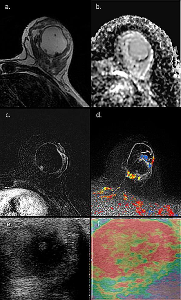
This composite diagnostic image illustrates a case of mammary duct ectasia in a 40-year-old lactating patient, presented through MRI and ultrasound (US) modalities. Panel (a) shows an axial T2-weighted MRI where a large, well-defined cystic structure exhibits high signal intensity. Panel (b) presents the corresponding Apparent Diffusion Coefficient (ADC) map, indicating a lack of internal diffusion restriction within the cyst. Panels (c) and (d) utilize Dynamic Contrast-Enhanced (DCE) and Computer-Aided Diagnosis (CAD) mapping, respectively, revealing distinct rim enhancement along the thick cystic walls amidst background parenchymal enhancement typical of lactation. Panel (e) shows a gray-scale ultrasound image of the 4 cm mass, characterized by thick boundaries and mixed internal echogenicity. Panel (f) provides a static ultrasound elastography map demonstrating significant tissue stiffness throughout the lesion. Together, these images highlight the morphological mimics of malignancy, such as inflammatory breast cancer or abscess, which require US-guided biopsy to confirm the benign diagnosis of duct ectasia in the clinical context of a palpable lactating breast mass.

This composite diagnostic image illustrates a case of mammary duct ectasia in a 40-year-old lactating patient, presented through MRI and ultrasound (US) modalities. Panel (a) shows an axial T2-weighted MRI where a large, well-defined cystic structure exhibits high signal intensity. Panel (b) presents the corresponding Apparent Diffusion Coefficient (ADC) map, indicating a lack of internal diffusion restriction within the cyst. Panels (c) and (d) utilize Dynamic Contrast-Enhanced (DCE) and Computer-Aided Diagnosis (CAD) mapping, respectively, revealing distinct rim enhancement along the thick cystic walls amidst background parenchymal enhancement typical of lactation. Panel (e) shows a gray-scale ultrasound image of the 4 cm mass, characterized by thick boundaries and mixed internal echogenicity. Panel (f) provides a static ultrasound elastography map demonstrating significant tissue stiffness throughout the lesion. Together, these images highlight the morphological mimics of malignancy, such as inflammatory breast cancer or abscess, which require US-guided biopsy to confirm the benign diagnosis of duct ectasia in the clinical context of a palpable lactating breast mass.

Reading File
Reading File
Reading File
Loading Image
Finding Sources
I now have comprehensive information from multiple authoritative textbooks. Here is a full review of duct ectasia:

Duct Ectasia (Mammary Duct Ectasia)

Definition

Duct ectasia is the dilation of the major (subareolar) milk ducts, classified as a disorder of involution and part of the spectrum of Aberrations of Normal Development and Involution (ANDI). The dilated ducts fill with inspissated secretions and debris, triggering periductal inflammation and progressive fibrosis.

Epidemiology

  • Most common in the 5th-6th decade of life, predominantly in multiparous women
  • Affects peri- and post-menopausal women most often
  • Not associated with cigarette smoking (this distinguishes it from squamous metaplasia of lactiferous ducts, which is smoking-related)

Pathogenesis

  1. Major milk ducts become abnormally dilated (>3 mm diameter)
  2. Stagnant secretions and debris accumulate ("inspissated secretions")
  3. Debris acts as a chemical irritant -> periductal inflammation
  4. Duct rupture releases contents -> chronic inflammatory reaction (lymphocytes, macrophages, plasma cells, and sometimes granulomas around cholesterol deposits)
  5. Progressive periductal fibrosis -> nipple retraction and palpable mass

Clinical Features

FeatureDetails
Nipple dischargeThick, white ("toothpaste-like"), brown, green, or mud-coloured; the characteristic sign
Palpable massPeriareolar, firm, irregular
Nipple retractionDue to duct foreshortening from fibrosis; may appear slit-like
Pain/erythemaUncommon
Skin changesRetraction possible from fibrosis (mimics carcinoma)
Key clinical point: The clinical and radiological findings of duct ectasia can closely mimic invasive carcinoma - this is its principal clinical significance.

Histopathology (Morphology)
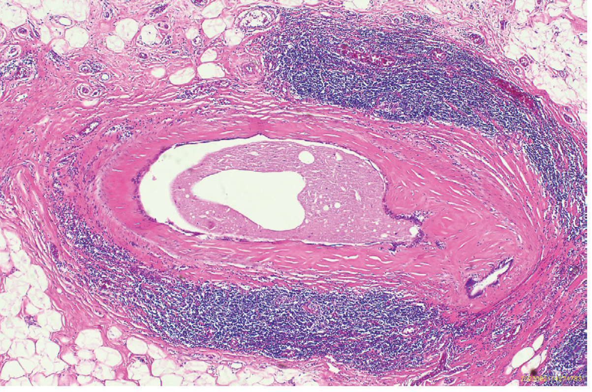
Duct ectasia histology - an ectatic duct filled with inspissated debris surrounded by dense chronic inflammation and fibrosis (Robbins Pathology, Fig. 23.4)
Fig. 23.4 from Robbins: Ectatic duct filled with inspissated debris, surrounded by a dense lymphocytic infiltrate and fibrosis. The fibrotic response can produce a firm, irregular mass mimicking invasive carcinoma.
Key microscopic features:
  • Dilated ducts filled with inspissated secretions and lipid-laden macrophages
  • Periductal chronic inflammation - lymphocytes, macrophages, plasma cells
  • Granulomas may form around cholesterol deposits
  • Periductal fibrosis producing irregular mass

Imaging

Ultrasound:
  • Dilated tubular ducts >3 mm in diameter (subareolar/periareolar)
  • May contain echogenic debris or inspissated material within the lumen
Ultrasound showing dilated duct with inspissated debris in duct ectasia
Mammography:
  • Branching tubular densities in a ductal distribution from the retroareolar region
  • Calcifications - characteristic "broken-needle" or "lead-pipe" appearance (tubular, periductal calcifications)
  • No suspicious pleomorphic microcalcifications
Mammogram showing dilated ducts with branching tubular densities characteristic of duct ectasia

Discharge Patterns (by type)

Discharge CharacterCause
Thick, toothpaste-like / coloured (brown, green, muddy)Duct ectasia (from multiple ducts)
Bloodstained (single duct)Intraduct papilloma, intraduct carcinoma
PurulentPeriductal mastitis
Serous/stickyDuct papilloma, ductal hyperplasia
(Bailey & Love's Surgery, 28th Ed.)

Differential Diagnosis

  • Invasive breast carcinoma - most important (same presentation: palpable mass, skin retraction, nipple retraction)
  • Periductal mastitis - may coexist; associated with smoking
  • Intraductal papilloma - causes bloodstained discharge
  • Subareolar abscess - may complicate duct ectasia

Management

Triple assessment (clinical examination + imaging + tissue biopsy) is the standard approach to exclude malignancy.
  1. Reassurance - if diagnosis is confirmed benign (especially peri/postmenopausal women)
  2. Antibiotics (if inflammation/infection is present):
    • Co-amoxiclav, flucloxacillin, ciprofloxacin, or cefixime
    • Plus anti-anaerobic cover with metronidazole or tinidazole for 2-3 weeks
  3. Surgery - Major mammary duct excision (Hadfield's operation) indicated for:
    • Profuse nipple discharge
    • Subareolar abscess
    • Diagnosis in doubt / to exclude malignancy

Classification

Duct ectasia is classified under nonproliferative disorders of the breast (Schwartz's Surgery classification), alongside cysts, apocrine metaplasia, mild ductal epithelial hyperplasia, calcifications, and fibroadenoma.

Sources:
  • [Robbins Pathologic Basis of Disease](Robbins, Cotran & Kumar, 10th Ed.), p. 947
  • [Bailey and Love's Short Practice of Surgery](28th Ed.), p. 951
  • [Berek & Novak's Gynecology](p. 994)
  • [Schwartz's Principles of Surgery](11th Ed.)
  • [Grainger & Allison's Diagnostic Radiology](Benign Microcalcifications chapter)

Real image

Searching Images

duct ectasia breast clinical photo nipple discharge
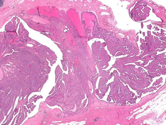
Imaging modality: light microscopy; specimen type: breast ductal tissue; technique: hematoxylin and eosin staining. The slide demonstrates a 1.5 cm intraductal papilloma arising within a central/subareolar duct with a palpable mass and associated bloody nipple discharge clinically. Histologically, the lesion consists of multiple papillary fronds protruding into a dilated duct lumen, each frond supported by a fibrovascular core and lined by a dual epithelial and myoepithelial layer. The epithelial component is typically columnar to cuboidal with uniform nuclei and minimal cytologic atypia; myoepithelial cells encircle the papillae, helping to distinguish benign intraductal papilloma from invasive carcinoma. The surrounding ductal epithelium shows mild ductal ectasia and focal hyperplasia but lacks invasion, high-grade atypia, or marked mitotic activity. The architecture is characteristic of benign papillary lesions of the breast, though larger papillomas can be solitary and clinically present as subareolar masses with Bloody discharge. The lesion may be associated with ductal dilation and mild stromal inflammation; no necrosis or stromal desmoplasia is evident. Clinically, this finding correlates with nipple discharge spectrum (bloody) and mass effect; management typically involves surgical excision to rule out atypia or malignant transformation and ensure complete removal of the papillary lesion.
Imaging modality: light microscopy; specimen type: breast ductal tissue; technique: hematoxylin and eosin staining. The slide demonstrates a 1.5 cm intraductal papilloma arising within a central/subareolar duct with a palpable mass and associated bloody nipple discharge clinically. Histologically, the lesion consists of multiple papillary fronds protruding into a dilated duct lumen, each frond supported by a fibrovascular core and lined by a dual epithelial and myoepithelial layer. The epithelial component is typically columnar to cuboidal with uniform nuclei and minimal cytologic atypia; myoepithelial cells encircle the papillae, helping to distinguish benign intraductal papilloma from invasive carcinoma. The surrounding ductal epithelium shows mild ductal ectasia and focal hyperplasia but lacks invasion, high-grade atypia, or marked mitotic activity. The architecture is characteristic of benign papillary lesions of the breast, though larger papillomas can be solitary and clinically present as subareolar masses with Bloody discharge. The lesion may be associated with ductal dilation and mild stromal inflammation; no necrosis or stromal desmoplasia is evident. Clinically, this finding correlates with nipple discharge spectrum (bloody) and mass effect; management typically involves surgical excision to rule out atypia or malignant transformation and ensure complete removal of the papillary lesion.

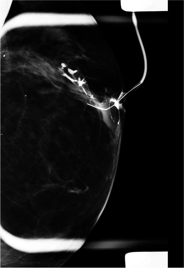
This diagnostic radiograph is a ductogram (galactogram) of the left breast, a specialized mammographic technique used to evaluate the ductal system. The image displays a retrograde injection of radiopaque contrast material through a cannula into a single lactiferous duct at the nipple. The primary findings include significant ductal dilatation (ectasia) of the main lactiferous duct. Distally, the contrast column terminates abruptly, demonstrating a clear ductal obstruction. Within the dilated segment, there is an irregular, intraluminal filling defect, suggesting the presence of an intraductal mass such as a papilloma or malignancy. The ductal wall appears irregular, and there is a noted displacement of adjacent ductal branches. This imaging modality is clinically indicated for the evaluation of pathologic nipple discharge when conventional mammography and ultrasound are inconclusive. The focal filling defect and distal obstruction are key diagnostic markers for identifying the location and character of potential intraductal lesions.
This diagnostic radiograph is a ductogram (galactogram) of the left breast, a specialized mammographic technique used to evaluate the ductal system. The image displays a retrograde injection of radiopaque contrast material through a cannula into a single lactiferous duct at the nipple. The primary findings include significant ductal dilatation (ectasia) of the main lactiferous duct. Distally, the contrast column terminates abruptly, demonstrating a clear ductal obstruction. Within the dilated segment, there is an irregular, intraluminal filling defect, suggesting the presence of an intraductal mass such as a papilloma or malignancy. The ductal wall appears irregular, and there is a noted displacement of adjacent ductal branches. This imaging modality is clinically indicated for the evaluation of pathologic nipple discharge when conventional mammography and ultrasound are inconclusive. The focal filling defect and distal obstruction are key diagnostic markers for identifying the location and character of potential intraductal lesions.

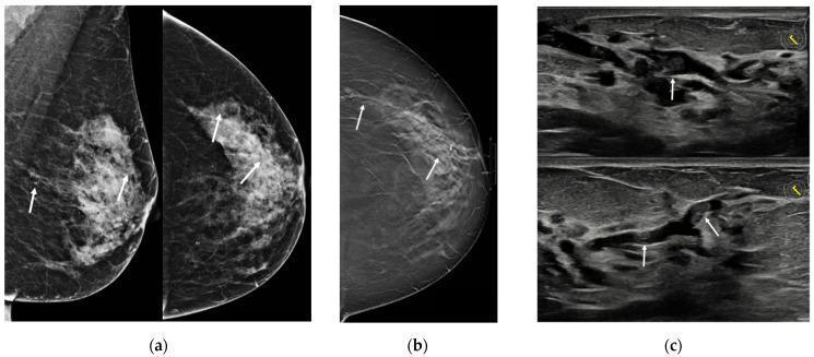
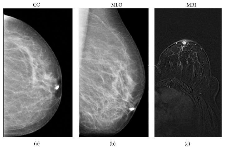
Multi-modal diagnostic imaging of the breast in a 55-year-old patient. (a) Conventional mammography in medio-lateral oblique (MLO) and cranio-caudal (CC) views showing scattered pleomorphic calcifications (indicated by white arrows) within fibroglandular tissue. (b) Digital breast tomosynthesis (DBT) slice in CC view providing enhanced visualization of multiple dilated ducts extending from the retroareolar region toward the outer quadrants. (c) High-resolution ultrasound (US) images depicting prominent, anechoic to hypoechoic dilated ducts containing heterogeneous, echogenic intraluminal material, which may represent solid components or thickened secretions. The findings are characteristic of ductal pathology, such as papillomatosis or duct ectasia, in the clinical context of unilateral bloody nipple discharge. The images illustrate the complementary roles of different imaging modalities in identifying subtle ductal changes and intraluminal contents.
Multi-modal diagnostic imaging of the breast in a 55-year-old patient. (a) Conventional mammography in medio-lateral oblique (MLO) and cranio-caudal (CC) views showing scattered pleomorphic calcifications (indicated by white arrows) within fibroglandular tissue. (b) Digital breast tomosynthesis (DBT) slice in CC view providing enhanced visualization of multiple dilated ducts extending from the retroareolar region toward the outer quadrants. (c) High-resolution ultrasound (US) images depicting prominent, anechoic to hypoechoic dilated ducts containing heterogeneous, echogenic intraluminal material, which may represent solid components or thickened secretions. The findings are characteristic of ductal pathology, such as papillomatosis or duct ectasia, in the clinical context of unilateral bloody nipple discharge. The images illustrate the complementary roles of different imaging modalities in identifying subtle ductal changes and intraluminal contents.

This diagnostic image is a craniocaudal (CC) view mammogram of the right breast. The breast tissue exhibits a scattered fibroglandular density pattern, where approximately 25% of the parenchyma consists of glandular tissue interspersed with fatty areas. Centrally, there is evidence of slight nipple inversion. A notable feature is the presence of increased radiodensity/echogenic material within the retroareolar lactiferous ducts, which appear prominent and tubular as they extend from the nipple into the deeper breast parenchyma. No discrete, spiculated masses or suspicious clustered pleomorphic microcalcifications are identified in this view. This image serves as an educational example of evaluating ductal changes in patients presenting with clinical symptoms such as nipple discharge or inversion, often correlating with conditions like duct ectasia or underlying occult malignancy.
This diagnostic image is a craniocaudal (CC) view mammogram of the right breast. The breast tissue exhibits a scattered fibroglandular density pattern, where approximately 25% of the parenchyma consists of glandular tissue interspersed with fatty areas. Centrally, there is evidence of slight nipple inversion. A notable feature is the presence of increased radiodensity/echogenic material within the retroareolar lactiferous ducts, which appear prominent and tubular as they extend from the nipple into the deeper breast parenchyma. No discrete, spiculated masses or suspicious clustered pleomorphic microcalcifications are identified in this view. This image serves as an educational example of evaluating ductal changes in patients presenting with clinical symptoms such as nipple discharge or inversion, often correlating with conditions like duct ectasia or underlying occult malignancy.

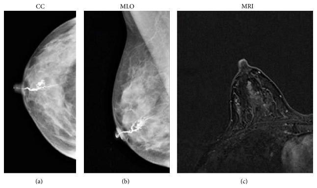
Multi-modal diagnostic imaging of the breast in a 53-year-old patient with nipple discharge, including galactography and MRI. Panel (a) and (b) present craniocaudal (CC) and mediolateral oblique (MLO) mammographic views during a galactogram, demonstrating a 'filling stop' in a cannulated duct located in the retroareolar region, a few millimeters from the nipple. Panel (c) shows a T1-weighted 3D FLASH axial MRI image of the same breast, highlighting a small, enhancing mass within the ductal system. The MRI finding is characterized by a round shape with sharp, well-defined margins in the retroareolar area. These combined findings are pathognomonic for an intraductal papilloma, a benign epithelial proliferation. This comparison illustrates the clinical utility of integrating ductography and contrast-enhanced MRI for localizing small intraductal lesions that may be difficult to characterize on standard mammography alone.
Multi-modal diagnostic imaging of the breast in a 53-year-old patient with nipple discharge, including galactography and MRI. Panel (a) and (b) present craniocaudal (CC) and mediolateral oblique (MLO) mammographic views during a galactogram, demonstrating a 'filling stop' in a cannulated duct located in the retroareolar region, a few millimeters from the nipple. Panel (c) shows a T1-weighted 3D FLASH axial MRI image of the same breast, highlighting a small, enhancing mass within the ductal system. The MRI finding is characterized by a round shape with sharp, well-defined margins in the retroareolar area. These combined findings are pathognomonic for an intraductal papilloma, a benign epithelial proliferation. This comparison illustrates the clinical utility of integrating ductography and contrast-enhanced MRI for localizing small intraductal lesions that may be difficult to characterize on standard mammography alone.

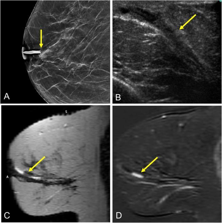
This composite of diagnostic breast imaging features a case of solitary intraductal papilloma in a 65-year-old woman. Image A is a craniocaudal ductogram (galactogram) showing a concave filling defect (arrow) within a contrast-filled duct, located approximately 2 cm posterior to the nipple. Image B is a grayscale ultrasound demonstrating ductal ectasia containing a corresponding hypoechoic intraductal lesion (arrow). Image C is a sagittal T2-weighted MRI revealing hyperintense signal within a dilated duct (ductal ectasia) containing a low-signal intraductal mass (arrow). Image D is a sagittal contrast-enhanced T1-weighted subtracted MRI showing the lesion (arrow) with focal, differential contrast enhancement compared to surrounding tissue. Collectively, these modalities illustrate the diagnostic approach to pathological nipple discharge, highlighting the appearance of a benign papillary lesion across x-ray, ultrasound, and magnetic resonance imaging platforms.

This composite of diagnostic breast imaging features a case of solitary intraductal papilloma in a 65-year-old woman. Image A is a craniocaudal ductogram (galactogram) showing a concave filling defect (arrow) within a contrast-filled duct, located approximately 2 cm posterior to the nipple. Image B is a grayscale ultrasound demonstrating ductal ectasia containing a corresponding hypoechoic intraductal lesion (arrow). Image C is a sagittal T2-weighted MRI revealing hyperintense signal within a dilated duct (ductal ectasia) containing a low-signal intraductal mass (arrow). Image D is a sagittal contrast-enhanced T1-weighted subtracted MRI showing the lesion (arrow) with focal, differential contrast enhancement compared to surrounding tissue. Collectively, these modalities illustrate the diagnostic approach to pathological nipple discharge, highlighting the appearance of a benign papillary lesion across x-ray, ultrasound, and magnetic resonance imaging platforms.

Searching Images

duct ectasia mammogram calcification lead pipe broken needle
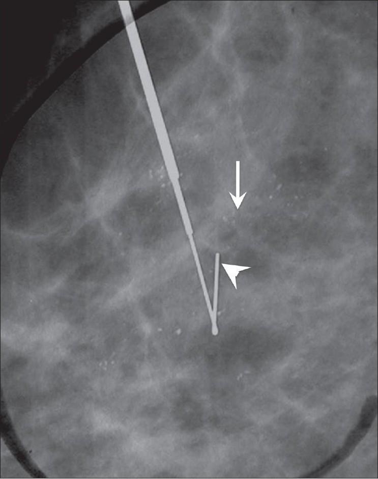
This diagnostic mammogram displays a magnified view of breast tissue featuring amorphous calcifications and a localization device. The image shows several hazy, indistinct, and tiny microcalcifications (indicated by a white arrow) distributed in a scattered, amorphous pattern. These calcifications lack a clearly defined shape or branching morphology. A metallic hook-wire localization needle (indicated by a white arrowhead) is visible, with its distal 'V-shaped' hook positioned adjacent to the cluster of calcifications to facilitate surgical excision or biopsy. The mammographic background shows fibroglandular density. This visual demonstrates a standard preoperative localization procedure for indeterminate or suspicious breast calcifications that are not palpable. The educational focus is on identifying amorphous calcification morphology and understanding the radiographic appearance of a hook-wire localization device used in breast imaging interventions.
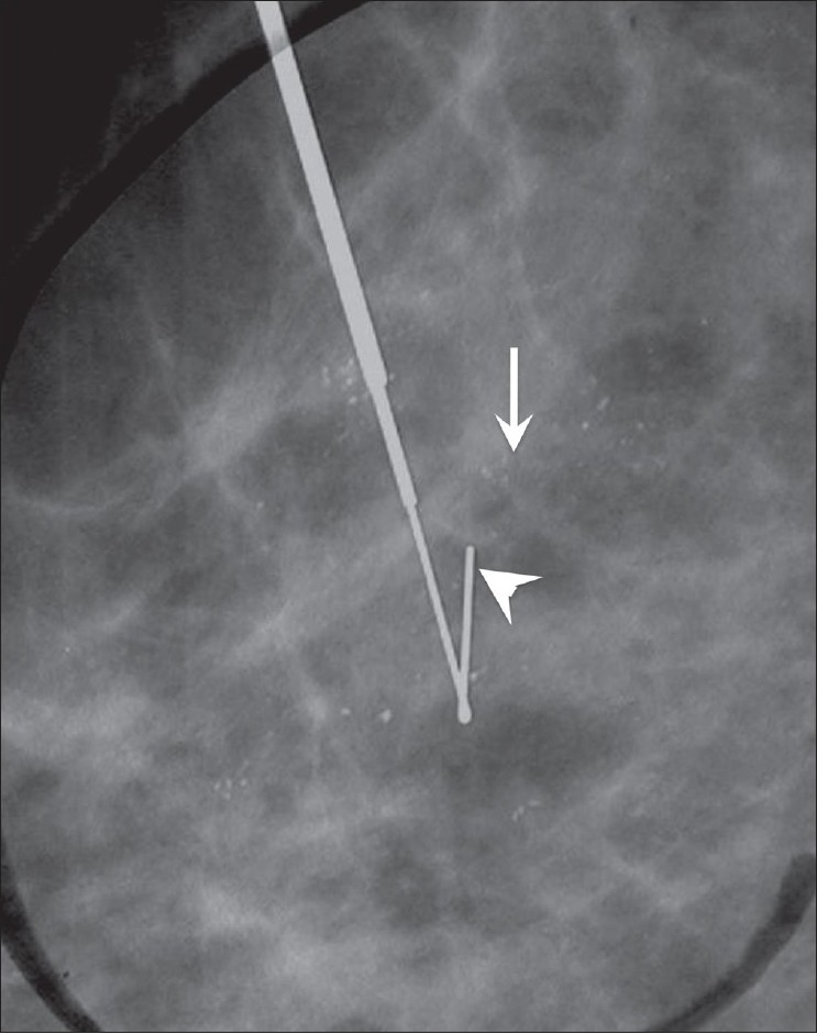
This diagnostic mammogram displays a magnified view of breast tissue featuring amorphous calcifications and a localization device. The image shows several hazy, indistinct, and tiny microcalcifications (indicated by a white arrow) distributed in a scattered, amorphous pattern. These calcifications lack a clearly defined shape or branching morphology. A metallic hook-wire localization needle (indicated by a white arrowhead) is visible, with its distal 'V-shaped' hook positioned adjacent to the cluster of calcifications to facilitate surgical excision or biopsy. The mammographic background shows fibroglandular density. This visual demonstrates a standard preoperative localization procedure for indeterminate or suspicious breast calcifications that are not palpable. The educational focus is on identifying amorphous calcification morphology and understanding the radiographic appearance of a hook-wire localization device used in breast imaging interventions.

**Imaging Modality:** Magnification view mammogram (digital mammography).

**Anatomical Region:** Breast parenchyma.

**Observed Pathology/Features:**
- **Calcifications:** A cluster of small, indistinct, amorphous microcalcifications is visible within the fibroglandular tissue (indicated by the white arrow). These calcifications lack a clearly defined shape or sharp margins.
- **Interventional Device:** A metallic hook-wire localization needle is present (indicated by the arrowhead), with the distal hook deployed in immediate proximity to the suspicious calcification cluster to guide surgical excision.

**Radiologic Characteristics:** 
The image demonstrates scattered fibroglandular densities. The primary finding is the morphological appearance of amorphous calcifications, which are categorized as "suspicious" according to BI-RADS descriptors, necessitating the illustrated preoperative localization procedure.

**Diagnostic Context:** 
Preoperative hook-wire localization for non-palpable breast lesions. The procedure aims to ensure accurate surgical sampling of the targeted area of interest within the breast tissue.
**Imaging Modality:** Magnification view mammogram (digital mammography). **Anatomical Region:** Breast parenchyma. **Observed Pathology/Features:** - **Calcifications:** A cluster of small, indistinct, amorphous microcalcifications is visible within the fibroglandular tissue (indicated by the white arrow). These calcifications lack a clearly defined shape or sharp margins. - **Interventional Device:** A metallic hook-wire localization needle is present (indicated by the arrowhead), with the distal hook deployed in immediate proximity to the suspicious calcification cluster to guide surgical excision. **Radiologic Characteristics:** The image demonstrates scattered fibroglandular densities. The primary finding is the morphological appearance of amorphous calcifications, which are categorized as "suspicious" according to BI-RADS descriptors, necessitating the illustrated preoperative localization procedure. **Diagnostic Context:** Preoperative hook-wire localization for non-palpable breast lesions. The procedure aims to ensure accurate surgical sampling of the targeted area of interest within the breast tissue.

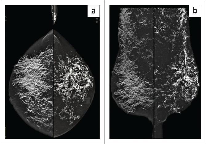
This diagnostic image consists of bilateral mammogram views, including Craniocaudal (CC, image a) and Mediolateral Oblique (MLO, image b) projections. The mammograms reveal extensive, high-density calcifications distributed throughout the parenchyma and subcutis of both breasts. These calcifications exhibit a distinct coarse, linear, and bizarre morphology characteristic of dystrophic calcification. The distribution is widespread and diffuse, significantly obscuring the normal glandular stroma. Notably, the pectoralis muscles seen on the MLO views are relatively spared, indicating the process is primarily confined to the breast tissue and superficial layers. This presentation is clinically significant for systemic conditions such as calcinosis cutis or calcification secondary to connective tissue disorders, such as ANCA-negative panniculitis, rather than typical breast malignancy.
This diagnostic image consists of bilateral mammogram views, including Craniocaudal (CC, image a) and Mediolateral Oblique (MLO, image b) projections. The mammograms reveal extensive, high-density calcifications distributed throughout the parenchyma and subcutis of both breasts. These calcifications exhibit a distinct coarse, linear, and bizarre morphology characteristic of dystrophic calcification. The distribution is widespread and diffuse, significantly obscuring the normal glandular stroma. Notably, the pectoralis muscles seen on the MLO views are relatively spared, indicating the process is primarily confined to the breast tissue and superficial layers. This presentation is clinically significant for systemic conditions such as calcinosis cutis or calcification secondary to connective tissue disorders, such as ANCA-negative panniculitis, rather than typical breast malignancy.

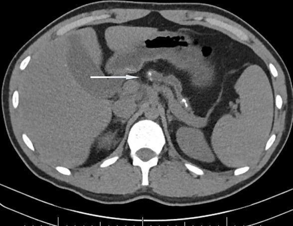
This axial abdominal CT scan demonstrates classic findings of severe chronic pancreatitis. The primary pathological finding is marked parenchymal atrophy of the pancreas, which is significantly reduced in size. Scattered throughout the remaining pancreatic tissue are multiple hyperdense, sub-centimeter calcifications. A white arrow points to a prominently dilated pancreatic duct (ductal ectasia) containing intraductal calculi (stones). Surrounding anatomy is clearly visible, including the liver in the right upper quadrant, the spleen in the left upper quadrant, and the stomach located anterior to the pancreas. The vertebral body and dorsal musculature are visible posteriorly. This imaging modality is essential for identifying structural changes associated with long-standing pancreatic inflammation, such as those found in hereditary or obstructive chronic pancreatitis. The presence of ductal dilation and diffuse calcification are hallmark diagnostic features of end-stage chronic pancreatic disease.

This axial abdominal CT scan demonstrates classic findings of severe chronic pancreatitis. The primary pathological finding is marked parenchymal atrophy of the pancreas, which is significantly reduced in size. Scattered throughout the remaining pancreatic tissue are multiple hyperdense, sub-centimeter calcifications. A white arrow points to a prominently dilated pancreatic duct (ductal ectasia) containing intraductal calculi (stones). Surrounding anatomy is clearly visible, including the liver in the right upper quadrant, the spleen in the left upper quadrant, and the stomach located anterior to the pancreas. The vertebral body and dorsal musculature are visible posteriorly. This imaging modality is essential for identifying structural changes associated with long-standing pancreatic inflammation, such as those found in hereditary or obstructive chronic pancreatitis. The presence of ductal dilation and diffuse calcification are hallmark diagnostic features of end-stage chronic pancreatic disease.

Here are the real clinical/radiological images of duct ectasia:

Duct Ectasia - Real Images

1. Ultrasound - Dilated Duct with Inspissated Debris

Ultrasound showing chronically dilated duct with echogenic debris inside - characteristic of duct ectasia
Longitudinal and cross-sectional ultrasound views showing a significantly dilated tubular duct (>3 mm). The lumen contains echogenic debris (inspissated secretions) - a hallmark of duct ectasia.

2. Mammogram - Dilated Retroareolar Ducts

Mammogram (CC view) showing branching tubular densities from the retroareolar region consistent with duct ectasia
Craniocaudal mammogram showing prominent branching tubular radiodensities extending from the retroareolar region - the classic mammographic appearance of duct ectasia. No suspicious masses or pleomorphic calcifications.

3. Multi-modal Imaging - Mammography + Tomosynthesis + Ultrasound

Multi-modal breast imaging showing dilated ducts on mammography, tomosynthesis, and ultrasound in a case of ductal pathology
Panel (a) - Conventional mammogram showing calcifications. Panel (b) - Digital breast tomosynthesis clearly showing dilated ducts extending from the retroareolar region. Panel (c) - Ultrasound showing prominent anechoic/hypoechoic dilated ducts with echogenic intraluminal material.

4. Mammogram - Nipple Inversion with Prominent Ducts

Mammogram showing nipple inversion with prominent retroareolar ductal densities characteristic of duct ectasia
CC view mammogram showing slight nipple inversion with increased radiodensity in the retroareolar lactiferous ducts - typical of duct ectasia causing nipple retraction via fibrosis.

5. Histology (H&E) - Ectatic Duct (Robbins Pathology)

H&E histology of duct ectasia - ectatic duct filled with inspissated debris surrounded by dense chronic lymphocytic inflammation and fibrosis
H&E section showing the ectatic duct lumen filled with pale inspissated secretions, surrounded by a dense ring of lymphocytic and plasma cell infiltrate, with outer fibrosis - this fibrotic cuff is what produces the firm palpable mass mimicking carcinoma on clinical examination.

Clinical image

Searching Images

duct ectasia breast nipple retraction clinical photograph
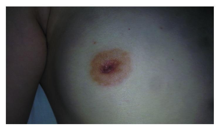
This clinical photograph shows a close-up view of a female right breast, primarily demonstrating nipple retraction (inversion). The nipple is clearly pulled inward into the breast tissue rather than projecting normally. The surrounding areola appears hyperpigmented (darker in color) relative to the adjacent skin but maintains a circular, regular border. The surrounding breast skin appears intact without visible erythema (redness), edema (swelling), Peau d'orange texture, or ulceration. There is no evidence of active nipple discharge or inflammatory changes on the skin surface. This finding is a significant clinical sign in breast pathology, often associated with underlying masses or architectural distortion, requiring diagnostic imaging such as ultrasound or mammography to rule out malignancy or benign fibroepithelial lesions.
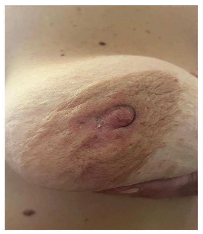
This clinical photograph shows a close-up view of a female right breast, primarily demonstrating nipple retraction (inversion). The nipple is clearly pulled inward into the breast tissue rather than projecting normally. The surrounding areola appears hyperpigmented (darker in color) relative to the adjacent skin but maintains a circular, regular border. The surrounding breast skin appears intact without visible erythema (redness), edema (swelling), Peau d'orange texture, or ulceration. There is no evidence of active nipple discharge or inflammatory changes on the skin surface. This finding is a significant clinical sign in breast pathology, often associated with underlying masses or architectural distortion, requiring diagnostic imaging such as ultrasound or mammography to rule out malignancy or benign fibroepithelial lesions.

This clinical photograph shows a close-up view of a female breast during a clinical examination, demonstrating significant indicators of breast malignancy. The primary visual findings include marked nipple retraction (inversion), where the nipple is pulled inward toward the breast tissue. The surrounding areola and periareolar skin exhibit extensive epidermal infiltration, characterized by a thickened, erythematous, and 'peau d'orange' (orange peel) texture. The skin appears indurated and leathery, with visible wrinkling and crusting near the nipple-areolar complex. These features are classic clinical signs of advanced or inflammatory breast cancer, where tumor infiltration of the Cooper's ligaments and the dermal lymphatic system leads to tethering and edema. The photograph serves as an educational example of the utility of Clinical Breast Examination (CBE) in identifying physical symptoms like skin dimpling, tethering, and nipple displacement which necessitate further diagnostic imaging such as mammography or ultrasound.
This clinical photograph shows a close-up view of a female breast during a clinical examination, demonstrating significant indicators of breast malignancy. The primary visual findings include marked nipple retraction (inversion), where the nipple is pulled inward toward the breast tissue. The surrounding areola and periareolar skin exhibit extensive epidermal infiltration, characterized by a thickened, erythematous, and 'peau d'orange' (orange peel) texture. The skin appears indurated and leathery, with visible wrinkling and crusting near the nipple-areolar complex. These features are classic clinical signs of advanced or inflammatory breast cancer, where tumor infiltration of the Cooper's ligaments and the dermal lymphatic system leads to tethering and edema. The photograph serves as an educational example of the utility of Clinical Breast Examination (CBE) in identifying physical symptoms like skin dimpling, tethering, and nipple displacement which necessitate further diagnostic imaging such as mammography or ultrasound.

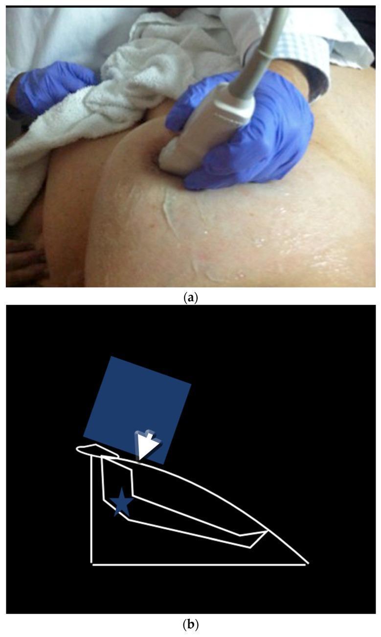
This composite educational resource illustrates the 'peripheral compression technique' for breast ultrasound, designed to improve visualization of the peripheral segments of retroareolar ducts. (a) Clinical photograph: A medical professional, wearing blue nitrile gloves, applies an ultrasound transducer to the breast of a supine patient. The probe is angled and positioned to apply pressure at the edge of the areola. (b) Anatomical diagram: A schematic representation of the maneuver showing the transducer (blue rectangle) applying a downward vector of force (white arrow). This compression displaces the nipple and flattens the underlying ductal structures (outlined in white), making the beam perpendicular to the duct for better resolution. A star marks the anatomical target within the retroareolar segment. This technique is clinically significant in breast imaging for overcoming the acoustic shadowing and anatomical irregularities inherent to the nipple-areolar complex, facilitating the detection of intraductal lesions or ductal ectasia.
This composite educational resource illustrates the 'peripheral compression technique' for breast ultrasound, designed to improve visualization of the peripheral segments of retroareolar ducts. (a) Clinical photograph: A medical professional, wearing blue nitrile gloves, applies an ultrasound transducer to the breast of a supine patient. The probe is angled and positioned to apply pressure at the edge of the areola. (b) Anatomical diagram: A schematic representation of the maneuver showing the transducer (blue rectangle) applying a downward vector of force (white arrow). This compression displaces the nipple and flattens the underlying ductal structures (outlined in white), making the beam perpendicular to the duct for better resolution. A star marks the anatomical target within the retroareolar segment. This technique is clinically significant in breast imaging for overcoming the acoustic shadowing and anatomical irregularities inherent to the nipple-areolar complex, facilitating the detection of intraductal lesions or ductal ectasia.

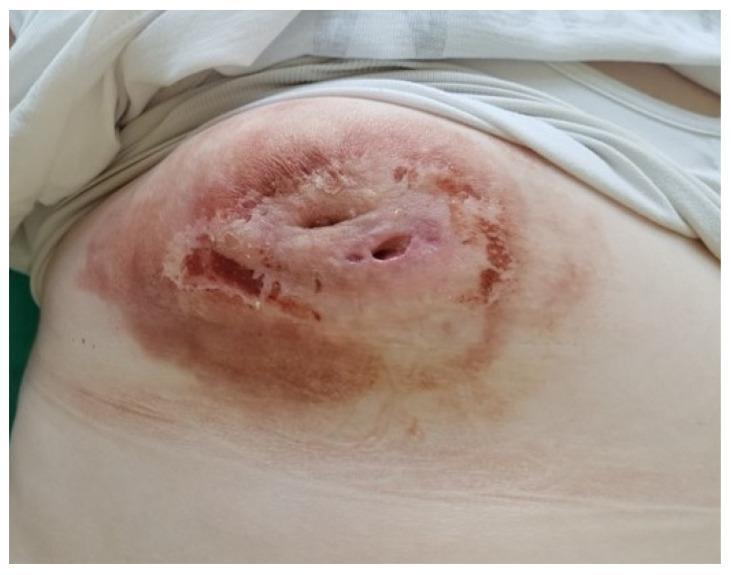
This clinical photograph shows the right breast of a patient following the treatment and evacuation of a purulent inflammatory lesion, such as a severe breast abscess or mastitis. The image demonstrates significant dermatological and structural changes: the central area exhibits persistent nipple retraction and several visible fistula tracts (openings in the skin) indicative of chronic inflammation or previous drainage sites. The skin across the breast displays generalized erythema (redness) and hyperpigmentation, with focal areas of desquamation or crusting near the periareolar region. The tissue appears thickened and uneven, reflecting the residual effects of inflammatory infiltration and subsequent drainage. This visual illustrates the clinical presentation of complicated breast infections, highlighting the importance of distinguishing inflammatory conditions from malignancy when structural distortions like nipple retraction and skin thickening are present.
This clinical photograph shows the right breast of a patient following the treatment and evacuation of a purulent inflammatory lesion, such as a severe breast abscess or mastitis. The image demonstrates significant dermatological and structural changes: the central area exhibits persistent nipple retraction and several visible fistula tracts (openings in the skin) indicative of chronic inflammation or previous drainage sites. The skin across the breast displays generalized erythema (redness) and hyperpigmentation, with focal areas of desquamation or crusting near the periareolar region. The tissue appears thickened and uneven, reflecting the residual effects of inflammatory infiltration and subsequent drainage. This visual illustrates the clinical presentation of complicated breast infections, highlighting the importance of distinguishing inflammatory conditions from malignancy when structural distortions like nipple retraction and skin thickening are present.

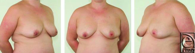
Clinical photograph set showing preoperative anterior and bilateral oblique views of a 38-year-old female patient's torso. The images demonstrate bilateral Grade 2 breast ptosis, characterized by the nipple-areola complex (NAC) positioned at or slightly below the level of the inframammary fold. Significant breast asymmetry is visible; the right breast exhibits post-surgical distortion with a flattened, less projected nipple and altered contour compared to the left. The patient has a history of multiple duct excision procedures for recurrent periductal mastitis and fistula formation, primarily affecting the right side. The lateral views highlight the downward slope of the breast tissue and the conical shape typical of secondary ptosis. Skin texture is largely intact, though surgical scarring and nipple retraction are evident on the right. This visual serves as a primary assessment for plastic and reconstructive surgery, specifically for evaluating volume, symmetry, and NAC position prior to potential mastopexy or breast reconstruction.
Clinical photograph set showing preoperative anterior and bilateral oblique views of a 38-year-old female patient's torso. The images demonstrate bilateral Grade 2 breast ptosis, characterized by the nipple-areola complex (NAC) positioned at or slightly below the level of the inframammary fold. Significant breast asymmetry is visible; the right breast exhibits post-surgical distortion with a flattened, less projected nipple and altered contour compared to the left. The patient has a history of multiple duct excision procedures for recurrent periductal mastitis and fistula formation, primarily affecting the right side. The lateral views highlight the downward slope of the breast tissue and the conical shape typical of secondary ptosis. Skin texture is largely intact, though surgical scarring and nipple retraction are evident on the right. This visual serves as a primary assessment for plastic and reconstructive surgery, specifically for evaluating volume, symmetry, and NAC position prior to potential mastopexy or breast reconstruction.

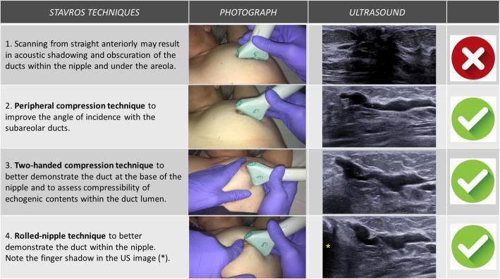
A comparison chart illustrating four Stavros ultrasound (US) techniques for evaluating the breast nipple-areolar complex and subareolar ducts. The chart is organized into four columns: technique description, clinical photograph of probe positioning, resulting ultrasound image, and a diagnostic value indicator (red 'X' or green checkmark).

1. Straight anterior scanning: Shows a probe placed vertically on the nipple, resulting in an ultrasound image with significant posterior acoustic shadowing that obscures the ducts.
2. Peripheral compression technique: Demonstrates the probe angled from the periphery toward the nipple. The US image shows improved visualization of longitudinal, hypoechoic subareolar ducts due to a better angle of incidence.
3. Two-handed compression technique: A photograph shows one gloved hand stabilizing/compressing the tissue while the probe is applied. This evaluates duct compressibility and echogenic lumen contents.
4. Rolled-nipple technique: Shows the nipple being manipulated/rolled by a finger while scanning. The US image reveals the duct within the nipple, marked by a characteristic finger shadow (yellow asterisk).

This educational visual is designed for radiology training to optimize the detection of intraductal pathologies like papillomas or ectasia.

A comparison chart illustrating four Stavros ultrasound (US) techniques for evaluating the breast nipple-areolar complex and subareolar ducts. The chart is organized into four columns: technique description, clinical photograph of probe positioning, resulting ultrasound image, and a diagnostic value indicator (red 'X' or green checkmark). 1. Straight anterior scanning: Shows a probe placed vertically on the nipple, resulting in an ultrasound image with significant posterior acoustic shadowing that obscures the ducts. 2. Peripheral compression technique: Demonstrates the probe angled from the periphery toward the nipple. The US image shows improved visualization of longitudinal, hypoechoic subareolar ducts due to a better angle of incidence. 3. Two-handed compression technique: A photograph shows one gloved hand stabilizing/compressing the tissue while the probe is applied. This evaluates duct compressibility and echogenic lumen contents. 4. Rolled-nipple technique: Shows the nipple being manipulated/rolled by a finger while scanning. The US image reveals the duct within the nipple, marked by a characteristic finger shadow (yellow asterisk). This educational visual is designed for radiology training to optimize the detection of intraductal pathologies like papillomas or ectasia.

Searching Images

mammary duct ectasia periareolar abscess fistula nipple discharge green brown
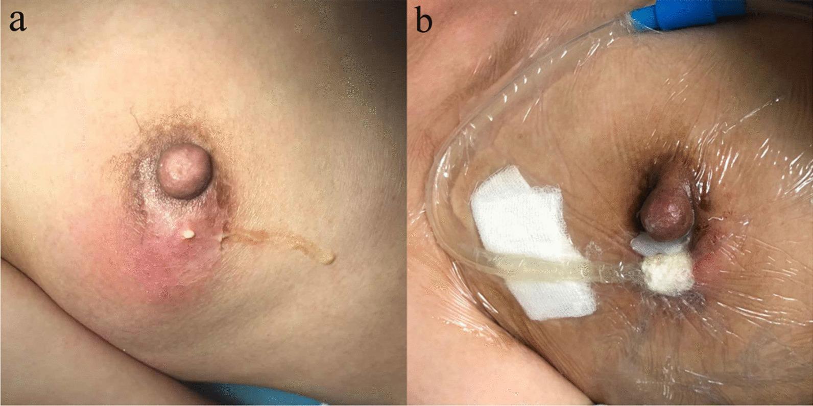
This clinical comparison photograph illustrates the management of a breast abscess using Vacuum Sealing Drainage (VSD). Image A (Preoperative) depicts an acute breast abscess involving the periareolar region. Key findings include prominent erythema, localized edema, and a spontaneous cutaneous fistula with visible purulent discharge. The nipple and areola show significant inflammatory changes consistent with suppurative mastitis. Image B (Post-intervention) shows the same anatomical site after 3 days of VSD treatment. The active medical setup consists of a specialized transparent semi-permeable adhesive film creating a hermetic seal over the breast, a drainage tube connected to a blue suction port, and white sterile dressing material positioned at the abscess site. There is a visible reduction in cutaneous erythema and swelling compared to the preoperative state. This comparison demonstrates the efficacy of negative pressure wound therapy in promoting the resolution of infection, managing exudate, and facilitating granulation tissue formation in complex soft tissue infections of the breast.
This clinical comparison photograph illustrates the management of a breast abscess using Vacuum Sealing Drainage (VSD). Image A (Preoperative) depicts an acute breast abscess involving the periareolar region. Key findings include prominent erythema, localized edema, and a spontaneous cutaneous fistula with visible purulent discharge. The nipple and areola show significant inflammatory changes consistent with suppurative mastitis. Image B (Post-intervention) shows the same anatomical site after 3 days of VSD treatment. The active medical setup consists of a specialized transparent semi-permeable adhesive film creating a hermetic seal over the breast, a drainage tube connected to a blue suction port, and white sterile dressing material positioned at the abscess site. There is a visible reduction in cutaneous erythema and swelling compared to the preoperative state. This comparison demonstrates the efficacy of negative pressure wound therapy in promoting the resolution of infection, managing exudate, and facilitating granulation tissue formation in complex soft tissue infections of the breast.

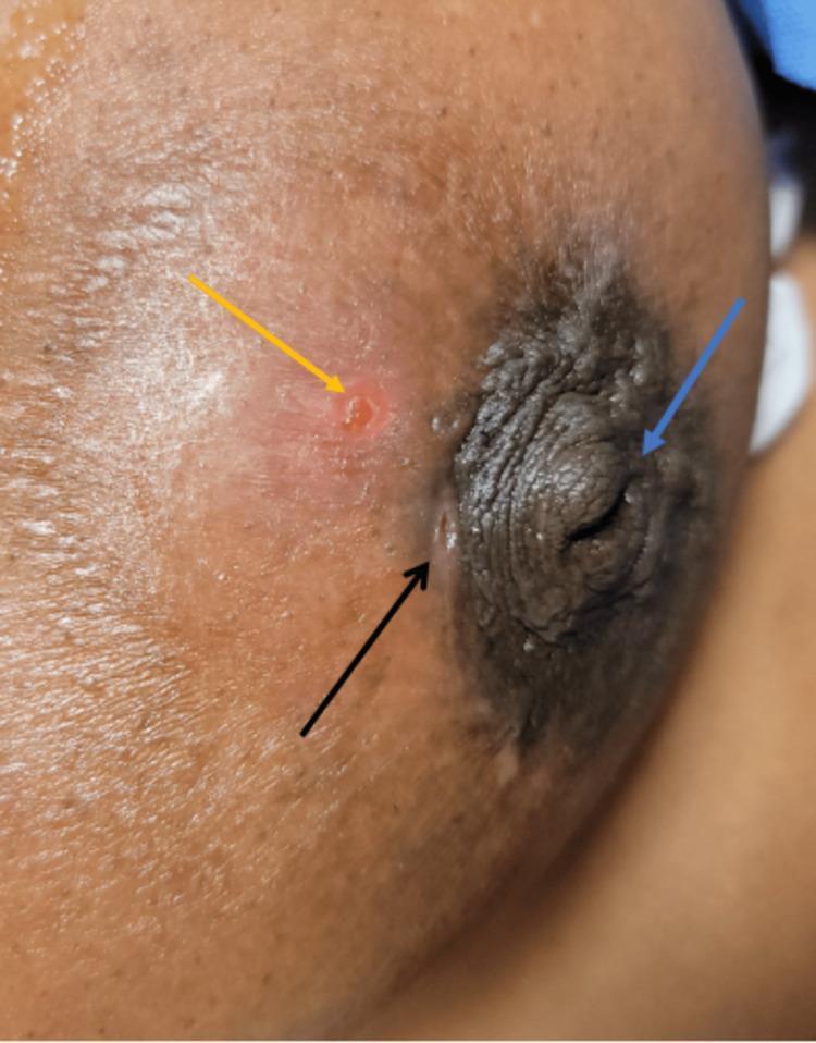
This clinical photograph displays a lateral view of a human breast, focusing on the nipple-areolar complex (NAC) to illustrate findings characteristic of Zuska's disease (recurrent subareolar abscess). Three key clinical features are highlighted with colored arrows: A blue arrow points to a significant nipple inversion, where the nipple is retracted into the areola. A black arrow indicates a periareolar draining fistula located at the vermillion border of the areola, appearing as a small, slit-like opening. A yellow arrow identifies a localized area of skin thinning and erythema peripheral to the areola, representing the site of an underlying subareolar abscess. The surrounding skin shows secondary signs of inflammation, including redness and swelling. This image serves as a textbook example of the triad of nipple inversion, recurrent abscess, and fistulization used in diagnosing chronic periductal mastitis and related breast infections.
This clinical photograph displays a lateral view of a human breast, focusing on the nipple-areolar complex (NAC) to illustrate findings characteristic of Zuska's disease (recurrent subareolar abscess). Three key clinical features are highlighted with colored arrows: A blue arrow points to a significant nipple inversion, where the nipple is retracted into the areola. A black arrow indicates a periareolar draining fistula located at the vermillion border of the areola, appearing as a small, slit-like opening. A yellow arrow identifies a localized area of skin thinning and erythema peripheral to the areola, representing the site of an underlying subareolar abscess. The surrounding skin shows secondary signs of inflammation, including redness and swelling. This image serves as a textbook example of the triad of nipple inversion, recurrent abscess, and fistulization used in diagnosing chronic periductal mastitis and related breast infections.

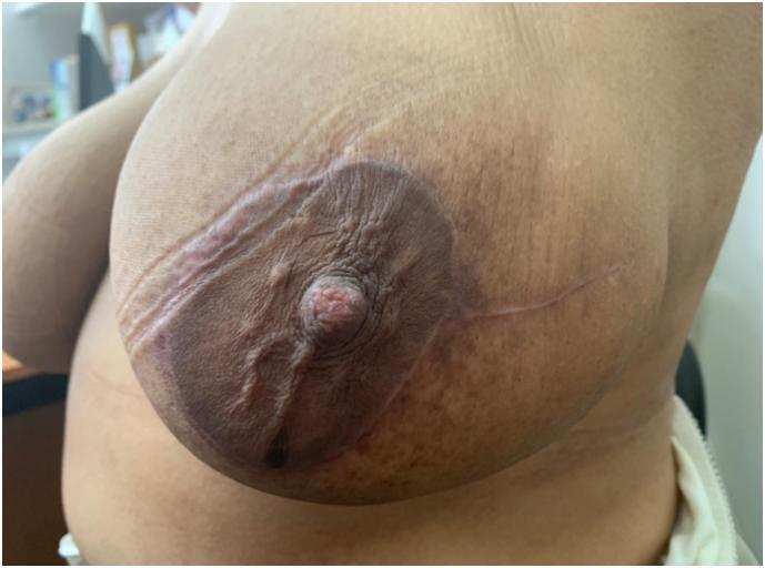
This clinical photograph displays post-operative scarring on a female breast following abscess excision and recentralization of the nipple-areola complex (NAC). The image demonstrates a combination of circumareolar (periareolar) and radial surgical scars. The periareolar scar encircles the hyperpigmented areola, exhibiting a slightly raised, hyperpigmented, and wrinkled texture that marks the site of a 'round block' incision. A prominent radial scar extends laterally from the areolar border toward the axillary region, representing the closure of a previous skin fistula or 'racquet' incision. The central nipple is intact but shows minor architectural distortion due to the underlying tissue remodeling and tension from the layered suture closure. These visual findings are characteristic of oncoplastic breast surgery techniques used to maintain breast symmetry and volume after significant tissue excision in the upper outer quadrant. The educational focus is on identifying common surgical scar patterns in breast reconstructive or corrective procedures.
This clinical photograph displays post-operative scarring on a female breast following abscess excision and recentralization of the nipple-areola complex (NAC). The image demonstrates a combination of circumareolar (periareolar) and radial surgical scars. The periareolar scar encircles the hyperpigmented areola, exhibiting a slightly raised, hyperpigmented, and wrinkled texture that marks the site of a 'round block' incision. A prominent radial scar extends laterally from the areolar border toward the axillary region, representing the closure of a previous skin fistula or 'racquet' incision. The central nipple is intact but shows minor architectural distortion due to the underlying tissue remodeling and tension from the layered suture closure. These visual findings are characteristic of oncoplastic breast surgery techniques used to maintain breast symmetry and volume after significant tissue excision in the upper outer quadrant. The educational focus is on identifying common surgical scar patterns in breast reconstructive or corrective procedures.

This composite diagnostic image showcases breast imaging findings in a 41-year-old patient with bloody nipple discharge, diagnosed histologically with papillomatosis. Panels (a) and (b) are galactographic (ductographic) images in craniocaudal (CC) and mediolateral oblique (MLO) projections, respectively. They demonstrate marked ductal ectasia (dilation) characterized by focal wall irregularities and intraductal filling defects within the contrast-enhanced mammary ducts. Panel (c) is an axial T1-weighted 3D FLASH MRI with contrast, showing prominent ductal enhancement and small hyperintense foci corresponding to the pathologic ductal changes. The image serves as an educational comparison between conventional galactography and contrast-enhanced MRI in the evaluation of pathological nipple discharge and intraductal lesions like papillomatosis. Key concepts illustrated include ductal morphology, contrast filling patterns, and the correlation between different imaging modalities in senology.

This composite diagnostic image showcases breast imaging findings in a 41-year-old patient with bloody nipple discharge, diagnosed histologically with papillomatosis. Panels (a) and (b) are galactographic (ductographic) images in craniocaudal (CC) and mediolateral oblique (MLO) projections, respectively. They demonstrate marked ductal ectasia (dilation) characterized by focal wall irregularities and intraductal filling defects within the contrast-enhanced mammary ducts. Panel (c) is an axial T1-weighted 3D FLASH MRI with contrast, showing prominent ductal enhancement and small hyperintense foci corresponding to the pathologic ductal changes. The image serves as an educational comparison between conventional galactography and contrast-enhanced MRI in the evaluation of pathological nipple discharge and intraductal lesions like papillomatosis. Key concepts illustrated include ductal morphology, contrast filling patterns, and the correlation between different imaging modalities in senology.

Here are the real clinical photographs related to duct ectasia and its complications:

Duct Ectasia - Clinical Images

1. Nipple Retraction (Classic Sign)

Clinical photograph of a female breast showing nipple retraction/inversion - a classic sign of duct ectasia caused by periductal fibrosis
Nipple retraction/inversion - the hallmark clinical sign of duct ectasia. The nipple is pulled inward due to periductal fibrosis and foreshortening of the affected ducts. Note the intact surrounding skin with no peau d'orange or ulceration (distinguishing features from malignancy).

2. Zuska's Disease - Periareolar Fistula (Complication of Duct Ectasia / Periductal Mastitis)

Clinical photograph showing the classic triad of Zuska's disease: nipple inversion (blue arrow), periareolar draining fistula (black arrow), and underlying subareolar abscess with erythema (yellow arrow)
Classic Zuska's disease (recurrent subareolar abscess / chronic periductal mastitis) - a complication of duct ectasia. Three clinical features are marked:
  • Blue arrow - nipple inversion
  • Black arrow - periareolar draining fistula at the areolar border
  • Yellow arrow - erythema and skin thinning over underlying abscess

3. Periareolar Abscess with Fistula Formation

Clinical photograph showing a periareolar abscess with spontaneous cutaneous fistula and purulent discharge - a complication of duct ectasia/periductal mastitis
Acute periareolar abscess - a direct complication of duct ectasia. Shows erythema, edema, and a spontaneous cutaneous fistula with purulent discharge at the periareolar region. This represents the spectrum from duct ectasia -> periductal mastitis -> abscess formation.

4. Post-surgical Changes After Recurrent Periductal Mastitis

Bilateral oblique clinical photograph of a patient with history of multiple duct excision procedures for recurrent periductal mastitis, showing nipple retraction and surgical distortion
Patient after multiple duct excisions for recurrent periductal mastitis (complication of duct ectasia). Note surgical scarring, nipple retraction, and breast contour changes on the right side - illustrating why surgical excision (Hadfield's operation) is used for recurrent/severe cases.

Clinical Spectrum Summary:
Duct ectasia -> periductal inflammation -> fibrosis (nipple retraction + palpable mass) -> if duct ruptures: abscess -> fistula (Zuska's disease)
This is a shared conversation. Sign in to Orris to start your own chat.