
Explain me 10 common heart diseases with ECG changes and ECG images of those diseases
Reading File
Finding Sources
Finding Sources
Searching Images
ECG electrocardiogram myocardial infarction ST elevation
Now let me search for more ECG images for different conditions simultaneously:
Searching Images
ECG atrial fibrillation irregular rhythm no P waves
Searching Images
ECG heart block first degree second degree third degree AV block
Searching Images
ECG left ventricular hypertrophy voltage criteria
Finding Sources
Finding Sources
Searching Images
ECG Wolff-Parkinson-White syndrome delta wave short PR interval
Searching Images
ECG pericarditis saddle-shaped ST elevation diffuse
Searching Images
ECG pulmonary embolism S1Q3T3 right heart strain
Searching Images
ECG ventricular tachycardia wide complex tachycardia
Searching Images
ECG hypertrophic cardiomyopathy deep Q waves ST changes
Searching Images
ECG long QT syndrome prolonged QTc torsades de pointes
Finding Sources
Finding Sources
I now have comprehensive ECG images and textbook data for all 10 conditions. Let me compile the full answer.Here is a comprehensive guide to 10 common heart diseases with their ECG changes and ECG images.
10 Common Heart Diseases with ECG Changes
1. ST-Elevation Myocardial Infarction (STEMI)
Overview: Acute complete occlusion of a coronary artery causing full-thickness myocardial ischemia and necrosis.
ECG Changes:
- Hyperacute T-waves (earliest sign) — tall, peaked, broad T-waves
- ST-segment elevation ≥ 1 mm in ≥2 contiguous leads (convex/tombstone morphology)
- Reciprocal ST depression in opposing leads
- Pathological Q waves — wide (>40 ms) and deep (>25% of R wave) develop over hours
- T-wave inversion in the infarcted territory (late sign)
Localisation:
| Territory | ST Elevation Leads | Artery |
|---|---|---|
| Anterior | V1–V4 | LAD |
| Inferior | II, III, aVF | RCA |
| Lateral | I, aVL, V5–V6 | LCx |
| Posterior | Tall R in V1, ST depression V1–V3 | RCA/LCx |
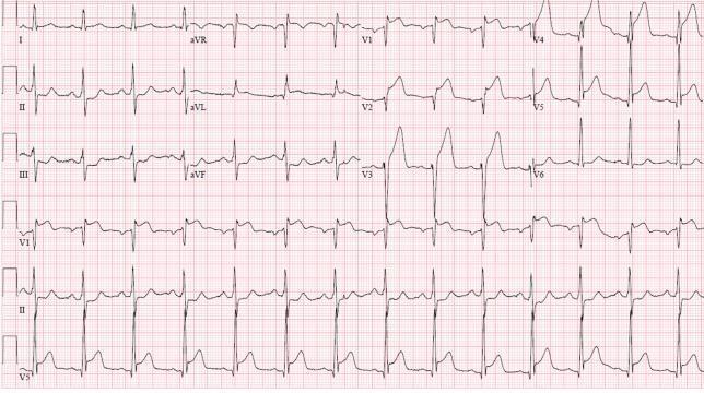
Anterolateral STEMI showing prominent ST elevation in V1–V6 and leads I, aVL, with reciprocal depression in II, III, aVF
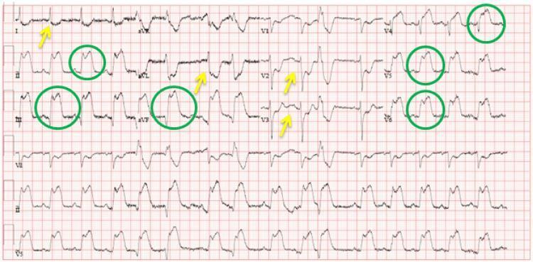
Inferolateral STEMI: ST elevations in II, III, aVF and V4–V6 (green circles) with reciprocal depressions in I, aVL, V2–V3 (yellow arrows)
2. Atrial Fibrillation (AF)
Overview: Disorganised electrical activity in the atria producing chaotic firing at 350–600 impulses/min, causing an irregularly irregular ventricular response.
ECG Changes:
- Absent P waves — replaced by irregular fibrillatory (f) waves, best seen in V1 and II
- Irregularly irregular R-R intervals (pathognomonic)
- Narrow QRS complexes (unless aberrant conduction or BBB coexists)
- Rate can be rapid (uncontrolled AF >100 bpm) or controlled (<100 bpm)
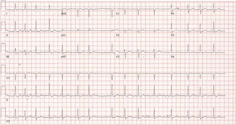
Classic AF: No organised P waves, fine fibrillatory baseline, irregularly irregular narrow QRS complexes
3. AV Heart Block (1st, 2nd, 3rd Degree)
Overview: Impaired conduction through the AV node, ranging from simple delay to complete dissociation.
ECG Changes:
| Degree | Key ECG Feature |
|---|---|
| 1st degree | PR interval > 200 ms, every P followed by QRS |
| 2nd degree Mobitz I (Wenckebach) | Progressive PR lengthening until a QRS is dropped |
| 2nd degree Mobitz II | Constant PR interval, then sudden dropped QRS |
| 3rd degree (Complete) | Complete AV dissociation — P waves and QRS complexes at independent rates |
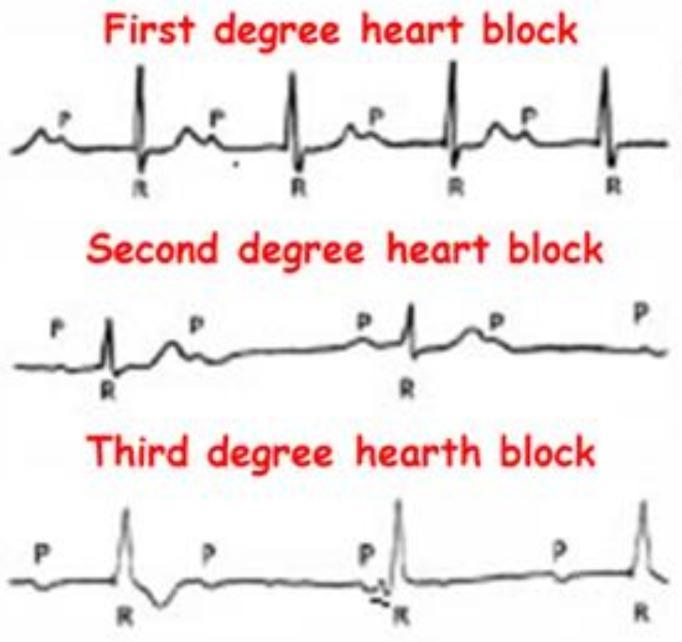
Educational comparison of all three degrees of AV block showing P-QRS relationships
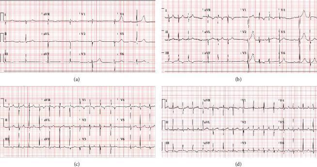
Clinical series showing (a) complete heart block, (b) Mobitz II, (c) Wenckebach, and (d) 1st-degree block in the same patient during treatment
4. Wolff-Parkinson-White (WPW) Syndrome
Overview: An accessory pathway (Bundle of Kent) bypasses the AV node, causing ventricular pre-excitation and predisposing to re-entrant tachyarrhythmias.
ECG Changes (classic triad):
- Short PR interval < 120 ms (bypass of AV node delay)
- Delta wave — slurred upstroke at the beginning of the QRS complex
- Widened QRS complex (> 120 ms) due to fusion of pre-excited and normal depolarisation
- Secondary ST/T changes (discordant to QRS)
- Risk of pseudo-infarction pattern (negative delta waves mimicking Q waves)
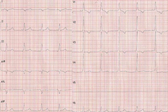
Classic WPW: Short PR interval, prominent delta waves visible in II, III, aVF and V2–V6, with secondary QRS widening
5. Acute Pericarditis
Overview: Inflammation of the pericardium (most often viral), producing characteristic diffuse ECG changes that evolve through 4 stages.
ECG Changes (4 Stages):
| Stage | Change |
|---|---|
| I (Acute) | Diffuse concave ("saddle-shaped") ST elevation in all leads except aVR/V1; PR segment depression (hallmark) |
| II (Days 1–2) | ST returns to baseline; PR depression persists |
| III (Days 2–3) | Widespread T-wave inversion |
| IV (Weeks later) | Normalisation |
Key differentiators from STEMI:
- Diffuse ST elevation (not territory-specific)
- PR depression (absent in STEMI)
- Concave (not convex) ST morphology
- No reciprocal changes (except aVR)
- Spodick's sign (downsloping TP segment)
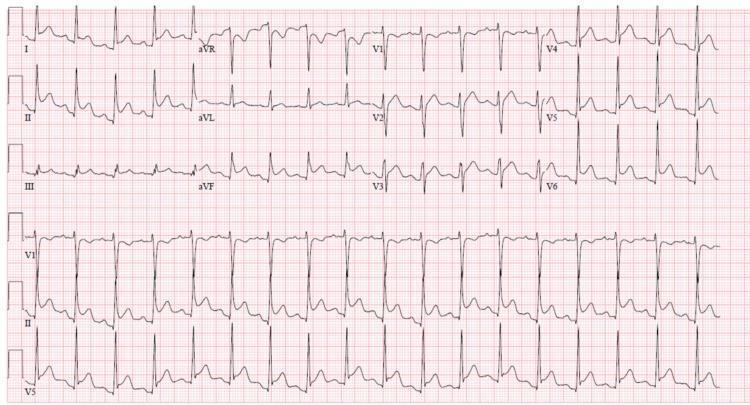
Pericarditis: Concave ST elevation in I, II, III, aVF, V2–V6; PR depression in II; reciprocal PR elevation and ST depression in aVR
6. Pulmonary Embolism (PE)
Overview: Thrombotic obstruction of the pulmonary vasculature causes acute right heart strain, reflected on ECG.
ECG Changes:
- Sinus tachycardia (most common, ~40% of cases)
- S1Q3T3 pattern (classic but present in only ~20%): Deep S in lead I + Q wave + T inversion in lead III
- Right bundle branch block (complete or incomplete)
- T-wave inversions V1–V4 (right ventricular strain pattern)
- Rightward axis deviation
- Atrial fibrillation may develop
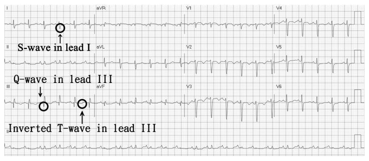
Classic S1Q3T3 (black circles/arrows): Deep S in I, Q wave in III, inverted T in III — hallmark of acute right heart strain from PE
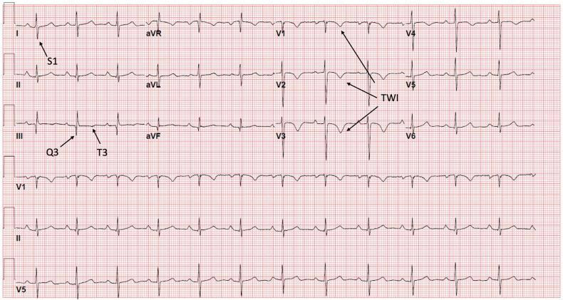
Massive PE: S1Q3T3 plus deep, symmetric T-wave inversions V1–V6 (McGinn-White sign)
7. Left Ventricular Hypertrophy (LVH)
Overview: Increased LV muscle mass from chronic pressure overload (e.g., hypertension, aortic stenosis), causing characteristic voltage and repolarisation changes.
ECG Changes:
- Increased QRS voltage (key diagnostic criteria):
- Sokolow-Lyon: S in V1 + R in V5 or V6 ≥ 35 mm
- Cornell: R in aVL + S in V3 > 28 mm (men) / >20 mm (women)
- Left axis deviation
- LV strain pattern: ST depression and asymmetric T-wave inversion in lateral leads (I, aVL, V5–V6)
- Prolonged QRS duration
- Left atrial enlargement (broad, notched P wave in II — "P mitrale")
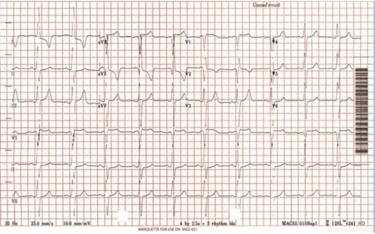
LVH: High-amplitude R in aVL and deep S in V3 meeting Cornell criteria; prominent T-wave inversions in V4–V6 (strain pattern)
8. Hypertrophic Cardiomyopathy (HCM)
Overview: Genetic disorder (sarcomere mutations) causing asymmetric septal hypertrophy, often with dynamic outflow obstruction. Leading cause of sudden cardiac death in young athletes.
ECG Changes:
- LVH voltage criteria (prominent R or S waves)
- Deep, narrow ("dagger-like") Q waves in lateral leads (I, aVL, V5–V6) and inferior leads — due to septal hypertrophy
- Giant T-wave inversions (particularly in apical HCM — Yamaguchi variant) in V3–V6, depth often >10 mm
- ST segment depression in lateral leads
- Atrial fibrillation (common in advanced disease)
- Absent septal Q waves in some variants
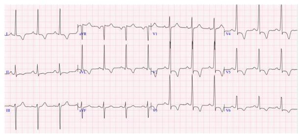
HCM (septal variant): High R-wave voltages V2–V4, deep symmetric T-wave inversions V2–V6 (strain pattern), positive T in aVR (arrhythmia risk marker)
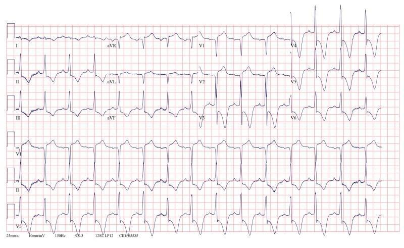
Yamaguchi (apical) HCM: Giant symmetric T-wave inversions deepest in V4–V5 — classic "spade" pattern
9. Ventricular Tachycardia (VT)
Overview: Three or more consecutive ventricular beats at >100 bpm originating below the Bundle of His. A life-threatening arrhythmia requiring urgent treatment.
ECG Changes:
- Wide complex tachycardia — QRS ≥ 120 ms (often >160 ms)
- Rapid regular rhythm — typically 100–250 bpm
- AV dissociation — P waves march through at a slower rate, independent of QRS (pathognomonic of VT)
- Fusion beats and capture beats (Dressler beats) — confirm VT
- Positive or negative concordance across precordial leads
- Brugada criteria used to differentiate VT from SVT with aberrancy
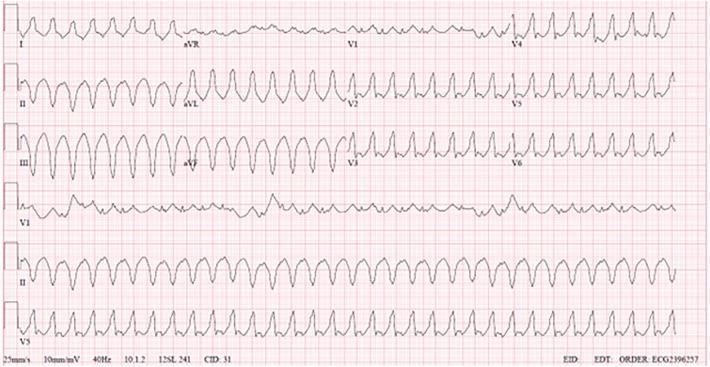
Monomorphic VT: Wide, bizarre QRS complexes with rapid regular rate, positive concordance in precordial leads, no visible P waves
10. Long QT Syndrome (LQTS)
Overview: A channelopathy (inherited or acquired) impairing ventricular repolarisation, predisposing to the deadly arrhythmia Torsades de Pointes (TdP) and sudden cardiac death.
ECG Changes:
- Prolonged QTc interval:
- Normal QTc: ≤ 440 ms (men), ≤ 460 ms (women)
- Borderline risk: 440–500 ms
- High risk / LQTS: > 500 ms
- Abnormal T-wave morphology: broad, notched, or biphasic T-waves
- T-wave alternans (beat-to-beat variation in T-wave amplitude — harbinger of TdP)
- Torsades de Pointes: polymorphic VT with "twisting" of QRS complexes around the isoelectric line
Common causes of acquired LQTS: Hypokalaemia, hypomagnesaemia, drugs (antiarrhythmics, antipsychotics, antibiotics such as azithromycin).
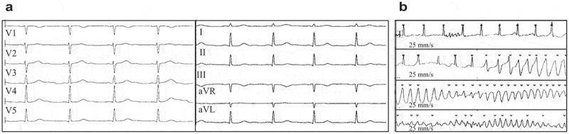
LQTS: Panel (a) sinus rhythm with QTc 478 ms; Panel (b) onset of Torsades de Pointes — twisting polymorphic VT
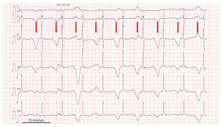
Extreme QTc 610 ms with macro T-wave alternans (red arrows) — electrical instability immediately preceding TdP
Quick Reference Summary
| # | Disease | Key ECG Finding |
|---|---|---|
| 1 | STEMI | ST elevation ≥1mm in ≥2 contiguous leads + Q waves |
| 2 | Atrial Fibrillation | Absent P waves + irregularly irregular rhythm |
| 3 | AV Heart Block | PR prolonged / dropped beats / AV dissociation |
| 4 | WPW Syndrome | Short PR + delta wave + widened QRS |
| 5 | Acute Pericarditis | Diffuse saddle ST elevation + PR depression |
| 6 | Pulmonary Embolism | Sinus tachycardia + S1Q3T3 + RV strain |
| 7 | LVH | High QRS voltage + lateral strain pattern |
| 8 | HCM | Deep Q waves + giant T inversions (apical type) |
| 9 | Ventricular Tachycardia | Wide complex tachycardia + AV dissociation |
| 10 | Long QT Syndrome | QTc >500 ms + T-wave alternans → Torsades |
*Sources: Braunwald's Heart Disease, Harrison's Principles of Internal Medicine 22E, Tintinalli's Emergency Medicine, Ganong's Review of Medical Physiology, Goldman-Cecil Medicine.*You've run out of free credits. Sign up for a free account to continue using Orris.
This is a shared conversation. Sign in to Orris to start your own chat.