
A baby was diagnosed to have diaphragmatic hernia antenatally. At birth child had respiratory distress and was cyanosed. What is the first step in immediate management? A. NG tube insertion B. Nasal cpap oxygen / Commence nasal prolong o2? C. ET tube insertion D. IV fluid E. CXR
congenital diaphragmatic hernia immediate management newborn first step NG tube
Answer: A. NG tube insertion
Why NG tube is the first step
"Resuscitation should begin with standard neonatal resuscitation guidelines and then proceed with endotracheal intubation and nasogastric tube insertion. Bag-valve mask should be avoided."
Why the other options are wrong (or lower priority):
| Option | Why it's not first |
|---|---|
| B. Nasal CPAP/O2 | CPAP/mask ventilation is actively contraindicated in CDH - positive pressure ventilation via mask forces air into herniated bowel, worsening compression. Ventilation support is needed but via ET tube, not mask/CPAP |
| C. ET tube | Definitive airway management is needed, but the NG tube comes first to decompress the gut before ventilation. Many sources list NG then ET as sequential immediate steps |
| D. IV fluids | Important but not the immediate first priority - gut decompression is more urgent |
| E. CXR | Useful for diagnosis confirmation, but the diagnosis is already known antenatally here, and delaying gut decompression to get a CXR would be harmful |
Key concept - Why bag-mask is forbidden in CDH:
- Mulholland & Greenfield's Surgery, 7th ed., Chapter 100 (Congenital Diaphragmatic Hernia - Treatment section)
- Supported by the CDH EURO Consortium guidelines and Nationwide Children's Hospital/CHOP CDH protocols
A 3 weeks old neonate admitted with a distended abdomen. He first passed meconium, 3 days after birth following a saline enema. Afterwards he passed stools only twice. His weight is now 200g more than his birth weight. On examination he had a distended abdomen with an umbilical hernia. What is the most useful investigation in aid of diagnosis? A. Colonoscopy B. Lower GI contrast studies C. Plain Xray abdomen D. Serum calcium E. TSH
Hirschsprung disease neonatal investigation contrast enema diagnosis delayed meconium
Hirschsprung disease contrast enema transition zone neonate
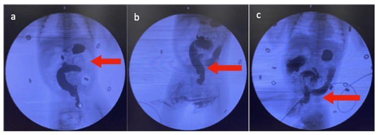
A series of three fluoroscopic images (a, b, and c) from a pediatric contrast enema study, likely in a neonate, evaluating for Hirschsprung's disease. The images demonstrate the retrograde flow of radiopaque contrast material through the distal colon. Image (a) shows the contrast filling the sigmoid and descending colon, with a notable distension of the abdomen visible. Image (b) highlights irregular wall contractions in the contrast-filled segment (red arrow). Image (c) clearly depicts a classic 'transition zone' (red arrow), where there is a sudden and marked change in bowel diameter between a distal, narrowed aganglionic segment and a more proximal, dilated segment of the colon. This radiographic finding is highly suggestive of intestinal obstruction secondary to Hirschsprung's disease. The imaging sequence captures the dynamic nature of bowel motility and the distinct transition point necessary for pediatric surgical diagnosis.
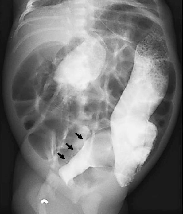
This abdominal contrast enema (fluoroscopy/X-ray radiography) of a neonate illustrates the classic radiological features of Hirschsprung disease. The image demonstrates a clear transition zone in the distal bowel. Specifically, black arrows highlight a narrowed caliber and constricted lumen within the rectosigmoid colon, representing the aganglionic segment. In contrast, the proximal descending colon appears dilated (megacolon) as it attempts to overcome the distal obstruction. The abdominal cavity shows diffuse gaseous distension of bowel loops superior to the pelvic cavity. This diagnostic imaging is used in pediatric gastroenterology to identify a 'rectosigmoid ratio' suggestive of functional obstruction. The clinical context involves a newborn presenting with delayed meconium passage, abdominal distension, and poor feeding, where the transition from a narrow rectum to a dilated proximal colon is the hallmark finding for identifying the transition zone before definitive surgical biopsy.
Answer: B. Lower GI Contrast Studies
Why this is Hirschsprung disease
| Feature | Significance |
|---|---|
| Delayed meconium passage (day 3, only after enema) | HD hallmark - >90% fail to pass meconium within 24 hrs |
| Sparse stools (only twice in 3 weeks) | Functional obstruction due to aganglionosis |
| Distended abdomen | Proximal colonic dilation above aganglionic segment |
| Umbilical hernia | Secondary to chronic raised intra-abdominal pressure |
| Poor weight gain (only 200g above birth weight at 3 weeks) | Normal gain is ~150-200g/week - severely underweight |
Why Lower GI Contrast Studies is the best investigation
"The diagnostic imaging study of choice in a newborn is a contrast enema. In Hirschsprung disease, aganglionosis of the distal rectum usually results in a narrow caliber with a transition zone and dilated, normal, proximal sigmoid colon. Failure to evacuate the instilled contrast medium completely after 24 hours strongly indicates the presence of Hirschsprung disease."
- Transition zone - abrupt change from narrow aganglionic distal segment to dilated proximal colon
- Reversed rectosigmoid ratio - the rectum is narrower than the sigmoid (normally rectum > sigmoid)
- Irregular contractions (saw-tooth pattern) in the aganglionic segment
- Contrast retention at 24 hours - failure to evacuate the colon
Why the other options are wrong
| Option | Why not |
|---|---|
| A. Colonoscopy | Invasive, technically difficult in a neonate, and provides no additional diagnostic benefit over contrast enema in the acute setting |
| C. Plain X-ray abdomen | Shows non-specific dilated loops and absent rectal gas - useful to screen, but cannot distinguish the cause of obstruction or identify the transition zone |
| D. Serum calcium | Would investigate hypercalcemia (e.g. Williams syndrome with constipation) - not the right diagnosis here |
| E. TSH | Hypothyroidism can cause constipation and umbilical hernia, and should be considered, but the delayed meconium + post-enema relief pattern + abdominal distension are far more specific for HD. TSH is a distant second at best |
The complete diagnostic pathway for HD:
- Lower GI contrast enema - first-line imaging (identifies transition zone)
- Rectal suction biopsy - definitive diagnosis (absence of ganglion cells + increased acetylcholinesterase staining)
- Anorectal manometry - confirmatory (absent rectoanal inhibitory reflex)
- Sabiston Textbook of Surgery, Chapter on Hirschsprung Disease
- Grainger & Allison's Diagnostic Radiology, Chapter on Intestinal Motility Disorders
- Mulholland & Greenfield's Surgery, 7th ed.
13-month-old baby boy presented with low body weight, two episodes of upper respiratory tract infections. Birth weight - 3 kg. He is breast fed on demand and two main meals of small quantity. His weight for length is between -1SD and -2SD. What is the best management option to gain to normal weight of this child? A. Advice to reduce the breast feeding B. Add milk fortification C. Provide thriposha D. Provide micronutrients and iron E. BP 100
BP-100 ready to use therapeutic food moderate acute malnutrition child nutrition management
thriposha Sri Lanka supplementary food moderate malnutrition child weight -1SD -2SD management
Sri Lanka child malnutrition -1SD -2SD weight for length management dietary counselling WHO guidelines
https://journals.sagepub.com/doi/pdf/10.1177/156482651203300…
https://pubmed.ncbi.nlm.nih.gov/23424891
Answer: C. Provide Thriposha
Step 1 - Classify the child's nutritional status
| Parameter | This child | Interpretation |
|---|---|---|
| Weight for length | Between -1SD and -2SD | At risk / mild undernutrition (NOT yet moderate or severe wasting) |
| Age | 13 months | Toddler, beyond 6 months - complementary feeding expected |
| Feeding | Breastfed on demand + 2 small meals | Inadequate dietary diversity and quantity |
| Weight gain | Very poor (not stated but implied by "low body weight") | Failing to thrive |
| Recurrent URTI x2 | Immune compromise from suboptimal nutrition | Supports undernutrition |
- Normal: WFL ≥ -1SD
- At risk / mild: WFL between -1SD and -2SD
- Moderate Acute Malnutrition (MAM): WFL between -2SD and -3SD
- Severe Acute Malnutrition (SAM): WFL < -3SD
Why Thriposha is the correct answer
- Children aged 6-59 months who are undernourished (WFL between -1SD and -2SD, or below)
- Pregnant and lactating mothers with low BMI
- Low weight for length (-1SD to -2SD)
- Inadequate dietary intake (only 2 small meals + breastfeed)
- Recurrent infections suggesting micronutrient deficiency and immunocompromise
Why the other options are wrong
| Option | Why incorrect |
|---|---|
| A. Reduce breastfeeding | WHO recommends continued breastfeeding up to 2 years and beyond - reducing it would WORSEN nutrition and immunity. Breastfeed on demand is correct at 13 months |
| B. Add milk fortification | This is used for SAM rehabilitation (F-75/F-100 therapeutic milks) - not indicated here; the child is not severely malnourished and is home-based |
| D. Micronutrients and iron only | Micronutrients are important co-interventions but treating inadequate energy/protein intake with only micronutrients will not restore weight - the primary deficit is caloric/macronutrient |
| E. BP 100 | BP-100 is a ready-to-use therapeutic food (RUTF) biscuit used for Severe Acute Malnutrition (SAM) - this child's WFL is -1SD to -2SD, which does NOT qualify for SAM treatment. BP-100 would be inappropriate and excessive for this degree of undernutrition |
Key principle: Matching intervention to severity
| Severity | WFL z-score | Management |
|---|---|---|
| At risk/mild | -1SD to -2SD | Thriposha + dietary counselling + continued breastfeeding |
| Moderate (MAM) | -2SD to -3SD | Thriposha + enhanced dietary counselling (or RUSF/corn-soya blend) |
| Severe (SAM) | < -3SD or MUAC <115mm | RUTF (BP-100/Plumpy'nut) - therapeutic food programme, may require hospitalisation |
-
Continue breastfeeding on demand (do NOT reduce)
-
Increase meal frequency from 2 to 4-5 small meals/day with energy-dense foods
-
Dietary counselling to improve quality and quantity of complementary feeds
-
Monitor growth monthly on the well baby chart
-
Investigate and treat the recurrent URTIs (possibly vitamin A/zinc supplementation)
-
Park's Textbook of Preventive and Social Medicine, 9th ed. (PEM section)
-
Sri Lankan national nutrition programme and Thriposha distribution guidelines
-
Thriposha supplementation in Sri Lankan preschool children - confirmed positive weight/height gain