
Kidney diagram , limphatic blood and general diagram for exam
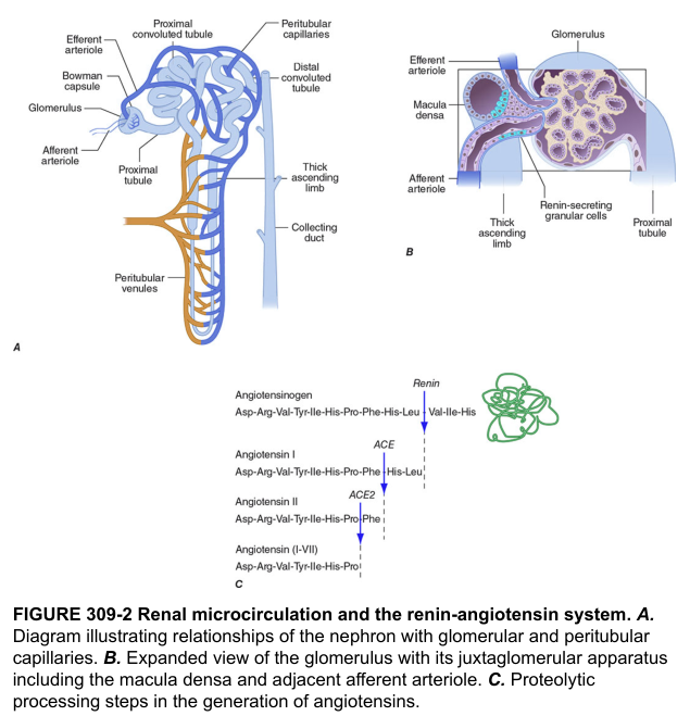
nephron diagram glomerulus proximal tubule loop of Henle collecting duct
lymphatic system diagram lymph nodes lymphatic vessels thoracic duct
urinary system overview kidneys ureters bladder urethra
Labeled educational diagram of the human kidney in coronal section (renal capsule, cortex, medulla, renal pyramids, renal columns, minor and major calyces, renal pelvis, ureter, renal artery and vein) with a labeled nephron beside it
Labeled educational diagram of the human lymphatic system: lymph nodes (cervical, axillary, inguinal), lymphatic vessels, thoracic duct, right lymphatic duct, spleen, thymus, tonsils, and drainage into subclavian veins
Labeled educational diagram of the general circulatory system showing heart, pulmonary and systemic circulation, arteries in red, veins in blue, capillary beds, and the lymphatic return to venous blood
Kidney, lymphatic and circulatory diagrams for exam
1. Kidney (coronal section) and nephron
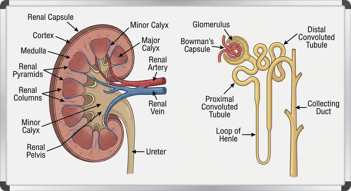
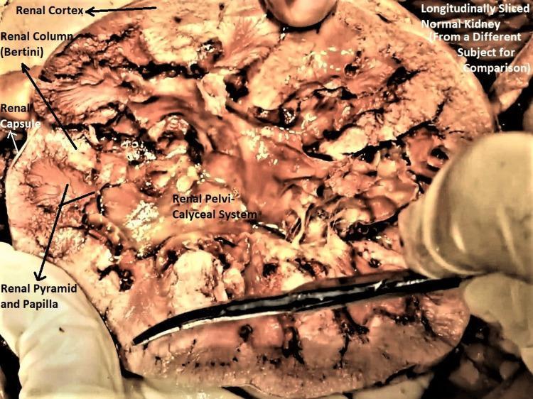
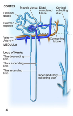
- Renal capsule
- Cortex, with the renal columns of Bertin dipping between the pyramids
- Medulla, made of pyramids ending in papillae
- Minor calyx, then major calyx, then renal pelvis, then ureter
- Renal artery and vein at the hilum
- Bowman's capsule and glomerulus
- Proximal convoluted tubule
- Descending limb of the loop of Henle
- Ascending limb of the loop of Henle
- Distal convoluted tubule
- Collecting duct, which opens at the papilla
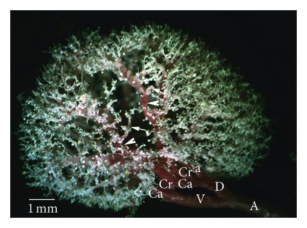
2. Lymphatic system
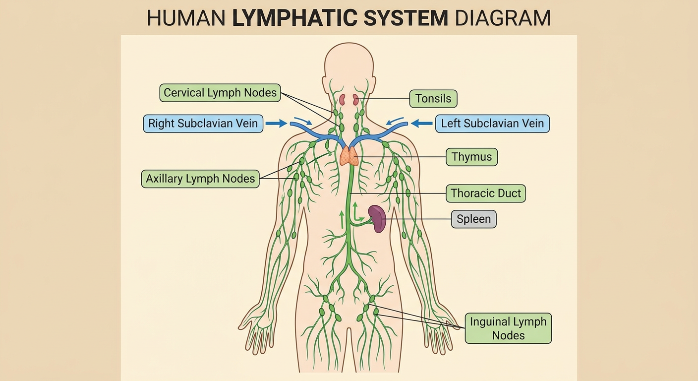
- Thoracic duct: it starts at the cisterna chyli, ascends through the aortic hiatus, and drains the whole body except the right upper quadrant. It empties at the left jugulosubclavian junction.
- Right lymphatic duct: it drains the right head and neck, the right arm and the right thorax. It empties into the right subclavian and internal jugular junction.
- Lymph node groups: cervical, axillary, inguinal, and para-aortic (lumbar) nodes.
- Lymphoid organs: thymus and spleen. The tonsils sit in the head and neck region.
- Functions:
- It returns interstitial fluid and protein to the blood.
- Lacteals absorb fat.
- It supports immune surveillance.
- Testicular lymph: it drains to the para-aortic nodes, not the inguinal nodes.
3. General circulation (blood and lymph return)
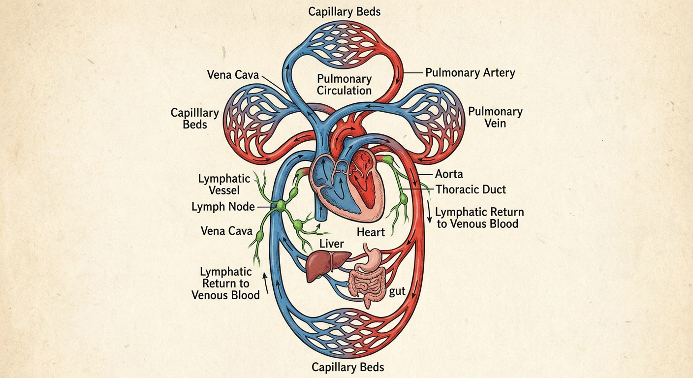
- Systemic circuit: left ventricle, aorta, arteries, arterioles, capillary beds, venules, veins, vena cava, right atrium.
- Pulmonary circuit: right ventricle, pulmonary artery (deoxygenated blood), lungs, pulmonary veins (oxygenated blood), left atrium.
- Portal circuit: gut capillaries, portal vein, liver sinusoids, hepatic veins, IVC.
- Lymph return: excess tissue fluid enters lymph capillaries, then lymph nodes, then the thoracic or right lymphatic duct, then the subclavian veins.
Exam drawing tips
- Use a pencil and label with straight, uncrossed lines.
- Use red for arteries and blue for veins.
- Draw arrows to show the direction of flow.
- Underline the structures that match the question's keywords.
Bro kidney lymphatis and its blood supply ? Diagram
renal lymphatic drainage kidney lymph nodes para-aortic
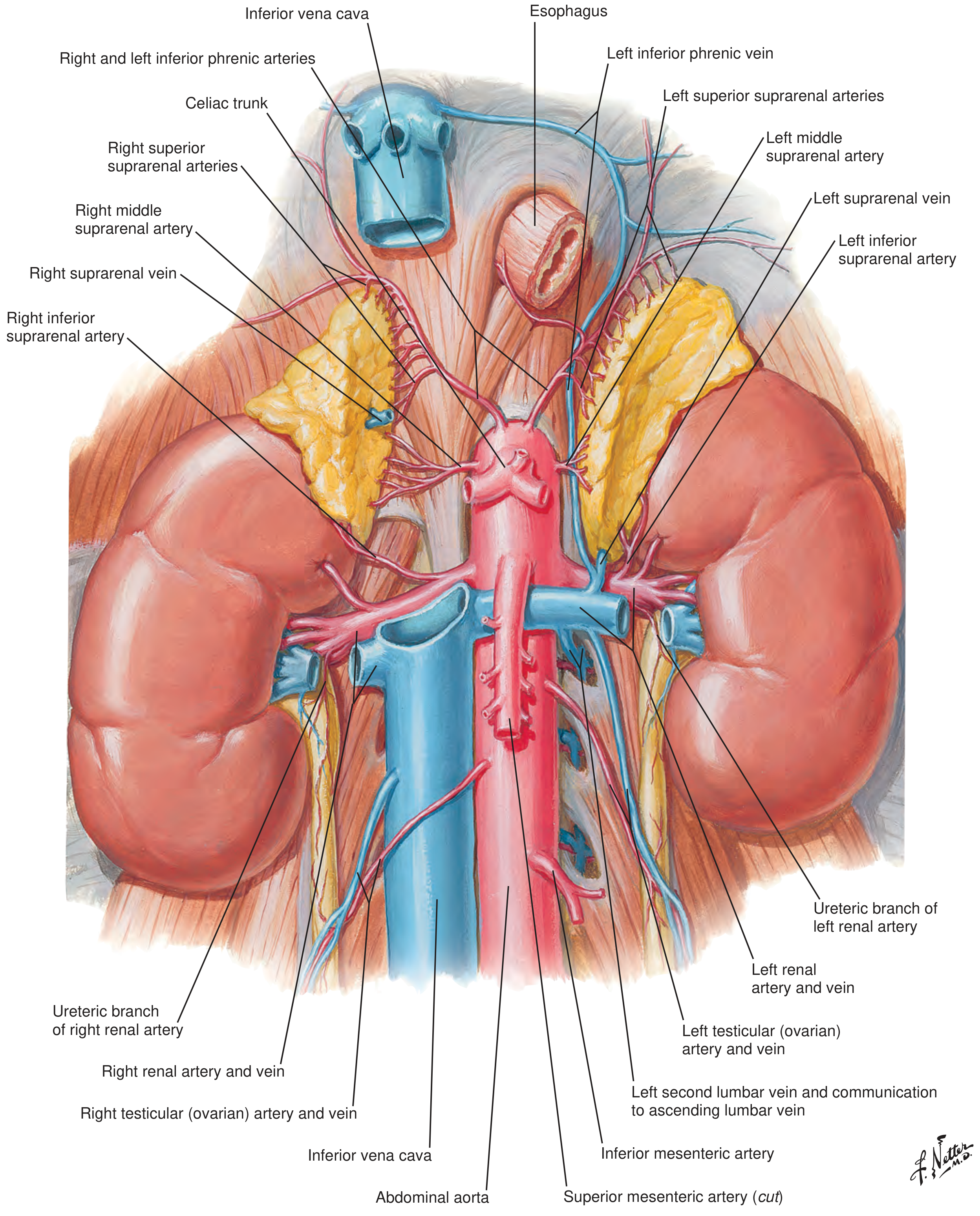
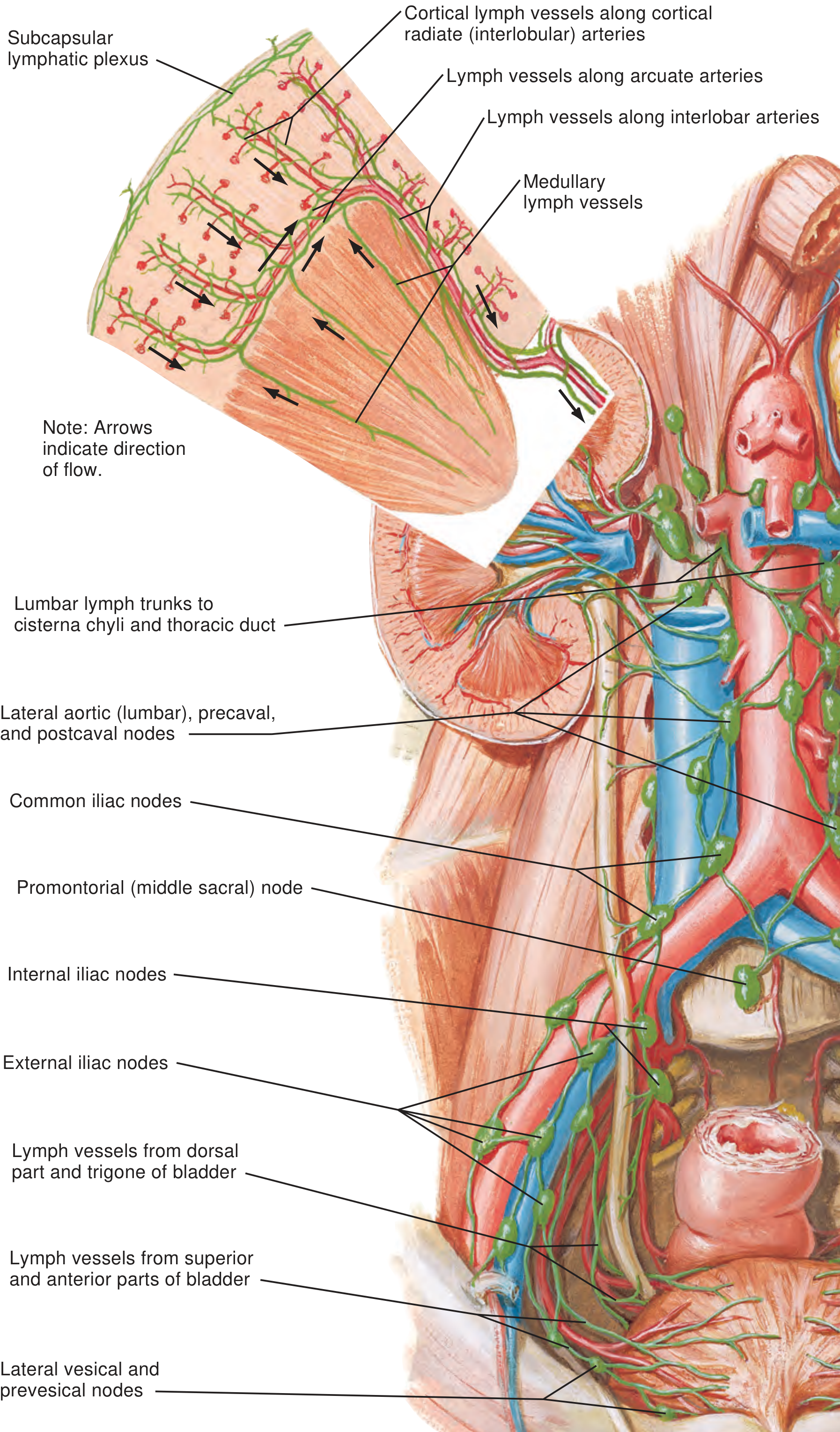
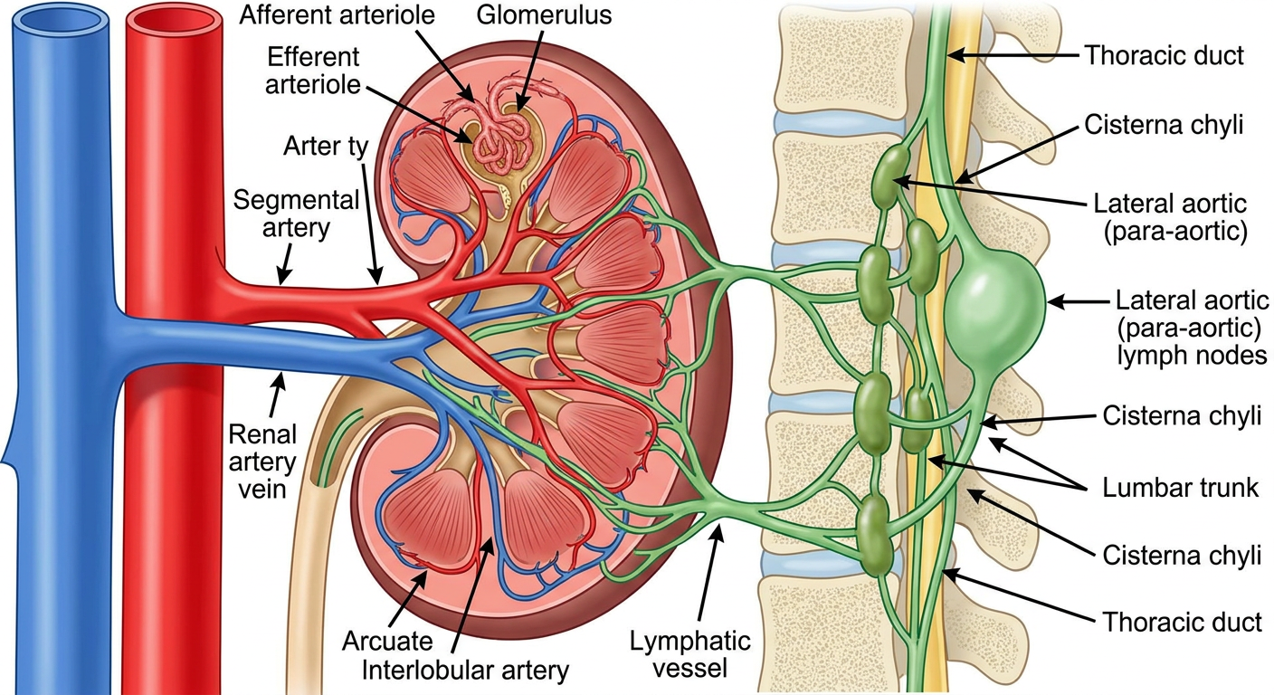
Labeled anatomical diagram of the kidney blood supply and lymphatic drainage: abdominal aorta, inferior vena cava, renal arteries and veins, segmental, interlobar, arcuate, interlobular arteries, afferent and efferent arterioles; green lymphatic vessels draining from the kidney hilum to lateral aortic (para-aortic) lymph nodes, then lumbar trunks, cisterna chyli and thoracic duct
Kidney: blood supply and lymphatic drainage
1. Diagrams
2. Blood supply (for exam)
- Segmental arteries are end arteries. There is no collateral flow, so blocking one causes segmental infarction.
- The renal artery gives off the inferior suprarenal and ureteric branches.
- The posterior segmental artery is usually the first branch of the main renal artery. It runs behind the renal pelvis (Campbell-Walsh Urology, Fig. 84.16).
- The five segments are apical, upper, middle, lower and posterior.
- The left renal vein is longer. It crosses between the superior mesenteric artery (in front) and the aorta (behind), and it receives the left gonadal, suprarenal and inferior phrenic veins.
- The left renal vein can be compressed here, which causes nutcracker syndrome.
- Anomalies are common. About 15% of people have supernumerary renal veins, and 2% to 16% have a circumaortic left renal vein (Campbell-Walsh Urology).
3. Lymphatic drainage (for exam)
- A superficial capsular network.
- A deeper hilar network, which follows the interlobular and arcuate arteries in the cortex. The arcuate vessels drain into the hilar vessels through interlobar lymphatics.
| Side | Main nodes |
|---|---|
| Left kidney | Left lateral aortic (para-aortic) nodes, between the inferior mesenteric artery and the diaphragm. Sometimes the retrocrural nodes or the thoracic duct directly. |
| Right kidney | Right interaortocaval and right paracaval nodes. Sometimes the retrocrural nodes or the left para-aortic nodes. |
4. Quick sketch plan for the exam
- Draw the aorta (red) and IVC (blue) as two vertical tubes.
- Draw a kidney on each side.
- Draw the renal artery as a horizontal red branch and the renal vein as a blue branch, with the left vein crossing in front of the aorta.
- Inside the kidney, draw the segmental, interlobar, arcuate and interlobular arteries.
- Draw green lymph vessels from the hilum to the para-aortic nodes beside the aorta, then up to the cisterna chyli and thoracic duct.
Femoral triangle Femoral sheath Popliteal fossa Knee joint Hip joint Great Saphenous Vein Foot drop Medial longitudinal Arch
Topic breakdown
- Femoral triangle: the boundaries are the inguinal ligament (base), sartorius (lateral) and adductor longus (medial). The floor is iliopsoas and pectineus. The roof is fascia lata, with the saphenous opening. From lateral to medial the contents are femoral nerve, artery, vein and lymphatics (NAVL).
- Femoral sheath: a funnel-shaped extension of the abdominal fascia (transversalis fascia and iliacus fascia) around the femoral vessels. It has 3 compartments: lateral (artery), intermediate (vein) and medial (femoral canal). The femoral nerve lies outside the sheath. The femoral canal is the site of femoral hernias.
- Popliteal fossa: a diamond-shaped space behind the knee. The boundaries are biceps femoris, semimembranosus and semitendinosus above, and the two heads of gastrocnemius below. Its contents are the tibial and common peroneal nerves, the popliteal vein and artery (the vein is superficial to the artery), small saphenous vein termination and lymph nodes. It is clinically important for popliteal aneurysm and Baker's cyst.
- Knee joint: a synovial hinge joint with some rotation. It has cruciate and collateral ligaments, menisci, bursae and the extensor mechanism. Movements are flexion, extension and a small amount of locked rotation. Key clinical points are the "unhappy triad" and the anterior drawer test.
- Hip joint: a ball-and-socket joint. It is stabilised by the labrum, the iliofemoral, pubofemoral and ischiofemoral ligaments, and the ligamentum teres. The blood supply is the medial and lateral circumflex femoral arteries, which matters for avascular necrosis after femoral neck fracture.
- Great saphenous vein: it starts at the medial end of the dorsal venous arch, passes in front of the medial malleolus, and ends in the femoral vein at the saphenofemoral junction. It has tributaries and perforators, and the saphenous nerve runs with it below the knee. It is used for varicose vein surgery and as a bypass graft.
- Foot drop: caused by common peroneal nerve injury, typically at the fibular neck. The result is loss of dorsiflexion and eversion, a high-stepping gait and sensory loss over the dorsum of the foot. Causes are fibular neck fracture, tight casts, L4-L5 radiculopathy and others.
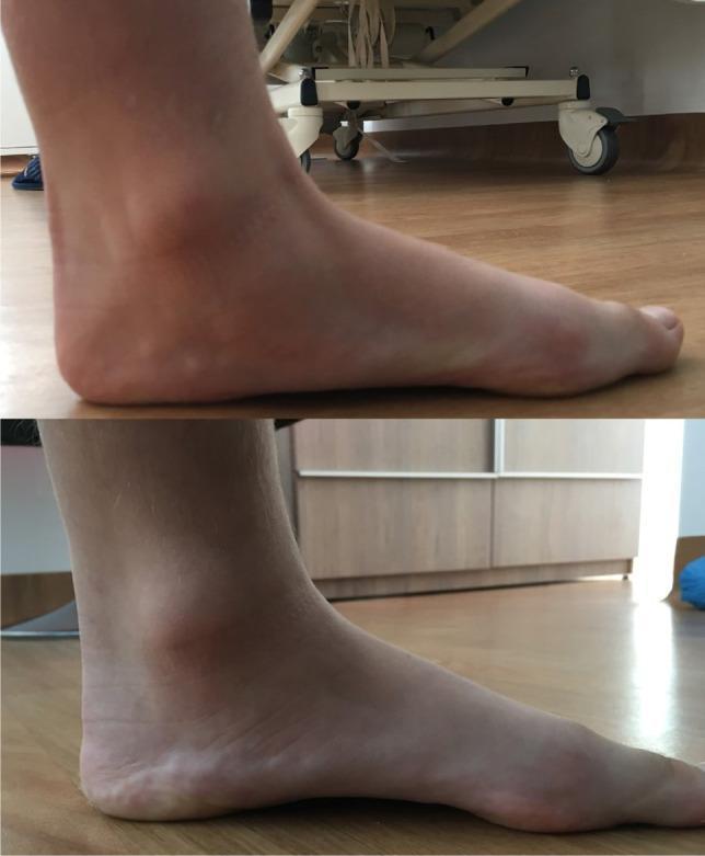
- Medial longitudinal arch: it is formed by the calcaneus, talus, navicular, three cuneiforms and the medial three metatarsals. It is supported statically by the spring ligament and plantar aponeurosis. The dynamic supports are tibialis posterior, flexor hallucis longus and tibialis anterior. Pes planus (flat foot) is the main clinical correlation.
Which topic should I cover in full detail first (diagram, textbook points and exam notes)?
Kidney, lymphatic and circulatory diagrams for exam
1. Kidney (coronal section) and nephron
- Renal capsule
- Cortex, with the renal columns of Bertin dipping between the pyramids
- Medulla, made of pyramids ending in papillae
- Minor calyx, then major calyx, then renal pelvis, then ureter
- Renal artery and vein at the hilum
- Bowman's capsule and glomerulus
- Proximal convoluted tubule
- Descending limb of the loop of Henle
- Ascending limb of the loop of Henle
- Distal convoluted tubule
- Collecting duct, which opens at the papilla
2. Lymphatic system
- Thoracic duct: it starts at the cisterna chyli, ascends through the aortic hiatus, and drains the whole body except the right upper quadrant. It empties at the left jugulosubclavian junction.
- Right lymphatic duct: it drains the right head and neck, the right arm and the right thorax. It empties into the right subclavian and internal jugular junction.
- Lymph node groups: cervical, axillary, inguinal, and para-aortic (lumbar) nodes.
- Lymphoid organs: thymus and spleen. The tonsils sit in the head and neck region.
- Functions:
- It returns interstitial fluid and protein to the blood.
- Lacteals absorb fat.
- It supports immune surveillance.
- Testicular lymph: it drains to the para-aortic nodes, not the inguinal nodes.
3. General circulation (blood and lymph return)
- Systemic circuit: left ventricle, aorta, arteries, arterioles, capillary beds, venules, veins, vena cava, right atrium.
- Pulmonary circuit: right ventricle, pulmonary artery (deoxygenated blood), lungs, pulmonary veins (oxygenated blood), left atrium.
- Portal circuit: gut capillaries, portal vein, liver sinusoids, hepatic veins, IVC.
- Lymph return: excess tissue fluid enters lymph capillaries, then lymph nodes, then the thoracic or right lymphatic duct, then the subclavian veins.
Exam drawing tips
- Use a pencil and label with straight, uncrossed lines.
- Use red for arteries and blue for veins.
- Draw arrows to show the direction of flow.
- Underline the structures that match the question's keywords.
1. Diagrams
2. Blood supply (for exam)
- Segmental arteries are end arteries. There is no collateral flow, so blocking one causes segmental infarction.
- The renal artery gives off the inferior suprarenal and ureteric branches.
- The posterior segmental artery is usually the first branch of the main renal artery. It runs behind the renal pelvis (Campbell-Walsh Urology, Fig. 84.16).
- The five segments are apical, upper, middle, lower and posterior.
- The left renal vein is longer. It crosses between the superior mesenteric artery (in front) and the aorta (behind), and it receives the left gonadal, suprarenal and inferior phrenic veins.
- The left renal vein can be compressed here, which causes nutcracker syndrome.
- Anomalies are common. About 15% of people have supernumerary renal veins, and 2% to 16% have a circumaortic left renal vein (Campbell-Walsh Urology).
3. Lymphatic drainage (for exam)
- A superficial capsular network.
- A deeper hilar network, which follows the interlobular and arcuate arteries in the cortex. The arcuate vessels drain into the hilar vessels through interlobar lymphatics.
| Side | Main nodes |
|---|---|
| Left kidney | Left lateral aortic (para-aortic) nodes, between the inferior mesenteric artery and the diaphragm. Sometimes the retrocrural nodes or the thoracic duct directly. |
| Right kidney | Right interaortocaval and right paracaval nodes. Sometimes the retrocrural nodes or the left para-aortic nodes. |
4. Quick sketch plan for the exam
- Draw the aorta (red) and IVC (blue) as two vertical tubes.
- Draw a kidney on each side.
- Draw the renal artery as a horizontal red branch and the renal vein as a blue branch, with the left vein crossing in front of the aorta.
- Inside the kidney, draw the segmental, interlobar, arcuate and interlobular arteries.
- Draw green lymph vessels from the hilum to the para-aortic nodes beside the aorta, then up to the cisterna chyli and thoracic duct.
Topic breakdown
- Femoral triangle: the boundaries are the inguinal ligament (base), sartorius (lateral) and adductor longus (medial). The floor is iliopsoas and pectineus. The roof is fascia lata, with the saphenous opening. From lateral to medial the contents are femoral nerve, artery, vein and lymphatics (NAVL).
- Femoral sheath: a funnel-shaped extension of the abdominal fascia (transversalis fascia and iliacus fascia) around the femoral vessels. It has 3 compartments: lateral (artery), intermediate (vein) and medial (femoral canal). The femoral nerve lies outside the sheath. The femoral canal is the site of femoral hernias.
- Popliteal fossa: a diamond-shaped space behind the knee. The boundaries are biceps femoris, semimembranosus and semitendinosus above, and the two heads of gastrocnemius below. Its contents are the tibial and common peroneal nerves, the popliteal vein and artery (the vein is superficial to the artery), small saphenous vein termination and lymph nodes. It is clinically important for popliteal aneurysm and Baker's cyst.
- Knee joint: a synovial hinge joint with some rotation. It has cruciate and collateral ligaments, menisci, bursae and the extensor mechanism. Movements are flexion, extension and a small amount of locked rotation. Key clinical points are the "unhappy triad" and the anterior drawer test.
- Hip joint: a ball-and-socket joint. It is stabilised by the labrum, the iliofemoral, pubofemoral and ischiofemoral ligaments, and the ligamentum teres. The blood supply is the medial and lateral circumflex femoral arteries, which matters for avascular necrosis after femoral neck fracture.
- Great saphenous vein: it starts at the medial end of the dorsal venous arch, passes in front of the medial malleolus, and ends in the femoral vein at the saphenofemoral junction. It has tributaries and perforators, and the saphenous nerve runs with it below the knee. It is used for varicose vein surgery and as a bypass graft.
- Foot drop: caused by common peroneal nerve injury, typically at the fibular neck. The result is loss of dorsiflexion and eversion, a high-stepping gait and sensory loss over the dorsum of the foot. Causes are fibular neck fracture, tight casts, L4-L5 radiculopathy and others.
- Medial longitudinal arch: it is formed by the calcaneus, talus, navicular, three cuneiforms and the medial three metatarsals. It is supported statically by the spring ligament and plantar aponeurosis. The dynamic supports are tibialis posterior, flexor hallucis longus and tibialis anterior. Pes planus (flat foot) is the main clinical correlation.
Bro for 6 marks explain with diagrams
femoral triangle boundaries contents femoral nerve artery vein
femoral sheath femoral canal compartments
popliteal fossa boundaries contents tibial nerve popliteal artery
knee joint ligaments anterior cruciate collateral menisci diagram
hip joint ligaments iliofemoral pubofemoral ischiofemoral anatomy
great saphenous vein course tributaries saphenofemoral junction
common peroneal nerve foot drop fibular neck
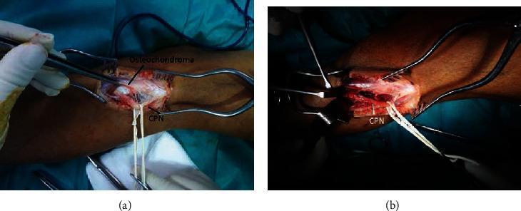
This dual-frame clinical photograph displays an intraoperative comparison of the common peroneal nerve (CPN) at the level of the fibular neck during surgical decompression. Panel (a) shows the initial surgical field where an osteochondroma, a benign bone exostosis, is directly compressing the CPN. In this state, the nerve appears pathologically flattened, thinned, and exhibits visual signs of mechanical displacement and secondary inflammation of the epineurium. Panel (b) illustrates the surgical site following the complete excision of the osteochondroma and subsequent neurolysis. In this postoperative view, the common peroneal nerve has regained a more physiological, rounded morphology and appears relieved from the localized pressure. The images are categorized under orthopedic surgery and neurosurgery, illustrating a common cause of peripheral nerve entrapment syndrome (foot drop) and the restorative effects of surgical decompression. The anatomical focus is on the lateral aspect of the knee and the relationship between bone lesions and adjacent neural structures.
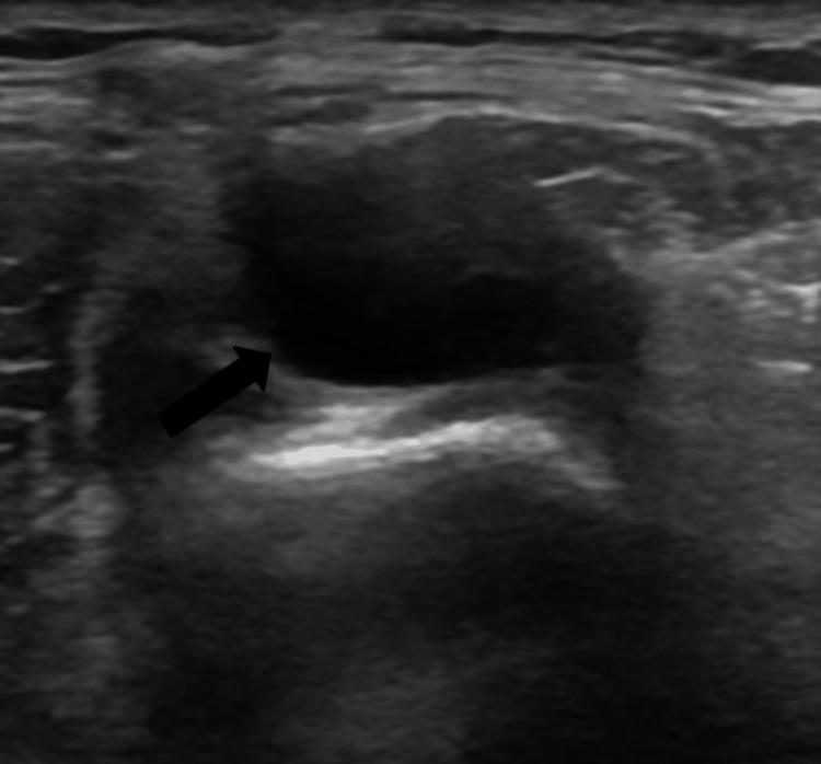
This grayscale diagnostic ultrasound image captures a transverse view of the right lower extremity at the level of the fibular tunnel. The image demonstrates a well-defined, heterogeneous soft tissue mass (indicated by a black arrow) characterized by mixed echogenicity. The mass contains a prominent anechoic (dark) cystic component, suggesting fluid collection, adjacent to hypoechoic and slightly hyperechoic solid components. This complex, cystic-solid morphology is located in close proximity to the common peroneal nerve and the fibular head. Anatomically, such findings are highly suggestive of an intraneural ganglion cyst, particularly when associated with clinical symptoms such as foot drop or pain in the fibular neck region. The high contrast between the anechoic fluid and the surrounding intermediate echotexture of the muscle and neural tissue facilitates the identification of the mass's borders. This visual is used to teach diagnostic imaging for peripheral nerve compression syndromes and musculoskeletal pathology.
medial longitudinal arch of foot bones ligaments spring ligament
Labeled anatomical diagram of the right femoral triangle (anterior thigh): inguinal ligament as base, sartorius lateral border, adductor longus medial border, femoral nerve, femoral artery, femoral vein, femoral canal with lymphatics (lateral to medial order N-A-V-L), floor iliopsoas and pectineus, great saphenous vein joining at saphenous opening
Labeled diagram of the femoral sheath in coronal section showing three compartments: lateral compartment with femoral artery, intermediate compartment with femoral vein, medial compartment femoral canal with lymphatics and lymph node of Cloquet; femoral nerve outside the sheath; femoral ring, inguinal ligament, fascia transversalis and fascia iliaca forming the sheath
Labeled anatomical diagram of the right popliteal fossa, diamond shaped: biceps femoris superolateral, semimembranosus and semitendinosus superomedial, lateral and medial heads of gastrocnemius inferior, plus contents: tibial nerve, common peroneal nerve, popliteal vein, popliteal artery (deepest), small saphenous vein, popliteal lymph nodes
Labeled diagram of the medial longitudinal arch of the foot, medial view of bones: calcaneus, talus (keystone), navicular, three cuneiforms, first to third metatarsals, with spring ligament, plantar aponeurosis, long plantar ligament, tibialis posterior tendon, flexor hallucis longus tendon and tibialis anterior tendon as supports
Lower limb anatomy: 8 topics for a 6-mark answer
1. Femoral triangle
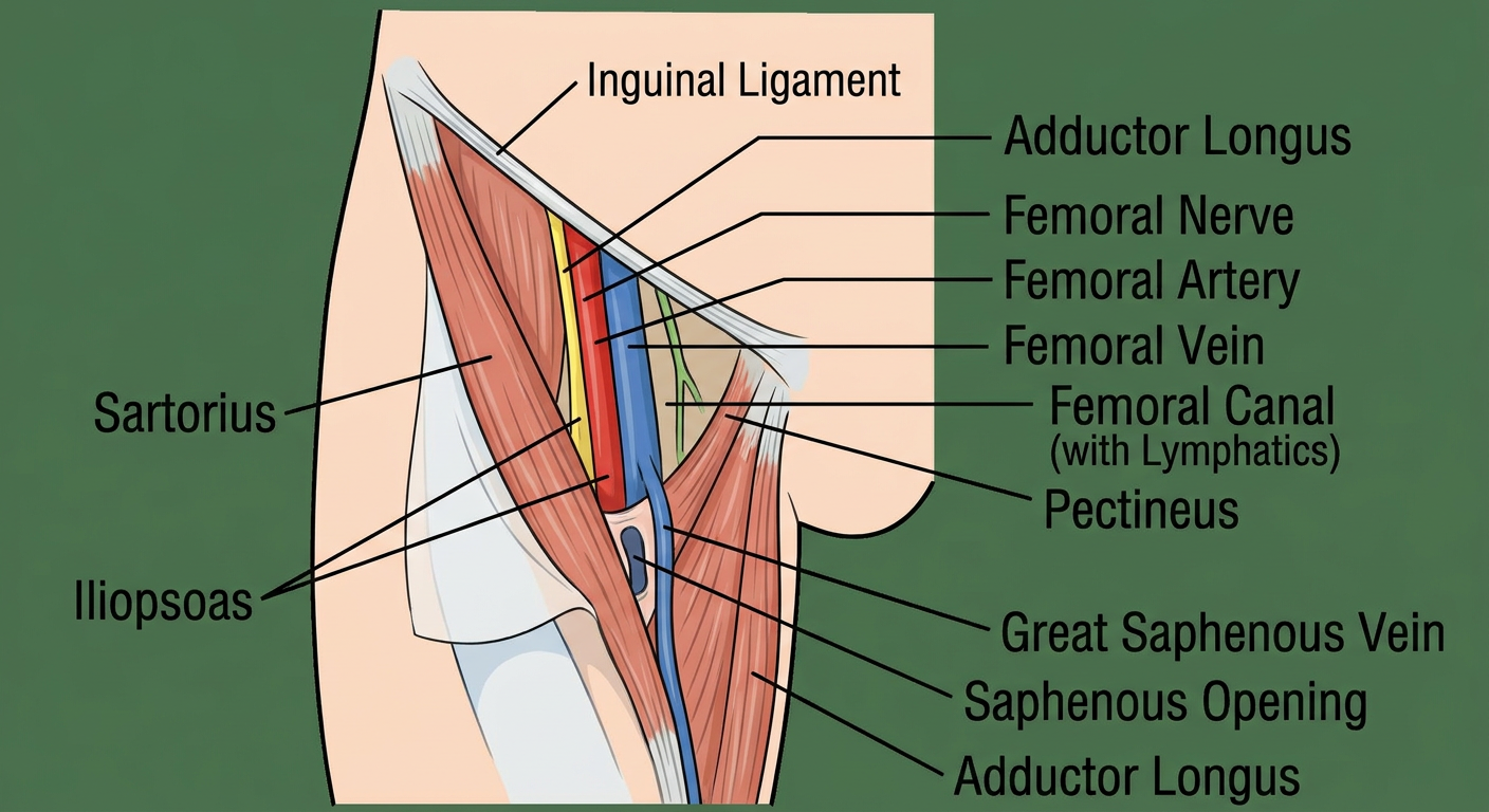
- Definition: a triangular depression in the upper anterior thigh.
- Boundaries:
- Base: inguinal ligament.
- Lateral: medial border of sartorius.
- Medial: medial border of adductor longus.
- Apex: where sartorius crosses adductor longus. It continues as the adductor canal.
- Roof: skin, superficial fascia (with superficial inguinal nodes and the great saphenous vein), then fascia lata with the saphenous opening.
- Floor (lateral to medial): iliopsoas, pectineus, adductor longus.
- Contents (lateral to medial): femoral Nerve, Artery, Vein, then the femoral canal with Lymphatics (mnemonic NAVL). The femoral nerve is outside the femoral sheath.
- Other contents:
- Femoral branch of the genitofemoral nerve.
- Deep inguinal nodes.
- Profunda femoris artery.
- Saphenofemoral junction.
- Clinical:
- The femoral pulse is felt at the mid-inguinal point.
- Femoral artery catheterisation.
- Femoral hernia.
- Femoral nerve block.
2. Femoral sheath
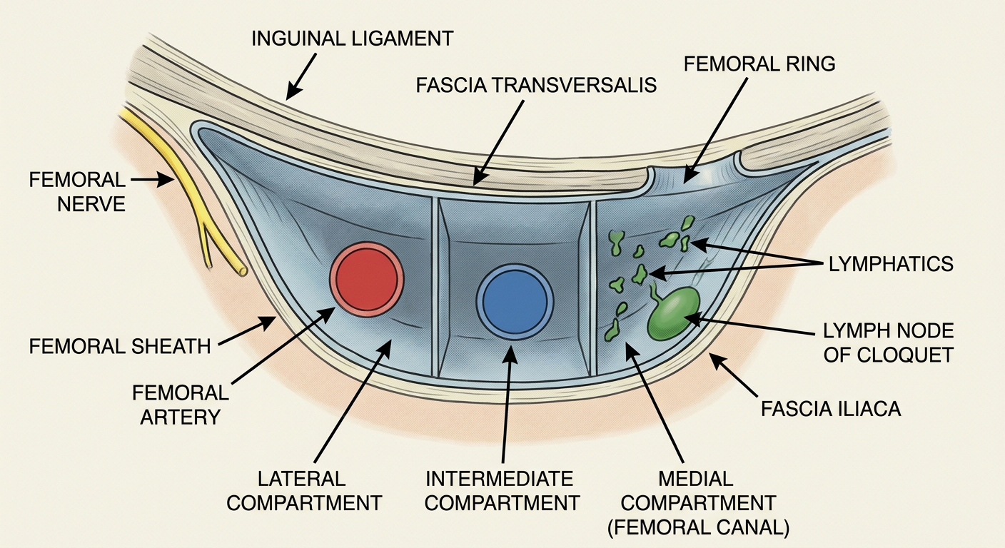
- Definition: a funnel-shaped fascial sleeve about 3 to 4 cm long around the femoral vessels, below the inguinal ligament.
- Formation: the anterior wall is fascia transversalis and the posterior wall is fascia iliaca (iliacus and psoas fascia). It ends by blending with the vessel adventitia.
- Three compartments:
- Lateral: femoral artery, plus the femoral branch of the genitofemoral nerve.
- Intermediate: femoral vein.
- Medial: femoral canal (about 1.25 cm long), containing lymphatics, the lymph node of Cloquet and fat.
- Femoral ring (upper opening of the canal):
- Anterior: inguinal ligament.
- Posterior: pectineal ligament and pectineus.
- Medial: lacunar ligament.
- Lateral: femoral vein.
- It is closed by the femoral septum.
- Functions: lets the vessels glide, and lets the femoral vein distend.
- Clinical: a femoral hernia passes through the ring into the canal. It is more common in women, lies below and lateral to the pubic tubercle, and strangulates easily because the neck is narrow and rigid.
3. Popliteal fossa
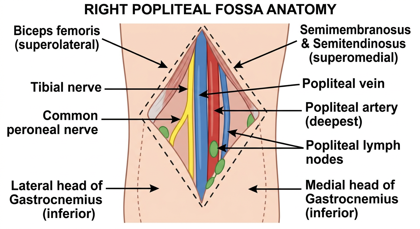
- Definition: a diamond-shaped intermuscular space behind the knee.
- Boundaries:
- Superolateral: biceps femoris.
- Superomedial: semimembranosus, overlapped by semitendinosus.
- Inferolateral: lateral head of gastrocnemius, with plantaris.
- Inferomedial: medial head of gastrocnemius.
- Roof: skin and popliteal fascia. The small saphenous vein pierces it.
- Floor: popliteal surface of the femur, oblique popliteal ligament, knee capsule, popliteus.
- Contents (superficial to deep):
- Tibial nerve.
- Popliteal vein.
- Popliteal artery, the deepest.
- Other contents:
- Common peroneal nerve.
- Small saphenous vein termination.
- Popliteal lymph nodes.
- Genicular vessels.
- Posterior cutaneous nerve of the thigh.
- Fat.
- Clinical:
- Popliteal aneurysm.
- Baker's cyst (semimembranosus bursa).
- Artery injury in knee dislocation.
- The popliteal pulse is best felt with the knee flexed.
4. Knee joint
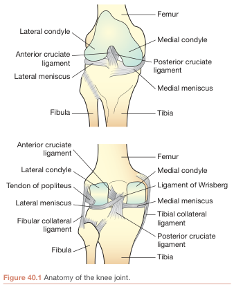
- Type: compound synovial joint, a modified hinge with some rotation. It has two condylar articulations (tibiofemoral) and one patellofemoral articulation.
- Intracapsular ligaments:
- ACL: prevents anterior displacement of the tibia.
- PCL: prevents posterior displacement, and matters when walking downhill.
- Extracapsular ligaments:
- Patellar ligament.
- Tibial collateral (medial).
- Fibular collateral (lateral).
- Oblique popliteal.
- Arcuate popliteal.
- Menisci:
- Medial: C-shaped, attached to the tibial collateral ligament, and injured more often.
- Lateral: O-shaped, more mobile.
- They deepen the joint and absorb shock.
- Movements:
- Flexion: hamstrings.
- Extension: quadriceps.
- Rotation: lateral by biceps femoris, medial by popliteus and the medial hamstrings.
- Locking: medial rotation of the femur on the tibia, and the popliteus unlocks it.
- Blood supply: genicular branches of the popliteal artery.
- Nerve supply: femoral, obturator, tibial and common peroneal nerves (Hilton's law).
- Clinical:
- "Unhappy triad": ACL, tibial collateral ligament and medial meniscus.
- Anterior drawer and Lachman tests.
- Prepatellar bursitis ("housemaid's knee").
5. Hip joint
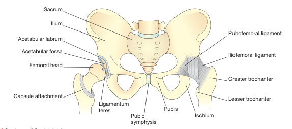
- Type: synovial ball-and-socket joint between the femoral head and the acetabulum.
- Stability:
- Deep socket.
- Acetabular labrum.
- Strong capsule.
- Transverse acetabular ligament.
- Ligaments:
- Iliofemoral (Y-shaped, strongest): limits hyperextension.
- Pubofemoral: limits excessive abduction.
- Ischiofemoral (weakest): limits medial rotation.
- Ligamentum teres: carries a small artery to the head.
- Movements and main muscles:
- Flexion: iliopsoas.
- Extension: gluteus maximus.
- Abduction: gluteus medius and minimus.
- Adduction: adductors.
- Medial rotation: gluteus minimus and tensor fasciae latae.
- Lateral rotation: short external rotators.
- Blood supply:
- Medial and lateral circumflex femoral arteries (the main supply).
- Artery of the ligamentum teres.
- Gluteal branches.
- Nerve supply: femoral, obturator, sciatic and superior gluteal nerves. Hip pain can be referred to the knee through the obturator nerve.
- Clinical:
- Posterior dislocation: limb flexed, adducted and medially rotated.
- Femoral neck fracture risks avascular necrosis of the head.
6. Great saphenous vein (GSV)
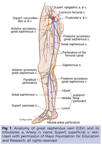
- Origin: the medial end of the dorsal venous arch of the foot.
- Course:
- In front of the medial malleolus.
- Up the medial leg, along the medial border of the tibia.
- A hand's breadth behind the medial border of the patella.
- Up the medial thigh.
- Through the saphenous opening, about 4 cm below and lateral to the pubic tubercle.
- Into the femoral vein at the saphenofemoral junction.
- Tributaries at the junction:
- Superficial circumflex iliac vein.
- Superficial epigastric vein.
- Superficial and deep external pudendal veins.
- Other tributaries: anterior and posterior accessory saphenous veins.
- Structure: it has 10 to 20 valves.
- Perforators: they join it to the deep veins (Hunter, Dodd, Boyd and Cockett).
- Relations: the saphenous nerve accompanies it below the knee.
- Clinical:
- Varicose veins (valve incompetence), treated with saphenofemoral ligation and stripping.
- Coronary bypass graft.
- Venous cutdown in front of the medial malleolus.
7. Foot drop
- Definition: inability to dorsiflex the foot, so the toes drag during the swing phase of gait.
- Anatomy: the common peroneal nerve comes off the sciatic nerve in the popliteal fossa. It winds round the fibular neck, where it lies superficial and is easily injured, and divides into:
- Superficial peroneal nerve: evertors (peroneus longus and brevis), plus sensation on the lower lateral leg and dorsum of the foot.
- Deep peroneal nerve: dorsiflexors (tibialis anterior, extensor hallucis longus, extensor digitorum longus, peroneus tertius), plus sensation in the first web space.
- Features:
- Loss of dorsiflexion and eversion.
- High-stepping (steppage) gait with foot slap.
- Sensory loss over the lateral leg and dorsum of the foot.
- Causes:
- Fibular neck fracture.
- Tight cast or habitual leg crossing.
- Knee dislocation.
- Ganglion cyst or tumour.
- L5 radiculopathy (this also weakens inversion and hip abduction).
- Sciatic nerve injury.
- Diabetic neuropathy.
- Management:
- Treat the cause.
- Ankle-foot orthosis.
- Nerve decompression or repair.
- Tibialis posterior tendon transfer.
8. Medial longitudinal arch
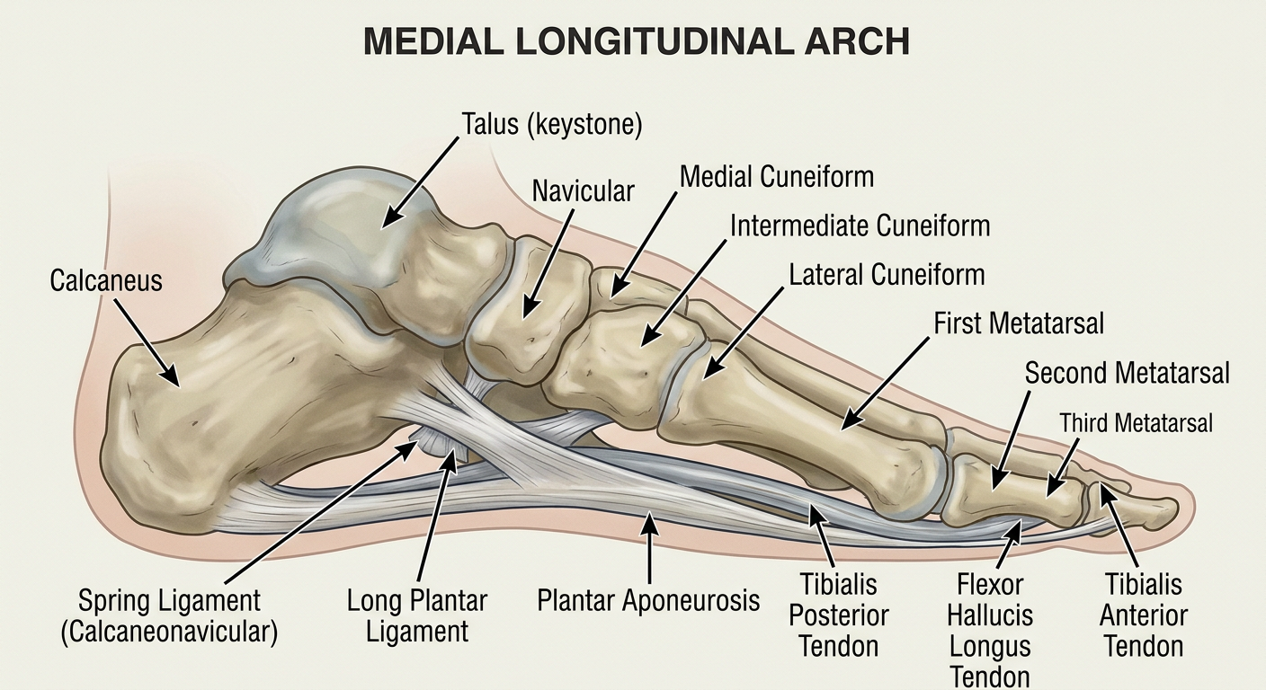
- Bones: calcaneus, talus (the keystone and highest point), navicular, three cuneiforms, medial three metatarsals.
- Pillars: the posterior pillar is the calcaneal tuberosity, and the anterior pillar is the head of the 1st metatarsal.
- Static supports:
- Shape of the bones.
- Spring (plantar calcaneonavicular) ligament, which supports the head of the talus.
- Plantar aponeurosis.
- Long and short plantar ligaments.
- Deltoid ligament.
- Dynamic supports:
- Tibialis posterior (the most important).
- Tibialis anterior.
- Flexor hallucis longus.
- Flexor digitorum longus.
- Abductor hallucis.
- Peroneus longus.
- Functions: shock absorption, weight transmission, a spring for push-off, and protection of the plantar vessels and nerves.
- Clinical:
- Flat foot (pes planus) from spring ligament or tibialis posterior failure.
- Plantar fasciitis.
- Pes cavus (high arch).
Exam tips
- Use a pencil, draw the outline first, and label with straight, uncrossed lines.
- For nerves, arteries and veins, use yellow, red and blue.
- For a 6-mark answer, aim for one diagram plus 6 or 7 key points: definition, boundaries or structure, contents or supports, and one clinical correlation.