

photo-1786443283901.jpg
Daignosis
I'll analyze this brain MRI image right away.
analyse_media
Brain MRI Analysis - Radiological Report
Disclaimer: This is an educational analysis only. Formal diagnosis must be made by a qualified radiologist or neurologist with access to the full DICOM dataset and clinical history.
Patient Details (from screen)
- Patient: Bishan Pandey, 75 years old
- Sequences visible: T2 FSE (axial) | FLAIR (axial) | DWI (axial)
Key Radiological Findings
1. Severe White Matter Hyperintensities (Leukoaraiosis)
- FLAIR & T2: Extensive, confluent, bilateral periventricular and deep white matter hyperintensities
- This pattern is consistent with Fazekas Grade 3 small vessel ischemic disease
- Classic features of chronic cerebrovascular disease in an elderly patient
2. Ventriculomegaly (Central Atrophy)
- Bilateral lateral ventricles are significantly and symmetrically enlarged
- Indicates parenchymal volume loss - cerebral atrophy appropriate to age but likely pathological given the degree
- Must be correlated clinically to exclude Normal Pressure Hydrocephalus (NPH) - the triad of gait disturbance, cognitive decline, and urinary incontinence
3. Cortical Sulcal Widening
- Prominent sulci seen bilaterally - consistent with generalized cortical atrophy
4. DWI Findings
- The DWI sequence needs correlation with ADC maps (not fully visible) to assess for acute ischemic stroke - no obvious bright DWI restriction lesion is apparent from the displayed image, but this requires formal review
Differential Diagnosis (Most to Least Likely)
| Rank | Diagnosis | Supporting Features |
|---|---|---|
| 1 | Chronic Small Vessel Disease / Vascular Dementia | Fazekas 3 white matter changes, age 75, symmetric |
| 2 | Alzheimer's Disease + SVD overlap | Cortical + central atrophy in elderly |
| 3 | Normal Pressure Hydrocephalus | Ventriculomegaly - needs clinical triad correlation |
| 4 | Subcortical Ischemic Vascular Disease (Binswanger's) | Confluent periventricular white matter changes |
| 5 | Acute-on-chronic infarct | DWI needed for confirmation |
Clinical Correlation Recommended
- History of hypertension, diabetes, hyperlipidemia, atrial fibrillation
- Cognitive screening (MMSE/MoCA)
- Gait assessment
- Blood pressure review
- Consider neurology/neuroradiology referral for complete reporting
Binswangers disease
Binswanger's disease (subcortical ischemic vascular dementia) is a chronic progressive condition caused by widespread small vessel disease leading to white matter damage. Let me pull detailed information from medical textbooks and imaging examples.
Searching sources...
Reading File
Finding Sources
Finding Sources
Searching Images
Binswanger disease subcortical leukoencephalopathy MRI white matter hyperintensity
Reading File
Reading File
Searching PubMed
Binswanger disease subcortical arteriosclerotic encephalopathy
Loading Image
Finding Sources
Reading File
Reading File
Here is a comprehensive summary of Binswanger's Disease drawn from multiple authoritative neurology and psychiatry textbooks.
Binswanger's Disease (Subcortical Arteriosclerotic Encephalopathy)
Gross Pathology
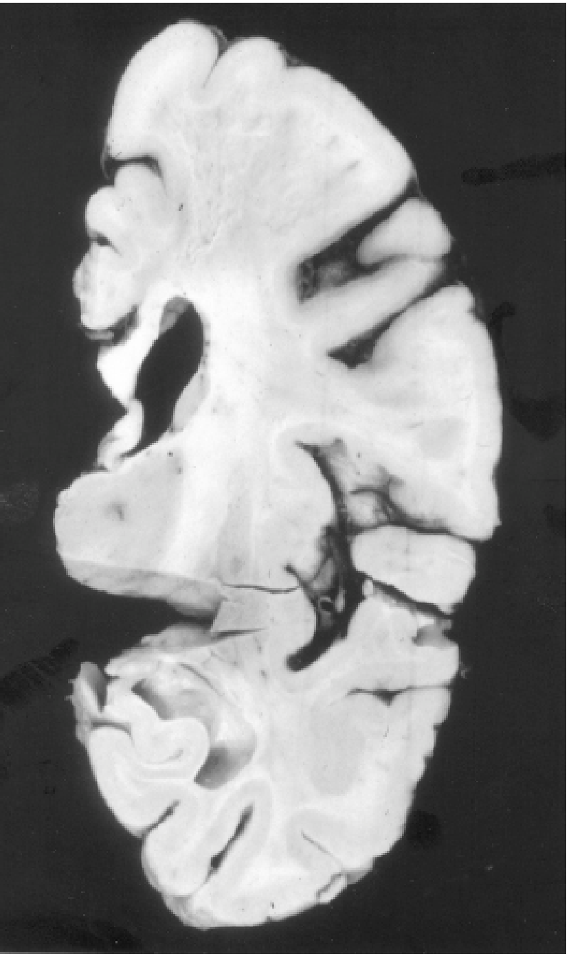
Below is the classic gross pathological cross-section from Kaplan & Sadock's Comprehensive Textbook of Psychiatry, demonstrating the hallmark finding - extensive subcortical white matter infarction with sparing of the overlying cortical gray matter:
Definition & Terminology
Binswanger's disease goes by several names:
- Subcortical Arteriosclerotic Encephalopathy (SAE)
- Leukoaraiosis (when referring to the imaging appearance)
- Subcortical Ischemic Vascular Dementia (SIVD)
It denotes a widespread degeneration of cerebral white matter occurring in the setting of hypertension, small vessel atherosclerosis, and multiple (often silent) strokes. - Adams and Victor's Principles of Neurology, 12th Ed.
Previously considered rare, MRI has revealed this condition is far more common than originally thought. - Kaplan & Sadock's Synopsis of Psychiatry
Pathophysiology
| Factor | Detail |
|---|---|
| Primary mechanism | Chronic ischemia of deep white matter from small arteriolar sclerosis |
| Vascular target | Small- and medium-sized cerebral vessels (arterioles) |
| Key paradox | Vessel lumens are often OPEN (patent) despite arteriolar sclerosis - Fisher, 1989 |
| White matter damage | Multiple small infarctions, gliosis, demyelination, increased water content |
| Cortex | SPARED - infarctions are subcortical |
The condition likely exists on a continuum with leukoaraiosis - many elderly individuals with white matter changes show cognitive impairment even without a formal Binswanger's diagnosis. - Adams and Victor's Principles of Neurology
Clinical Features (The Triad)
| Feature | Description |
|---|---|
| Dementia | Gradual cognitive deterioration, subcortical pattern - executive dysfunction, slowed processing |
| Pseudobulbar state | Dysarthria, dysphagia, emotional lability |
| Gait disorder | Frontal-pattern gait, small-stepped, wide-based ("marche à petits pas") |
Additional features:
- Hemiparesis
- TIAs
- Lacunar or cortical strokes (often admixed)
- Urinary incontinence (when combined with gait disorder - NPH overlap)
MRI Findings (Key Imaging Features)
From Grainger & Allison's Diagnostic Radiology and Harrison's Principles of Internal Medicine (2025):
FLAIR sequence is the best sequence for detecting small-vessel white matter disease.
| Finding | Description |
|---|---|
| White matter hyperintensities (FLAIR/T2) | Confluent, bilateral, periventricular and deep white matter |
| Distribution | Predominantly frontal and parietal deep white matter |
| Spared areas | U-fibres (subcortical arcuate fibres) and temporal lobes |
| Lacunes | Multiple small hypointense cavities (basal ganglia, centrum semiovale) |
| T1 | Severe lesions become T1 hypointense (tissue destruction) |
| Basal ganglia & pons | Frequently involved |
| DWI | Recent white matter lesions can be bright (acute lacunar infarction) |
Extremely severe diffuse white matter vascular injury is what defines Binswanger's - Harrison's Principles of Internal Medicine, 2025
Fazekas Grading (White Matter Lesion Severity)
| Grade | Description |
|---|---|
| 0 | No lesions |
| 1 | Punctate foci |
| 2 | Beginning confluence |
| 3 | Large confluent areas = consistent with Binswanger's |
The MRI in the patient (Bishan Pandey, 75y) showed Fazekas Grade 3 - the most severe category.
Differential Diagnosis
| Condition | Key Distinguishing Feature |
|---|---|
| CADASIL | Genetic (Notch-3 mutation), younger onset, temporal lobe involvement, family history |
| Multiple Sclerosis | Involves U-fibres and temporal lobes, periventricular lesions are more perpendicular ("Dawson's fingers") |
| Normal Pressure Hydrocephalus | Ventriculomegaly disproportionate to atrophy, classic triad (gait, cognition, incontinence) |
| Alzheimer's Disease | Cortical/hippocampal atrophy predominant, less white matter change |
| CARASIL | Recessive, alopecia, lumbar spondylosis |
Risk Factors
- Hypertension (most important)
- Diabetes mellitus
- Hyperlipidaemia
- Atrial fibrillation
- Smoking
- Advancing age (predominantly 60s-80s)
- Male sex
Management
There is no disease-specific treatment. Management is focused on:
- Aggressive vascular risk factor control
- Antihypertensives (target BP <130/80 mmHg)
- Statins
- Antiplatelet therapy (if stroke/TIA present)
- Glycaemic control
- Cognitive rehabilitation
- Symptomatic treatment of dementia (cholinesterase inhibitors have limited but sometimes used role)
- Fall prevention and physiotherapy for gait disorder
- Carer support and advance care planning
Relation to the Scanned Patient
The MRI findings seen in Bishan Pandey (75y) - confluent bilateral periventricular white matter T2/FLAIR hyperintensities (Fazekas 3), ventriculomegaly, and cortical atrophy - are classic for Binswanger's disease / subcortical ischemic vascular dementia. Clinical correlation with hypertension history, cognitive screening (MoCA/MMSE), and gait assessment is advised.
Sources: Adams and Victor's Principles of Neurology 12th Ed. | Kaplan & Sadock's Synopsis of Psychiatry | Grainger & Allison's Diagnostic Radiology | Harrison's Principles of Internal Medicine 2025
This is a shared conversation. Sign in to Orris to start your own chat.