
Create me a real life case scenario of ARDS due to pneumonia in Indian scenario
"ARDS" AND "pneumonia" AND "India"
Clinical Case Scenario: ARDS Secondary to Community-Acquired Pneumonia
Setting
Patient Profile
- Poorly controlled Type 2 Diabetes Mellitus (on irregular metformin, no monitoring)
- Chronic tobacco chewer (>20 years)
- Possible undiagnosed COPD (heavy agricultural dust exposure)
- Nutritional deficiency - BMI 17.8 kg/m² (chronic undernutrition)
History of Present Illness
- His wife (Lalitha, 48) provided history as the patient was too dyspneic to speak in full sentences
- She reported he had been working in waterlogged paddy fields for the past 3 weeks during the kharif season (monsoon, August)
- He self-medicated for the first 2 days with a local ayurvedic preparation and delayed seeking formal care
- No hospital record of prior HRCT or PFT
- He missed his metformin for the past 10 days because the ration shop was closed
Examination on Arrival (Day 1 - Emergency)
| Parameter | Value |
|---|---|
| BP | 94/60 mmHg |
| Heart Rate | 118 bpm (regular) |
| Temperature | 39.4°C (axillary) |
| RR | 32 breaths/min |
| SpO2 | 84% on room air; 92% on 10 L/min O2 via non-rebreather mask |
| GCS | 13/15 (E3V4M6) - mildly confused |
Initial Investigations (Day 1)
| Investigation | Value | Reference |
|---|---|---|
| Hb | 10.4 g/dL | Low |
| TLC | 18,400 cells/µL | Elevated (neutrophilia 86%) |
| Platelet | 98,000/µL | Thrombocytopenia |
| Blood glucose (random) | 462 mg/dL | Markedly elevated |
| HbA1c | 11.2% | |
| Serum creatinine | 2.1 mg/dL | Elevated (baseline unknown) |
| Serum Na+ | 131 mEq/L | Hyponatremia |
| Serum K+ | 3.2 mEq/L | Low |
| Total bilirubin | 1.9 mg/dL | Slightly elevated |
| Albumin | 2.4 g/dL | Low (hypoalbuminaemia) |
| CRP | 218 mg/L | Very high |
| Procalcitonin (PCT) | 18.4 ng/mL | Sepsis range |
| ABG (on 10L NRM) | pH 7.28, PaCO2 52 mmHg, PaO2 58 mmHg, HCO3 22, SpO2 89% | |
| PaO2/FiO2 ratio | ~88 mmHg (FiO2 ~0.65) | Severe ARDS |
| LDH | 680 U/L | High |
| Ferritin | 1,240 ng/mL | High |
Mild: 200-300 | Moderate: 100-200 | Severe: <100 - Ramaiah: ~88 = SEVERE ARDS
ARDS Diagnosis - Berlin Criteria (2012) Checklist
| Criterion | Finding in Ramaiah |
|---|---|
| Timing | Onset within 5 days of pneumonia |
| Bilateral opacities on CXR | Bilateral diffuse infiltrates |
| Not fully explained by cardiac failure/fluid overload | Echo EF 58%, no fluid overload signs |
| PaO2/FiO2 ≤100 with PEEP ≥5 | P/F ratio 88, requires PEEP |
ICU Admission and Management (Day 1-2)
Step 1: Intubation & Lung-Protective Ventilation
| Parameter | Setting | Rationale |
|---|---|---|
| Mode | Volume-Assist Control | |
| Tidal Volume | 6 mL/kg IBW (IBW = ~54 kg → Vt = 324 mL) | Lung-protective; avoids volutrauma |
| PEEP | 12 cm H2O | Alveolar recruitment; avoid de-recruitment |
| FiO2 | 0.80 initially, titrated down | Target SpO2 88-95% |
| RR | 22 breaths/min | |
| Plateau Pressure | 28 cm H2O | Target ≤30 cm H2O |
| Target PaO2 | 55-80 mmHg | Permissive hypoxemia accepted |
Step 2: Antibiotics
- Injection Piperacillin-Tazobactam 4.5g IV q8h (empirical broad-spectrum)
- Injection Azithromycin 500 mg IV OD (atypical coverage / anti-inflammatory effect)
- De-escalated on Day 4 to Injection Ampicillin-Sulbactam once culture showed pan-sensitive S. pneumoniae
Step 3: Glycaemic Control
- Insulin sliding scale → target blood glucose 140-180 mg/dL
- Stopped metformin (contraindicated - renal impairment + ICU setting)
Step 4: Fluid Management
- Conservative fluid strategy (avoid fluid overload which worsens ARDS)
- Norepinephrine infusion initiated for vasopressor support (MAP target >65 mmHg)
- Intravenous fluid: Normal saline 30 mL/kg over 3 hours for septic shock
Step 5: Prone Positioning (Day 3)
Clinical Course
Day 3-5
- P/F ratio improved from 88 → 128 after prone positioning
- Norepinephrine dose reduced
- Sputum culture confirmed: Streptococcus pneumoniae, sensitive to penicillin, erythromycin
- Blood culture: S. pneumoniae bacteraemia confirmed (pneumococcal sepsis)
- Antibiotics de-escalated
- Creatinine rising: 2.1 → 3.4 mg/dL → Nephrology consulted. CRRT considered but held (BP improving)
- Family counselled by resident and senior registrar in the ward corridor. Wife: "Doctor saab, kya baayega mera aadmi?" (Will my husband survive, doctor?)
Day 6-8
- Gradual improvement in lung compliance (static compliance 28 → 38 mL/cm H2O)
- P/F ratio 160 → moved to supine
- FiO2 reduced to 0.45
- Renal function stabilizing (creatinine 2.6 mg/dL)
- Daily spontaneous breathing trials (SBT) started
Day 10: Extubation
- SBT passed with 30-minute T-piece trial
- Extubated to HFNC 30 L/min, FiO2 0.35
- Transferred out of ICU to High-Dependency Unit (HDU) - Bed needed urgently for post-op patient
Day 14: General Ward
- SpO2 94% on 2L nasal prongs
- Oral diet resumed (hospital khichdi, started with soft diet)
- Physiotherapy started: incentive spirometry, graded ambulation
- HbA1c counselling done with diabetes educator
- Referred to district diabetes clinic for follow-up
Complications Encountered
| Complication | Day | Management |
|---|---|---|
| Ventilator-Associated Pneumonia (VAP) risk | Ongoing | VAP bundle: HOB 30-45°, oral care with chlorhexidine, subglottic suctioning |
| Stress ulcer | Day 4 | Pantoprazole IV |
| DVT prophylaxis | Day 2 onwards | Enoxaparin SC (dose-adjusted for renal impairment) |
| ICU-acquired weakness | Day 8 | Early physiotherapy |
| Hyperglycaemia | Persistent | Insulin infusion protocol |
| Delirium (ICU) | Day 5 | ABCDEF bundle; Haloperidol 1mg IM |
| Pressure sore (sacrum, Grade 1) | Day 6 | Pressure-relieving mattress, position changes |
Social and Systemic Challenges (Indian Context)
- Language barrier: Ramaiah spoke only Gondi/Telugu. No interpreter in ICU. Wife acted as interpreter with limited medical understanding.
- Financial distress: Despite Aarogyasri (state health scheme) covering most costs, family spent ~₹8,000 on transport, food during hospital stay, and informal payments. Daily wage loss of ~₹400/day for both patient and wife who stayed nearby.
- Delayed presentation: 5 days before seeking formal care. Self-medication is common in this agricultural community.
- ICU resource constraints: Limited ventilators (6 in the ICU), one ABG machine with intermittent reagent stock-outs (ABG run every 12 hours instead of 4 hours as ideally required).
- Prone positioning: Done manually (no specialized proning team or commercial proning beds). Improvised using rolled blankets and mattress padding.
- Lab delays: Bronchoalveolar lavage not feasible (no bronchoscope in district hospital). Culture sensitivity delayed 72 hours.
- Post-discharge gap: No pulmonary rehabilitation program at district level. Referred to PHC with written instructions and a follow-up card.
Outcome
- SpO2 97% on room air
- CXR: Resolving bilateral infiltrates, residual right lower lobe haziness
- Creatinine: 1.6 mg/dL (partial recovery of AKI)
- Fasting glucose: 138 mg/dL (improving glycaemic control)
- Instructions: metformin resumed (once renal function stable), follow-up at district OPD in 2 weeks, pneumococcal vaccination recommended (23-valent PPSV23), influenza vaccination
- Severe ARDS secondary to Severe Community-Acquired Pneumonia (S. pneumoniae bacteraemia)
- Septic Shock (resolved)
- Acute Kidney Injury (partial recovery)
- Uncontrolled Type 2 Diabetes Mellitus
- Protein-Energy Malnutrition
Teaching Points from This Case
| Concept | Clinical Pearl |
|---|---|
| Berlin Definition | Bilateral opacities + non-cardiogenic + P/F <100 on PEEP = Severe ARDS |
| Lung-protective ventilation | 6 mL/kg IBW, plateau ≤30 cm H2O, PEEP titration - the ARDSNet strategy is the cornerstone |
| Prone positioning | Only ventilatory adjunct with proven mortality benefit; indicated when P/F ≤150 |
| Direct vs. indirect ARDS | Pneumonia = direct/pulmonary cause; sepsis = indirect. Both present in this case |
| Comorbidity interaction | Diabetes (hyperglycaemia) + malnutrition + delayed care = worse outcomes |
| Indian-specific delay | Rural patients delay care by 3-7 days on average due to geographic barriers, self-medication, lack of awareness |
| Conservative fluids | Avoid fluid overload - worsens ARDS. Diuresis once haemodynamically stable |
| VAP bundle | Critical in resource-limited ICUs; prolonged ventilation in Indian ICUs carries high VAP risk |
| Mortality | Severe ARDS mortality is 40-45% (Berlin definition); sepsis as trigger carries highest mortality |
- Berlin Definition criteria and severity grading - Current Surgical Therapy 14e, p. 960
- ARDS lung-protective ventilation (ARDSNet protocol, 22% mortality reduction) - Barash Clinical Anesthesia 9e, p. 4843-4844
- Prone positioning evidence and P/F ≤150 threshold - Barash Clinical Anesthesia 9e, p. 4844
- ARDS mortality by precipitant (sepsis highest) - Murray & Nadel's Respiratory Medicine, p. 3300
- Complications (VAP in 30-65% of ARDS) - Murray & Nadel's Respiratory Medicine, p. 3316
- Mechanical ventilation strategy in ARDS - Goldman-Cecil Medicine, p. 1065-1067
Give me a valid chest x ray according to above clinical scenario
ARDS bilateral diffuse alveolar infiltrates chest x-ray pneumonia severe
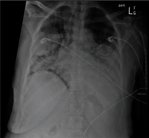
This diagnostic image is an anteroposterior (AP) chest X-ray demonstrating severe, diffuse, and bilateral patchy opacities throughout the lung fields. The findings are highly suggestive of multifocal pneumonia or Acute Respiratory Distress Syndrome (ARDS). Specifically, there are extensive irregular consolidations visible in both the upper and lower lung zones, indicating significant alveolar filling by inflammatory exudate or fluid. The heart borders are partially obscured by the adjacent pulmonary infiltrates. Additionally, multiple medical support devices are visible, including external EKG monitor leads and cables traversing the thoracic region, which indicate the patient is receiving intensive care monitoring. This radiological presentation is characteristic of severe pulmonary infections, such as COVID-19 pneumonia or secondary bacterial coinfection, correlating with acute hypoxemic respiratory failure.
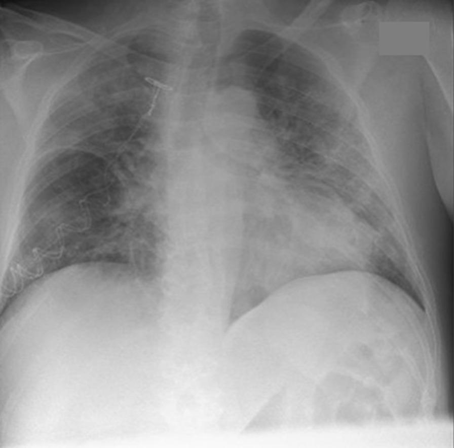
This diagnostic image is a frontal (anteroposterior) chest X-ray demonstrating severe pulmonary pathology. The primary findings are diffuse, bilateral lung opacities characterized by a mix of hazy ground-glass appearance and patchy consolidations. These infiltrates are widespread, showing a predisposition for the mid and lower lung zones, with relative sparing of the extreme apices. Such radiographic patterns are indicative of extensive alveolar and interstitial involvement, frequently seen in viral pneumonia, specifically SARS-CoV-2 (COVID-19), or acute respiratory distress syndrome (ARDS). Anatomically, the cardiac silhouette and mediastinal borders are partially obscured by the overlying parenchymal density. A radiopaque medical device, consistent with a central venous catheter or similar vascular access line, is visible in the upper right thoracic region, extending toward the superior vena cava. This visual is educationally significant for demonstrating the radiographic manifestation of severe lower respiratory tract infection and the progression to bilateral pulmonary infiltrates requiring intensive clinical management.
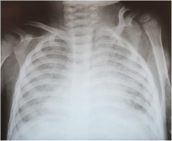
Anteroposterior (AP) pediatric chest X-ray taken following extubation, demonstrating significant bilateral lung pathology. The image reveals diffuse, bilateral interstitial and alveolar infiltrates across all lung zones, consistent with a primary pulmonary process such as acute respiratory distress syndrome (ARDS) or extensive pneumonia. Despite the diffuse opacities, there are no classic radiological signs of cardiogenic pulmonary edema, such as cardiomegaly or prominent cephalization of pulmonary vessels. The trachea is centrally positioned and patent. Visible skeletal structures include the clavicles, ribs, and scapulae, which appear intact and age-appropriate. This diagnostic image illustrates the clinical progression of severe pulmonary infection, specifically tuberculosis-related ARDS, highlighting the persistent radiological findings even as clinical oxygenation parameters (P/F ratio) improve. The radiograph serves as an educational example of managing complex pediatric respiratory failure in an intensive care setting.
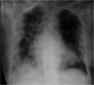
This diagnostic image is an anteroposterior (AP) chest X-ray demonstrating a case of viral pneumonia complicated by Acute Respiratory Distress Syndrome (ARDS). The radiograph reveals diffuse, bilateral, patchy alveolar opacities and ground-glass infiltrates. The abnormalities are primarily concentrated in the peripheral and basal regions of both lung fields, a hallmark distribution often seen in COVID-19 pneumonia. There is evidence of more confluent consolidation in the left lung compared to the right, particularly involving the lower lobe. The cardiac silhouette appears moderately enlarged, and the mediastinal contours are visible despite the overlapping pulmonary infiltrates. This image serves as an educational example of progressive pulmonary parenchymal involvement in severe respiratory infections, illustrating the typical radiographic progression from localized opacities to multi-lobar consolidation characteristic of clinical ARDS.
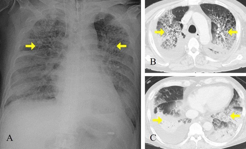
This composite diagnostic image features a chest X-ray (A) and axial CT scans (B, C) of a patient with Acute Respiratory Distress Syndrome (ARDS). (A) An anteroposterior chest radiograph demonstrates diffuse, bilateral alveolar infiltrates and patchy opacification across both lung fields, notably sparing the upper zones, with a normal cardiac silhouette and no signs of cardiomegaly. Yellow arrows highlight dense consolidation in the mid-lung regions. (B, C) Axial CT images at different thoracic levels reveal extensive, bilateral ground-glass opacities and dense consolidation, particularly in the dependent posterior regions of the lower lobes. Associated reticular patterns suggest interstitial involvement. The mediastinal structures and great vessels appear normal, and there is no evidence of significant pleural effusion. The imaging findings are classically consistent with non-cardiogenic pulmonary edema and diffuse alveolar damage typical of severe viral pneumonia (e.g., H1N1 influenza) progressing to ARDS. This material is suitable for training in intensive care medicine and radiology, focusing on the visual differentiation between cardiogenic and non-cardiogenic pulmonary opacities.
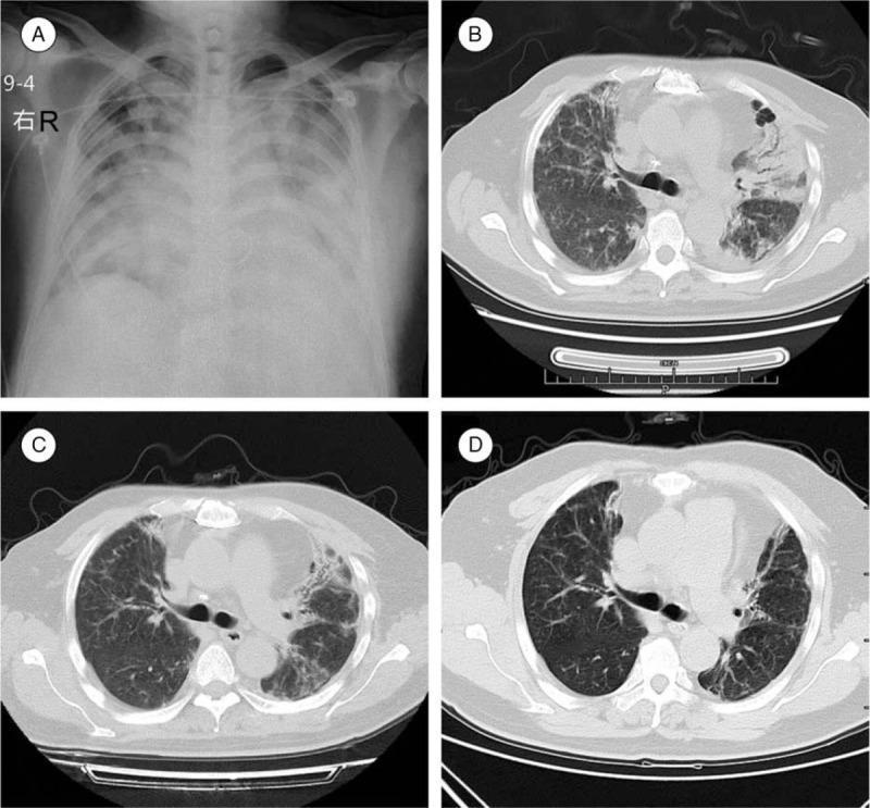
This composite figure presents a longitudinal imaging study of a patient with severe pulmonary infection and ARDS, likely due to H7N9 influenza. (A) Anteroposterior chest X-ray at admission shows diffuse, bilateral alveolar infiltrates and ground-glass opacities, consistent with widespread inflammation. (B) Axial chest CT at admission demonstrates extensive bilateral consolidation, ground-glass opacities, and early fibrotic changes; surgical clips and a mechanical valve reflect a history of cardiac surgery. (C) Follow-up axial CT at discharge shows significant interval resolution of consolidations, though persistent reticular opacities suggest moderate residual fibrosis and chronic inflammation. (D) A 1-year follow-up axial CT reveals stable, irreversible fibrotic changes, architectural distortion, and signs of chronic lung injury. The sequence illustrates the progression and partial resolution of severe viral pneumonia, highlighting the transition from acute exudative inflammation to chronic pulmonary fibrosis and long-term structural sequelae in a post-surgical cardiac patient.
CXR Report - Ramaiah Bhukya | Day 2 ICU (Post-Intubation)
Radiological Findings (Systematic Reading)
| Zone | Finding |
|---|---|
| Trachea & Mediastinum | Trachea central. Endotracheal tube tip visible in mid-trachea (to be confirmed at 3-5 cm above carina). Mediastinum not widened. |
| Cardiac silhouette | Heart borders partially obscured by bilateral infiltrates. Cardiothoracic (CT) ratio difficult to assess precisely - appears within normal limits (~0.5). No cardiomegaly. |
| Right lung | Dense, irregular consolidation in right lower and mid zones with air bronchograms. Blunting of right costophrenic angle (small pleural reaction). Patchy alveolar opacification in upper zone. |
| Left lung | Diffuse patchy alveolar opacities throughout all zones. Ground-glass appearance predominant. Less dense than right but clearly involved bilaterally. |
| Bones | Ribs and clavicles visible and intact. No fractures. |
| Soft tissue | No subcutaneous emphysema. |
| Lines/Tubes | ECG monitoring electrodes (bilateral chest leads). Right internal jugular / subclavian central venous catheter tip in SVC (appropriate position). Nasogastric tube present (seen on left). |
Key Distinguishing Features from Cardiogenic Oedema
| Feature | This CXR (ARDS) | Cardiogenic Pulmonary Oedema |
|---|---|---|
| Heart size | Normal | Enlarged (CTR >0.5) |
| Distribution of infiltrates | Patchy, bilateral, peripheral | Perihilar "bat-wing" |
| Kerley B lines | Absent | Present |
| Pleural effusions | Minimal (right only) | Bilateral, larger |
| Vascular redistribution (upper lobe diversion) | Absent | Present |
| Air bronchograms | Present | Usually absent |
| Clinical PCWP | Normal (<18 mmHg) | Elevated (>18 mmHg) |
Radiology Impression (as it would appear on a district hospital report)
"AP chest radiograph shows bilateral diffuse alveolar infiltrates involving all zones, right greater than left. Dense consolidation with air bronchograms in the right lower lobe. Blunting of right costophrenic angle. Cardiac silhouette appears normal in size. Trachea central. ETT in situ, CVC in situ. No evidence of pneumothorax. Findings are consistent with bilateral pneumonia / Acute Respiratory Distress Syndrome. Clinical correlation advised. Cardiogenic oedema appears less likely."