attachment

Isme mujhe ye bata ye V6 me x kaha se aa gya jab hame pata hai ki V1 phle positive ko r bole jo septal activity hai aur use q bole V6 me kyu negative hai

Reading File
Finding Sources
Finding Sources
Finding Sources
Reading File
Loading Image
attachment

Isme mujhe ye bata ye V6 me x kaha se aa gya jab hame pata hai ki V1 phle positive ko r bole jo septal activity hai aur use q bole V6 me q negative hai ab fir free wall matlab ventricular dikhaya gya hai dono vector me V1 me s se deep kyunki V1 se current opposite flow kar raha left hypertrophy se waise hi V6 me positive dikhaya R se kyunki ussi direction me hai ok itni baat samjh aa gyi ab dikkat ye hai ki V1 me toh base line pe aa gya kyu sab kaam toh ho gya lekin issi tarah V6 me kyu nhi gya wo deep kyu hua jisko X arrow se dikha raha wo kaha se aa gya special

Bilkul seedha point pe aata hoon. Ye bahut achha question hai aur confusion common hai.

V6 mein woh "X" (deep downward deflection) kahan se aaya?

Image mein V6 ka waveform dekho - pehle ek tall R wave hai, phir wo neeche jaata hai (X), phir thoda aur dip. Ye S wave hai V6 mein.

Step by step samjho - 3 vectors hain V6 ke liye

Vector 1 - Septal (Left → Right)

  • Current V6 se door ja raha hai (V6 left side pe hai, current right ja raha)
  • V6 mein small q (negative, neeche) - ye already samajh aa gaya tumhe ✓

Vector 2 - Main Ventricular (LV dominant, Left + Posterior)

  • Current V6 ki taraf aa raha hai
  • V6 mein tall R wave (positive, upar) - ye bhi samajh aa gaya ✓

Vector 3 - BASAL depolarization (yahi X ka source hai)

Yahan asli confusion hai. Ventricular depolarization apex se base ki taraf travel karta hai last mein.
Sequence:
  1. Pehle septum depolarize hota hai (left → right)
  2. Phir main free wall / apex (left + posterior dominant)
  3. Sabse end mein - basal portion of ventricles depolarize hota hai
Ye basal portion ka vector upar aur thoda right ki taraf oriented hota hai. V6 ke perspective se ye vector V6 se door ja raha hota hai.
Isliye V6 mein R ke baad ek small S wave dikhti hai - yahi wo "X" hai.

V1 mein S ke baad baseline pe kyun aa gaya?

V1 mein:
  • r (septal - V1 ki taraf) ✓
  • S (main LV vector - V1 se door) ✓
  • Basal vector thoda right oriented hai, matlab V1 ki taraf hai - but itna chota hota hai ki V1 pe ek negligible positive deflection hoti hai jo practically baseline jitni dikhti hai
Isliye V1 pe S ke baad baseline pe return dikh raha hai - basal vector V1 ke angle pe itna significant nahi.

LVH mein X (S wave in V6) aur deep S in V1 kyun zyada pronounced?

Ventricular depolarization vectors
LVH mein LV muscle mass bada ho jaata hai. Jab LV depolarize hota hai:
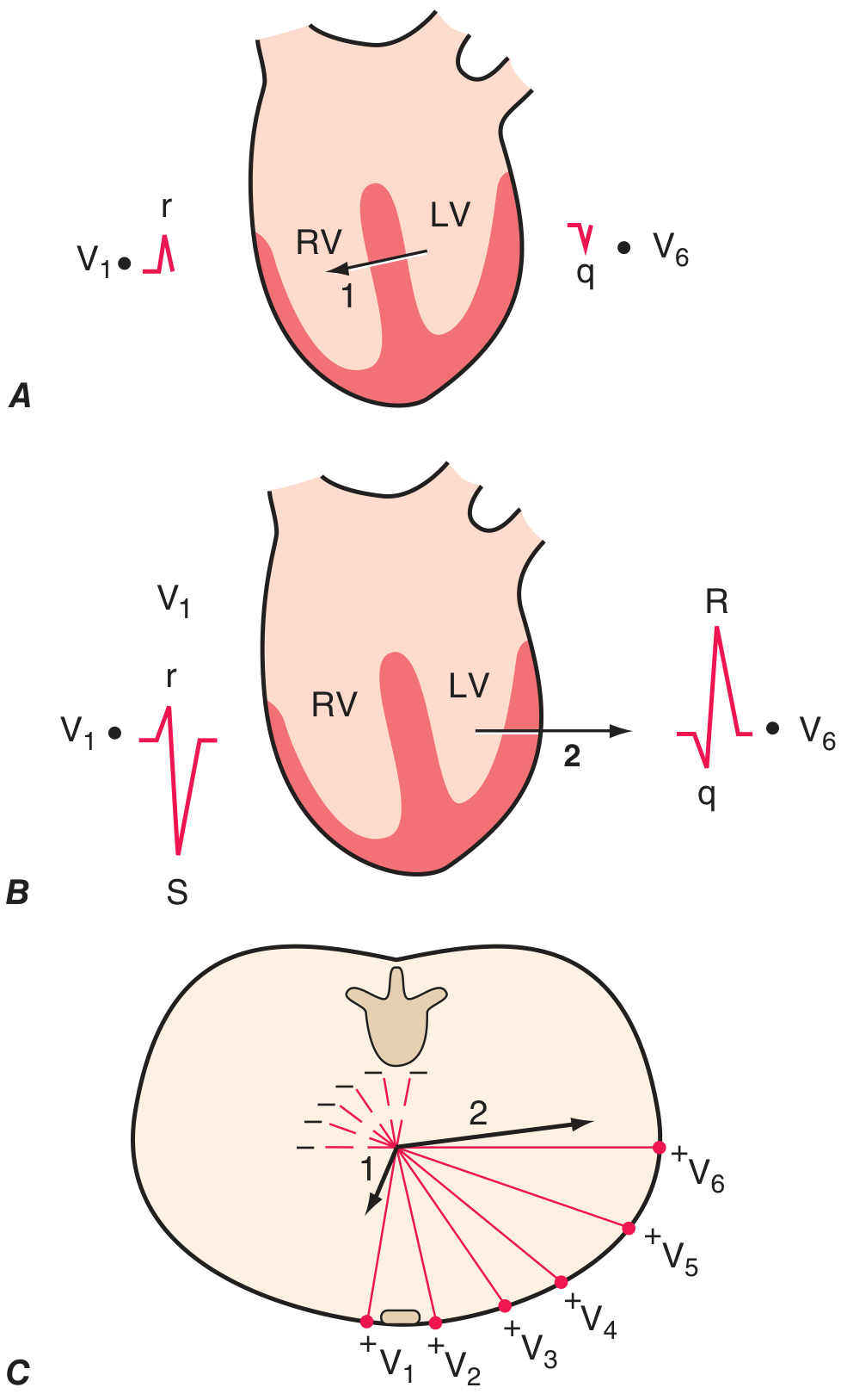
  • Vector 2 (main LV vector) bahut powerful ho jaata hai
  • V1 mein ye vector door ja raha hai → deeper S in V1 (Sv1 increases)
  • V6 mein ye vector aata hai → taller R in V6 (Rv6 increases)
Aur LVH formula yaad karo jo image mein likha hai: Sv1 + Rv5/v6 > 35 mm - dono mila ke measure karte hain kyunki dono usi ek hypertrophied vector ki wajah se badh rahe hain.

Summary ek line mein

X (S wave in V6) = Basal ventricular depolarization ka vector, jo V6 se door ja raha hai, isliye negative deflection aati hai R ke baad. V1 mein ye same basal vector V1 ke liye itna significant nahi hota isliye V1 baseline pe aa jaata hai. - Harrison's Principles of Internal Medicine, 22E

Konse book me acche se diya hai

Finding Sources
Finding Sources
Finding Sources
Finding Sources
Finding Sources
Reading File
Reading File
Tumhare sawal ke liye 3 books library mein hain jo ECG explain karti hain, aur ek specially tumhari confusion clear karti hai:

Best Book for This Topic

1. Ganong's Review of Medical Physiology (26th Edition)

Yeh sabse acchi hai tumhare question ke liye. Isme ek line mein clearly likha hai:
"...there is a large R wave (septal and left ventricular depolarization) followed in V1 and V5 by a moderate S wave (late depolarization of the ventricular walls moving back toward the AV junction)"
Matlab wo S wave / X jo V6 mein dikhta hai - woh late/basal ventricular wall depolarization hai jo AV junction ki taraf wapas jaata hai. Yahi tumhara answer hai - clearly physiology language mein. Ganong ka Chapter 29 (NORMAL ECG section) padho.

2. Harrison's Principles of Internal Medicine (22nd Ed, 2025)

Figures bahut acchi hain - 2 vector diagram (jo maine pehle image dikhaya tha) clearly septal + main ventricular vectors explain karta hai. Clinical angle zyada hai.

3. Goldman-Cecil Medicine

ECG leads ka concept accha explain kiya hai, but thoda clinical/dense hai.

Short Recommendation

BookKyu Padho
Ganong PhysiologyMechanism step-by-step, S wave ka origin clearly explain
Harrison'sDiagrams + LVH criteria + clinical context
Goldman-CecilReference ke liye, detail mein ECG leads
Tumhari specific doubt ke liye - Ganong Chapter 29 sabse pehle padho. Wahan V1 ka S aur V6 ka S wave dono ka origin ek saath explain kiya gaya hai - AV junction ki taraf late basal depolarization ka vector.

Maine Harrison padh usme toh aisa kuch hai hi nhi

Reading File
Loading Image
Sahi pakda - Harrison mein 2-vector model hai, 3rd vector (basal) explicitly explain nahi kiya. Ab Ganong ka exact relevant paragraph seedha dikhata hoon jo ye X explain karta hai:

Ganong mein exact explanation - Line by Line

Ganong's Review of Medical Physiology, 26th Ed, Chapter 29:
V1 ke baare mein likha hai:
"The wave of excitation first moves across the midportion of the septum from left to right toward the exploring electrode (= small r in V1)"
"Then moves down the septum and into the left ventricle AWAY from the electrode (= large S in V1)"
"Finally, it moves BACK along the ventricular wall TOWARD the electrode - producing the return to isoelectric line in V1"
Aur V6 ke baare mein:
"...there is a large R wave (septal and left ventricular depolarization) followed in V5 and V6 by a moderate S wave (late depolarization of the ventricular walls moving back toward the AV junction)"

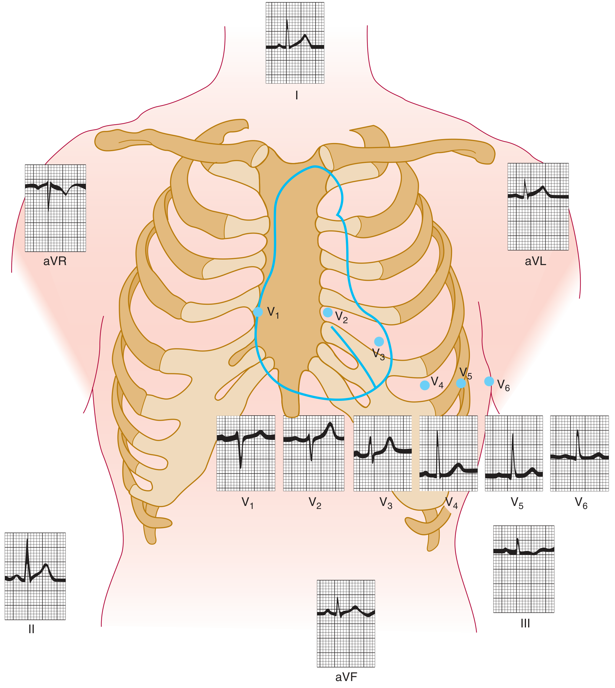
Ye image dekho - Normal ECG from Ganong:

Normal ECG lead positions - Ganong
V5 aur V6 mein clearly small S wave dikh raha hai R ke baad - yahi wo X hai.

To Seedha Answer

X (S wave in V6) ka origin = "Late depolarization of ventricular walls moving back toward AV junction"
Matlab ventricular depolarization jab apex se complete ho jaata hai, tab basal part (jo AV junction ke paas hota hai) sabse last mein depolarize hota hai. Ye basal activity ka vector upar ki taraf (toward AV junction) hota hai - jo V6 se door hai. Isliye R ke baad ek negative deflection (S wave / X) aati hai V6 mein.
Harrison mein ye 3rd phase explicitly nahi diya - isliye wahan se samajh nahi aata. Ganong padho iske liye. - Ganong's Review of Medical Physiology, 26th Ed, Chapter 29

Mujhe ecg basic se advance tak smajhao Harrison se sab kuch matlab tumse pdhne ke baad mujhe ab kav aur kuch ecg ka nhi padhna padd

Asking for Preferences
Finding Sources
Finding Sources
Finding Sources
Reading File
Reading File
Loading Image
Reading File
Ye raha poora ECG basic to advance - Harrison 22E se directly.
Ek important note: Arrhythmias (Harrison Chapter 252-255) ek alag bada topic hai - usmein sinus tachycardia, SVT, AF, VT, heart blocks sab aata hai. Agar wo bhi chahiye toh bolo, woh alag session mein detail mein cover karte hain.
Koi bhi specific topic - jaise LBBB vs RBBB aur unka difference, ya MI localization ka detail, ya axis calculation step-by-step - jo bhi confuse kar raha ho, seedha poocho.
This is a shared conversation. Sign in to Orris to start your own chat.