
Chest wall necrotizing fascitis management
I'll read the medical library skill first, then search in parallel.
Reading File
Now running parallel searches across the library and PubMed:
Finding Sources
Finding Sources
Searching PubMed
necrotizing fasciitis chest wall management
Good hits. Now reading the most relevant sections in parallel - Rosen's Emergency Medicine (general NF management), Campbell's Operative Orthopaedics (surgical management), Harrison's (with the chest wall image), and a broader PubMed search:
Reading File
Reading File
Reading File
Searching PubMed
necrotizing fasciitis chest wall surgical debridement
Let me now read the Campbell's surgical management section and Harrison's NF section more fully, and fetch the CT image from Harrison's:
Reading File
Reading File
Loading Image
Loading Image
Now let me read the key treatment section from Campbell's and search Harrison's for the NF treatment section:
Reading File
Reading File
Finding Sources
Excellent. Now let me read Harrison's dedicated NF + streptococcal treatment section and Tintinalli's full NF section for the most complete management details:
Reading File
Reading File
I now have comprehensive material. Let me also quickly check Goldman-Cecil's treatment table and fetch the NF CXR image:
Reading File
Loading Image
Searching the Web
chest wall necrotizing fasciitis management debridement reconstruction 2023 2024
I now have comprehensive, authoritative data from multiple textbooks and current literature. Here is the complete management guide:
Chest Wall Necrotizing Fasciitis - Management
Necrotizing fasciitis (NF) of the chest wall is a rapidly progressing, life-threatening deep soft-tissue infection. Spread along fascial planes to the chest wall - particularly from upper extremity NF - carries a mortality rate up to 75%. Overall NF mortality ranges from 23-76%, with delays in diagnosis and treatment being the primary driver of death.
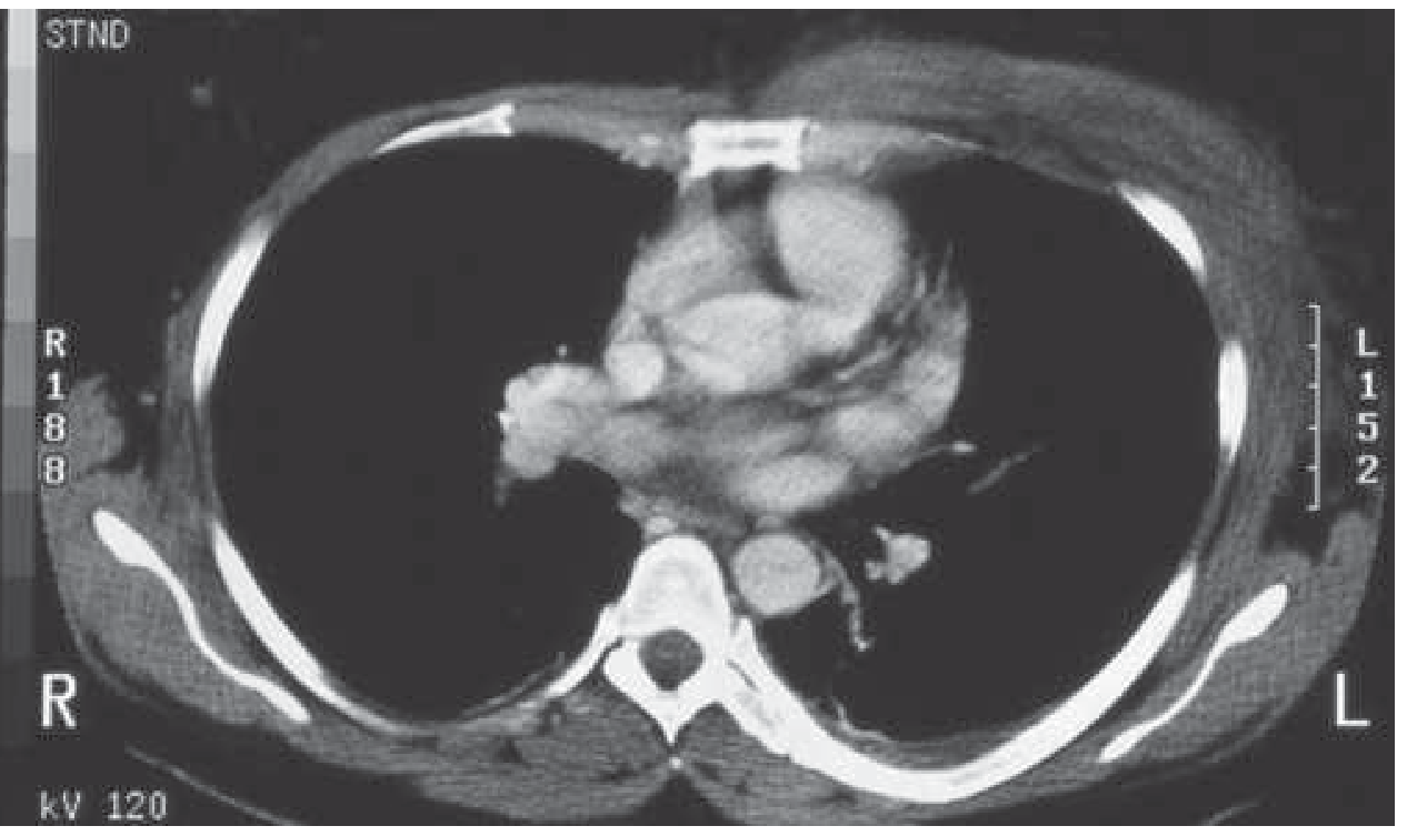
1. Microbiology
NF is classified into four groups (Campbell's Operative Orthopaedics):
| Type | Organisms |
|---|---|
| Type I (Polymicrobial) | Mixed non-GAS streptococci + aerobic/anaerobic organisms (most common in chest wall/trunk) |
| Type II (Monomicrobial) | Group A Streptococcus (GAS) alone, or Staphylococcus (including MRSA) |
| Type III | Gram-negative marine organisms (e.g., Vibrio species) |
| Type IV | Fungal (Candida, Zygomycetes) - immunocompromised or severe trauma |
Risk factors: Diabetes (most common comorbidity), IV drug use, smoking, trauma, immunosuppression (AIDS, chemotherapy), peripheral vascular disease, alcoholism, obesity, hypertension.
2. Diagnosis
Clinical Features
- Rapid progression from erythema/swelling to severe nonpitting edema, skin bullae, and blue-black discoloration
- Pain out of proportion to local findings - a hallmark sign
- Advanced cases: deep pain with surface hypesthesia, crepitus, and hemorrhagic bullae
- Systemic toxicity: fever, septic shock, multiorgan failure
- Lymphangitis/lymphadenopathy and fever may paradoxically be absent early
Investigations
- Labs: WBC >15,400/mm³ and serum Na <135 mmol/L together have 90% sensitivity, 76% specificity. CRP >150 mg/L is highly suspicious.
- LRINEC Score (≥6 = 96% positive predictive value):
| Variable | Score |
|---|---|
| CRP >150 mg/L | 4 |
| WBC 15-25 × 10³/mm³ | 1; >25 × 10³ = 2 |
| Hb <13.5 g/dL | 1-2 |
| Na <135 mmol/L | 2 |
| Creatinine >1.6 mg/dL | 2 |
| Glucose >180 mg/dL | 1 |
Note: LRINEC does not correlate with severity or outcome but helps exclude NF. The final diagnosis remains clinical.
-
Imaging:
- Plain X-ray: subcutaneous gas (pathognomonic when present)
- CT chest (98% sensitive for descending NF): asymmetric fascial thickening, gas tracking along fascial planes, fluid collections. CT is the study of choice for defining extent.
- MRI: sensitive for fascial involvement but not totally specific; useful adjunct when CT is inconclusive
- Imaging must never delay surgical exploration when clinical suspicion is high
-
Frozen section/intraoperative biopsy: useful for confirming diagnosis; aspiration of leading edge has low yield (~20% positive cultures)
-
Definitive diagnosis: Open surgical inspection remains the gold standard
3. Management - The Three Pillars
PILLAR 1: Urgent Surgical Debridement (LIFE-SAVING)
This is the cornerstone. Drainage and debridement are central - antibiotics are a critical adjunct, but surgery is life-saving (Harrison's 22E).
Timing:
- Debridement within 24 hours = 93% survival
- Delayed to 48 hours = survival drops to 75%
- Any further delay markedly increases mortality
Surgical principles (Campbell's Operative Orthopaedics 15th Ed):
- Wide aggressive debridement of all necrotic tissue, especially liquefying fascia. At surgery, the infection extends beyond the apparent surface involvement - always debride beyond visible margins.
- Use extensile incisions to preserve skin flaps and protect viable underlying structures.
- Obtain cultures for anaerobic organisms from areas of worst involvement; Gram stain guides initial antibiotic selection.
- Leave wounds open - do not close primarily.
- Return to OR for repeat inspection and debridement every 24-48 hours until all necrotic tissue is removed and healthy granulating wound is achieved.
- Fascial planes, not muscle, are the primary infection corridor - but myonecrosis occurs in ~50% of GAS cases and requires debridement of involved muscle.
Wound zones (Singapore General Hospital technique):
- Zone 1 (frankly necrotic): complete excision
- Zone 2 (infected but potentially salvageable): cut back as necessary, preserve viable tissue
- Zone 3 (non-infected skin): leave alone
Special chest wall considerations:
- Spread from the upper extremity to the chest wall carries 75% mortality - treat as urgent thoracic surgical emergency
- May require cardiothoracic surgery involvement if spread involves pleural or pericardial spaces
- Descending NF from the neck (Ludwig's angina) can reach the chest - CT neck + chest is mandatory in such cases
Wound care post-debridement:
- Non-occlusive dressings with gauze as secondary layer
- Negative pressure wound therapy (NPWT/VAC): valuable adjunct once hemostasis is secured, but avoid at first debridement as suction may increase postoperative bleeding. Use after second debridement onward.
PILLAR 2: Broad-Spectrum Antibiotic Therapy
Initiated empirically while cultures are pending. Choice depends on whether monomicrobial or polymicrobial NF is suspected.
Empirical therapy for polymicrobial/unknown NF (Goldman-Cecil Medicine; Rosen's Emergency Medicine):
Cover Gram-positive (including MRSA), Gram-negative, and anaerobes:
| Agent Class | Example Regimen |
|---|---|
| Anti-MRSA agent | Vancomycin 15-20 mg/kg IV q8-12h OR Linezolid 600 mg IV/PO q12h OR Tedizolid 200 mg daily |
| + | |
| Broad-spectrum beta-lactam | Piperacillin-tazobactam 3.375 g IV q6h OR Carbapenem (imipenem 500 mg IV q6h / meropenem) OR Ceftriaxone 2 g IV daily |
| + (if GAS suspected/confirmed) | Clindamycin 600-900 mg IV q8h (anti-toxin effect, protein synthesis inhibitor - active regardless of bacterial growth phase) |
Tintinalli's triple antibiotic approach: penicillin/cephalosporin + aminoglycoside + clindamycin
Confirmed GAS (Type II) NF (Harrison's 22E; Goldman-Cecil):
- High-dose penicillin G 4 million units IV q4h + clindamycin 600-900 mg IV q8h for 10-14 days
- Then oral step-down based on clinical course
- Rationale: At high bacterial density, beta-lactams lose efficacy (Eagle effect) - protein synthesis inhibitors (clindamycin, linezolid) retain activity at all growth phases
- If clindamycin resistance suspected: substitute linezolid
Duration: Continue IV antibiotics until systemic toxicity resolves and no further surgical debridement is required; total duration guided by clinical course.
Infectious disease consultation strongly recommended.
PILLAR 3: Intensive Supportive Care
- ICU admission - ideally in a regional burn center or trauma center
- Aggressive fluid resuscitation: early goal-directed therapy
- Vasopressors for refractory septic shock
- Nutritional support (hyperalimentation): early enteral or parenteral nutrition significantly aids wound healing and infection combat
- Management of coagulopathy: NF can cause DIC; monitor coagulation and treat accordingly
- Organ support: renal replacement therapy, mechanical ventilation as needed
4. Adjunctive Therapies
Hyperbaric Oxygen (HBO)
- May be beneficial with difficult anaerobic infections - increases tissue oxygen tension, inhibits anaerobic growth, enhances leukocyte killing (Campbell's Operative Orthopaedics; Barash's Clinical Anesthesia)
- Does not replace emergent surgical debridement; used as adjunct at specialized centers
- Access to HBO should never delay surgery
IVIG (Intravenous Immunoglobulin)
- Considered in streptococcal toxic shock syndrome complicating GAS NF
- Neutralizes streptococcal superantigens and exotoxins
- Evidence is limited but may reduce mortality in TSS
5. Wound Closure and Reconstruction
Once infection is controlled and healthy granulating tissue is present:
- Direct primary closure: if defect is small
- Split-thickness skin grafts (STSG): most common for larger defects
- Pedicled or free flaps: for deep or complex chest wall defects, particularly where ribs/sternum are exposed
- Artificial skin substitutes: when donor sites are limited (large BSA defects)
- Reconstruction planning begins at first debridement (skin-sparing approach where possible)
- Typically requires at least 2 debridements before reconstruction
6. Complications and Prognosis
| Complication | Notes |
|---|---|
| Septic shock | Major cause of death |
| Multiorgan failure | Renal, respiratory, hepatic |
| DIC | Monitor and treat |
| Abscess formation | Liver, spleen, lungs, brain |
| Empyema/mediastinitis | In chest wall NF with mediastinal spread |
| Amputation | 18-28% in extremity NF; may extend to chest wall tissue excision |
Mortality: 30-60% even with appropriate treatment. Delayed diagnosis raises mortality to 70-85%. The single most modifiable factor is time to first debridement.
7. Summary Algorithm
Suspected Chest Wall NF
        ↓
Labs (LRINEC) + CT Chest (do not delay if high clinical suspicion)
        ↓
Immediate surgical consult + ICU admission
        ↓
Broad-spectrum empirical antibiotics (vancomycin + pip-tazo + clindamycin)
        ↓
URGENT SURGICAL DEBRIDEMENT (< 24 hrs)
        ↓
Serial debridements q24-48h until clean wound
        ↓
Nutritional support + HBO (if anaerobic) + NPWT
        ↓
Wound closure / reconstruction (STSG / flap)
Sources:
- Harrison's Principles of Internal Medicine 22E (2025), Chapters 134 & 153
- Campbell's Operative Orthopaedics 15th Ed (2026), Chapter 84
- Goldman-Cecil Medicine, Chapter on Streptococcal Infections
- Rosen's Emergency Medicine, Chapter 107
- Tintinalli's Emergency Medicine, Chapter 152
- Recent case series (PMID 34934455, 39277787) confirm that timely debridement + broad-spectrum antibiotics + fluid resuscitation remain the pillars of chest wall NF management
This is a shared conversation. Sign in to Orris to start your own chat.