
Explain skeleton system in detail for teaching
human skeleton diagram labeled bones
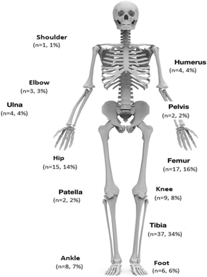
This infographic presents an anatomical diagram of the human skeleton used as a data visualization tool to map the distribution of orthopedic infections across various skeletal sites. The diagram provides quantitative data for 107 cases, labeling specific bones and joints with the number of patients (n) and the corresponding percentage of the total study population. Labeled anatomical regions include the upper extremity (shoulder, humerus, elbow, and ulna), the axial/pelvic region (pelvis), and the lower extremity (hip, femur, knee, patella, tibia, ankle, and foot). The visualization highlights the tibia as the most frequent site of infection (n=37, 34%), followed by the femur (n=17, 16%) and hip (n=15, 14%). This clinical illustration is designed for orthopedic and infectious disease education, effectively demonstrating the anatomical prevalence of conditions such as fracture-related infection (FRI), chronic osteomyelitis (COM), and prosthetic joint infection (PJI) within a clinical cohort.
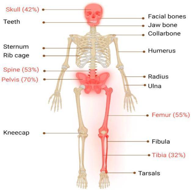
Anatomical diagram of a full human skeleton in an anterior view, illustrating the distribution and incidence rates of Paget's Disease of Bone (PDB). The diagram uses color-coding to highlight the most frequently affected skeletal sites. The primary regions highlighted in red include the skull (42%), lumbar spine (53%), pelvis (70%), femur (55%), and tibia (32%), demonstrating the disease's predilection for the axial skeleton and long bones. Other anatomical structures labeled for reference include the facial bones, jaw bone, collarbone, humerus, sternum, rib cage, radius, ulna, kneecap, fibula, and tarsals. This infographic serves as an educational tool to visualize the epidemiological patterns of PDB, emphasizing that while the disorder can be polyostotic, certain landmarks like the pelvis and femur show significantly higher involvement rates. The visual contrast between the highlighted red areas and the neutral-toned bone helps medical students and clinicians identify common sites for clinical and radiological investigation.
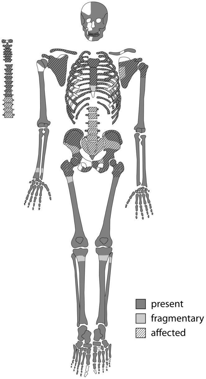
This anatomical diagram is a skeletal inventory and pathology map used in bioarchaeology and paleopathology to document the preservation and disease state of human remains (Skeleton Sk244-8). The graphic uses a standardized human skeletal template with a three-part coding system: dark gray indicates 'present' (full preservation), light gray indicates 'fragmentary' areas, and diagonal hatching denotes bones 'affected' by pathological changes. The map reveals that the axial skeleton—including the sternum, ribs, and most of the vertebral column—is heavily affected by lytic lesions. Both scapulae and the pelvic girdle (innominate bones and sacrum) also show significant involvement with hatching. The appendicular skeleton, specifically the long bones like the humeri, femora, and tibiae, is largely preserved but exhibits localized pathological involvement at the proximal joints. The skull shows a mix of full and fragmentary preservation. This visual summary is essential for illustrating the distribution of systemic skeletal diseases, such as metastatic carcinoma or multiple myeloma, in ancient populations.
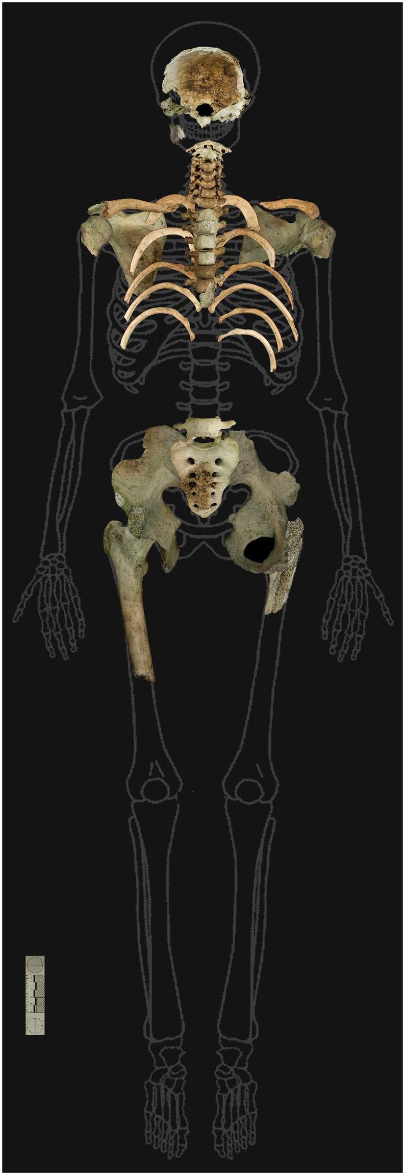
This forensic anatomical diagram displays skeletal remains overlaid on a human body outline to indicate the presence and condition of recovered bones. The image depicts a partially complete skeleton exhibiting significant thermal destruction and weathering. Key visible elements include the cranial vault (skull fragments), several cervical and thoracic vertebrae, clavicles, a partial rib cage, and a nearly complete pelvis with the sacrum. A single right femur is present, showing a distal fracture. The bones display distinctive brownish and blackened discoloration, characteristic of environmental exposure and fire damage (thermal destruction). Anatomically, the remains are arranged in a supine position, highlighting the absence of the upper and lower extremities, including the hands, feet, and most of the long bones. This visual serves as a forensic tool for documenting skeletal representation, estimating biological profile (male, approximately 18-25 years), and reconstructing taphonomic events such as burning in a forest environment.
bone histology structure compact spongy
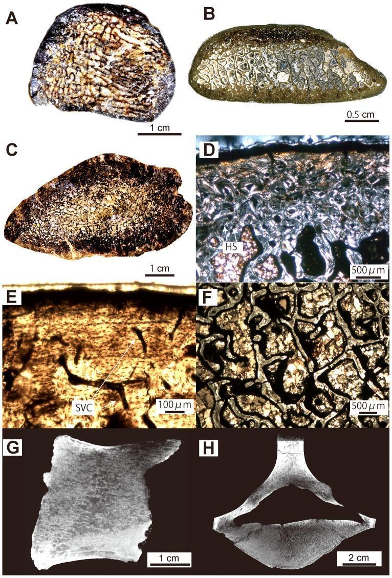
This composite educational image illustrates the microanatomy and histology of mammalian bone across multiple scales. Panels A–C show macro-scale transverse sections of long bones (femur and rib), highlighting a dense, cancellous inner structure with an extremely thin compact cortical layer and an extensive network of trabeculae instead of a central medullary cavity. Panels D–F present high-resolution light microscopy of the bone matrix. Panel D, under polarized light, demonstrates birefringent Haversian Systems (HS) and secondary osteons, indicative of intense remodeling. Panel E, viewed with natural light, reveals the primary cortical bone matrix containing simple vascular canals (SVC). Panel F provides a detailed view of the interconnected trabecular struts within the medullary region. Panels G and H represent virtual radiographic sections (sagittal and transverse) of a vertebral centrum, displaying a characteristic cancellous organization with small intertrabecular spaces and a minimal surrounding cortical shell. These visuals are pedagogically significant for studying bone density variations, pachyosteosclerosis, and the microscopic markers of bone remodeling (Haversian systems and Lines of Arrested Growth).
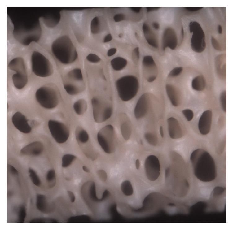
This clinical imaging specimen shows a high-magnification view (40x) of trabecular bone architecture. The image displays a complex, interconnected framework of dense bone trabeculae forming a porous, lattice-like structure. These trabeculae delimit numerous communicating cavities characterized by a variety of ovoid and spherical shapes. The morphology of the bone tissue appears well-preserved, exhibiting a uniform surface texture and a defined spatial arrangement. This visual is significant in orthopedics and histology for demonstrating the natural micro-architecture of cancellous (spongy) bone, illustrating the relationship between dense structural supports and the marrow-containing cavities they define. The specimen represents an experimental group focusing on bone health, regeneration, or collagen distribution, providing a reference for normal versus pathological bone density and structure.
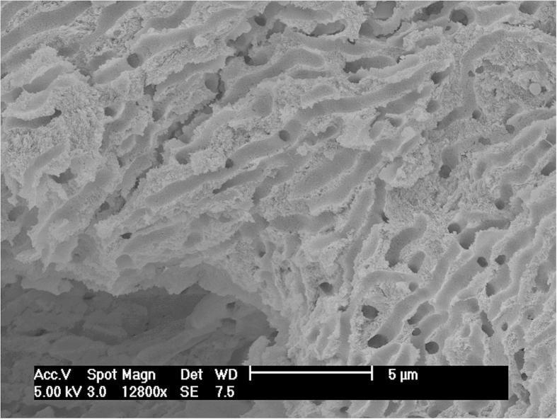
High-resolution Scanning Electron Microscopy (SEM) image of human compact bone at 12800x magnification. The visual shows the intricate micro-architecture of the inner bone lattice from an adult female humerus. The structure is characterized by well-preserved, interconnected trabecular-like patterns and uniform pore spaces, demonstrating a healthy or well-preserved solid bone matrix. The textured surface appears clean, with no visible soil contaminants, particulate matter, or cinnabar (mercury sulfide) inclusions despite high systemic mercury levels (137.41 μg/g). A 5 μm scale bar is provided for reference. This diagnostic imaging serves as a pathological and archaeological reference for assessing bone integrity and potential post-mortem environmental contamination versus ante-mortem metabolic accumulation of trace elements like mercury.
synovial joint anatomy diagram
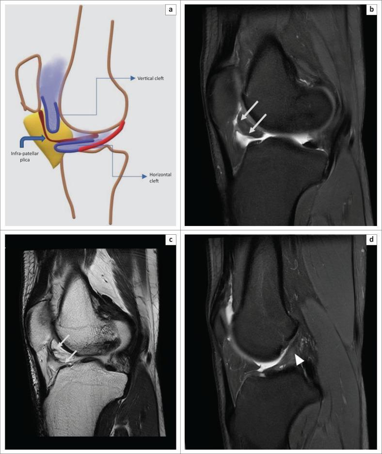
This composite educational resource illustrates the anatomy of normal synovial clefts in Hoffa’s fat pad and a common musculoskeletal pathology. Image (a) is a schematic diagram of the knee in a sagittal orientation, labeling the vertical (supra-Hoffotic) cleft, horizontal (infra-Hoffotic) cleft, and the infrapatellar plica (IPP). Images (b) and (c) provide radiological correlation using oblique sagittal Proton Density Fat-Saturated (PDFS) and T2-Weighted (T2WI) MRI sequences, respectively. They demonstrate hyperintense, smoothly marginated, fluid-filled synovial clefts (white arrows) within the infrapatellar fat pad that are contiguous with the joint fluid, representing normal anatomical variations rather than pathology. Image (d) is a sagittal PDFS MRI highlighting a mild sprain of the anterior cruciate ligament (ACL). A white arrowhead indicates a region of increased signal intensity within the middle third of the ACL, signifying ligamentous injury. This image set is designed for musculoskeletal radiology education, focusing on distinguishing normal synovial structures from intra-articular ligamentous sprains.
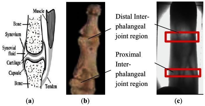
This figure presents a multi-part educational visual detailing the anatomy and imaging characteristics of human phalangeal joints. Panel (a) is an anatomical diagram of a diarthrodial joint, labeling the bone, articular cartilage, synovium, synovial fluid within the articular cavity, joint capsule, muscle, and tendon. It illustrates the spatial relationship between these connective tissues and the skeletal structure. Panel (b) shows a clinical photograph of a phalanx specimen, highlighting the distal and proximal joint locations. Panel (c) is a near-infrared (NIR) finger vein diagnostic image. It demonstrates the clinical significance of joint anatomy in biometric imaging; the distal and proximal inter-phalangeal joint regions are highlighted with red rectangles. These areas appear brighter due to the lower density of synovial fluid compared to bone, allowing greater NIR light penetration. This visual is designed for medical education regarding hand anatomy, musculoskeletal structures, and the application of physiological properties in diagnostic or biometric imaging systems.
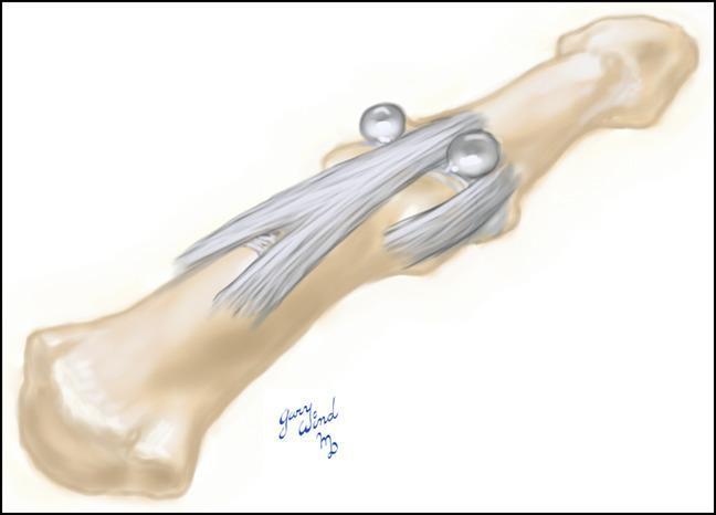
This anatomical diagram illustrates a distal interphalangeal (DIP) joint, depicting the underlying osseous structures and the dorsal extensor mechanism. The illustration highlights a bilateral pathological formation known as a 'dumbbell cyst,' which is a clinical manifestation of a digital mucous cyst. The cyst is shown with two distinct spherical, translucent lobes connected across the dorsal midline. Anatomically, these lobes are positioned symmetrically on both the radial and ulnar sides of the extensor tendon. They occupy the interval between the central slip of the extensor mechanism and the respective collateral ligaments of the DIP joint. The diagram emphasizes the relationship between the synovial cyst and the joint capsule, demonstrating how the lesion can emerge through these anatomical spaces. This visual is designed for orthopedic and dermatological education, specifically regarding the surgical anatomy of mucous cysts and their potential for multicompartmental presentation at the finger joint.
long bone anatomy periosteum epiphysis diaphysis
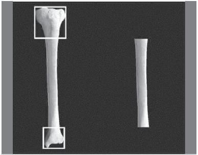
This clinical photograph displays a comparative view of long bone specimen preparation for orthopedic research. On the left, a complete tibia is shown with the proximal and distal epiphyses highlighted by white squares, demonstrating the application of Heim's square to identify the epiphysis-diaphysis transition. On the right, the resulting isolated diaphyseal segment is shown following osteotomy at those precise levels. The specimens exhibit a smooth, off-white surface texture, indicating thorough removal of soft tissue and the periosteum. The image illustrates the standardized anatomical methodology used to obtain uniform diaphyseal bone segments for clinical research, such as biomechanical testing or ultrasonometric analysis of fracture fixation methods. The focus is on the anatomical landmarks and the geometric precision required to isolate the shaft while maintaining consistency across experimental samples.
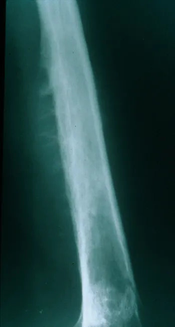
Plain Radiography of a long bone diaphysis demonstrates an aggressive, ill-defined osteolytic lesion with cortical destruction and periosteal reaction. The radiograph shows a moth-eaten destruction pattern with diffuse bone loss extending along a substantial portion of the shaft. Periosteal elevation produces Codman triangle in the swept cortex, while layered deposition of reactive new bone beneath the periosteum can create an onion-skin appearance; in some cases perpendicular radiating spicules or sunburst-like projections may be seen. The lesion often breaches the cortex or expands to form a soft tissue mass that extends beyond the bone. Although ultrasound or CT can delineate the soft tissue component, CT and MRI best characterize the extent, relation to surrounding structures, and marrow involvement. The radiographic pattern is highly suggestive of an aggressive small round blue cell tumor, most notably Ewing sarcoma, particularly in children and adolescents. Differential considerations include osteosarcoma, osteomyelitis, lymphoma, metastasis, or eosinophilic granuloma, but the combination of an diaphyseal, permeative lesion with onion-skin periosteal reaction strongly favors Ewing. Clinical correlation with pain, fever, raised inflammatory markers, and prompt biopsy is essential to confirm diagnosis and guide treatment, which typically combines chemotherapy, limb-sparing surgery, and possibly radiotherapy.
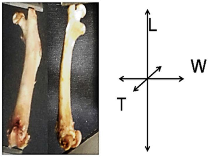
Anatomical anatomical photograph of the left femur from a murine model (rat), presented in two orientations: anterior and lateral views. The image illustrates the gross morphology of a long bone, identifying key landmarks including the proximal epiphysis (head and greater trochanter), the elongated diaphysis (shaft), and the distal epiphysis. To the right of the specimens, a Cartesian coordinate system provides a visual reference for morphometric analysis: 'L' indicates the longitudinal axis (length) from the proximal to distal epiphyses; 'W' represents the transverse axis (width) at the mid-diaphysis; and 'T' signifies the sagittal axis (thickness/depth) at the mid-diaphysis. The visual demonstrates differences in bone texture and coloration, ranging from pinkish-white to beige-yellow, which are used to evaluate bone health, mineral density, and cortical development in nutritional or pharmacological studies. This specimen-based visual is designed for educational use in orthopedic research, bone microstructure analysis, and comparative anatomy.
The Skeletal System - A Complete Teaching Guide
1. Overview
| Division | Bone Count | Components |
|---|---|---|
| Axial Skeleton | 80 bones | Skull (cranium), vertebral column, ribs, sternum |
| Appendicular Skeleton | 126 bones | Bones of the upper and lower limbs + pectoral and pelvic girdles |
Teaching tip: The word axial comes from "axis" - think of it as the central axis or core of the body. Appendicular refers to the appendages (limbs) that hang off it.
2. Functions of the Skeletal System
- Support - Provide structural support for the body
- Protection - Protect vital organs (skull → brain; ribcage → heart & lungs; vertebral column → spinal cord)
- Mineral Reservoir - Store calcium and phosphorus, which are released when needed by the body
- Mechanical Levers - Act as levers on which muscles pull to produce movement
- Haematopoiesis - Contain bone marrow, the site of blood cell production (red blood cells, white blood cells, platelets)
- Fat Storage - Yellow marrow stores fat (inactive marrow, approximately 80% fat)
3. Composition of Bone
- An intercellular calcified matrix (calcium hydroxyapatite crystals)
- Collagen fibers embedded within the matrix
- Several specialized cell types
3.1 Types of Bone Tissue
| Type | Description | Location |
|---|---|---|
| Compact (Cortical) Bone | Dense, solid outer shell of all bones | Outer layer of every bone |
| Spongy (Cancellous/Trabecular) Bone | Network of bony spicules (trabeculae) enclosing cavities filled with bone marrow | Interior of bones, epiphyses of long bones |
3.2 Bone Cells
| Cell | Function |
|---|---|
| Osteoblasts | Bone-forming cells; secrete bone matrix and calcium |
| Osteocytes | Mature bone cells; maintain bone matrix; communicate via canaliculi |
| Osteoclasts | Bone-resorbing cells; responsible for bone remodeling and calcium release |
3.3 Bone Vasculature and Innervation
- Each bone receives a nutrient artery (usually one per bone) that enters the internal cavity and supplies the marrow, spongy bone, and inner compact bone layers.
- The outer layers of compact bone are supplied by vessels from the periosteum.
- The periosteum (fibrous connective tissue membrane covering all bone surfaces except at joints) is richly supplied with sensory nerves - this is why periosteal damage is very painful.
- Bone itself has very few sensory nerve fibers.
4. Classification of Bones by Shape
| Shape | Description | Examples |
|---|---|---|
| Long | Tubular; longer than wide | Humerus, femur, tibia, fibula, radius, ulna |
| Short | Cuboidal; roughly equal dimensions | Carpal bones (wrist), tarsal bones (ankle) |
| Flat | Two plates of compact bone sandwiching spongy bone | Skull bones, scapula, sternum |
| Irregular | Complex shapes that don't fit other categories | Vertebrae, facial bones, hip bone |
| Sesamoid | Round/oval bones that develop within tendons | Patella (largest), sesamoids of thumb and big toe |
Teaching tip: Sesamoid bones function to change the direction of a tendon's pull and protect tendons from wear. The patella is the largest sesamoid in the body.
5. Structure of a Long Bone (Anatomy)
| Region | Description |
|---|---|
| Diaphysis | The shaft; made of thick compact bone with a medullary canal |
| Epiphysis (x2) | Expanded ends; contain spongy bone and are covered by articular cartilage |
| Metaphysis | Transition zone between epiphysis and diaphysis |
| Epiphyseal (Growth) Plate | In growing bones; cartilaginous plate where bone lengthening occurs |
| Periosteum | Fibrous membrane covering outer bone surface; essential for bone growth and repair |
| Endosteum | Thin membrane lining the medullary cavity |
| Medullary Cavity | Hollow center; contains yellow (fat) marrow in adults, red marrow in children |
| Articular Cartilage | Hyaline cartilage covering epiphyseal joint surfaces; reduces friction |
6. The Axial Skeleton in Detail
6.1 Skull (Cranium)
- 22 bones total: 8 cranial bones (protect the brain) + 14 facial bones
- Key cranial bones: frontal, parietal (x2), temporal (x2), occipital, sphenoid, ethmoid
- Bones joined by immovable joints called sutures
6.2 Vertebral Column (Spine)
- 33 vertebrae total, organized into 5 regions:
| Region | Count | Features |
|---|---|---|
| Cervical | 7 | Neck; C1 (atlas) and C2 (axis) allow head rotation |
| Thoracic | 12 | Articulate with ribs |
| Lumbar | 5 | Largest vertebrae; bear most body weight |
| Sacral | 5 (fused → sacrum) | Fused in adults; part of pelvic girdle |
| Coccygeal | 4 (fused → coccyx) | Vestigial tail |
- Between each vertebra (except C1-C2 and fused segments) sit intervertebral discs made of fibrocartilage - acting as shock absorbers
6.3 Thoracic Cage
- 12 pairs of ribs:
- Ribs 1-7: True ribs (attach directly to sternum via costal cartilage)
- Ribs 8-10: False ribs (attach indirectly via costal cartilage of rib 7)
- Ribs 11-12: Floating ribs (no anterior attachment)
- Sternum: Manubrium + body + xiphoid process
- Functions: Protects heart and lungs; facilitates breathing
7. The Appendicular Skeleton in Detail
Upper Limb (64 bones total, both sides)
| Segment | Bones |
|---|---|
| Pectoral girdle | Clavicle, scapula |
| Arm | Humerus |
| Forearm | Radius (lateral), ulna (medial) |
| Wrist | 8 carpal bones (mnemonic: "Some Lovers Try Positions That They Can't Handle" - Scaphoid, Lunate, Triquetrum, Pisiform, Trapezium, Trapezoid, Capitate, Hamate) |
| Hand | 5 metacarpals + 14 phalanges (thumb has 2, each finger has 3) |
Lower Limb (62 bones total, both sides)
| Segment | Bones |
|---|---|
| Pelvic girdle | Hip bone (ilium + ischium + pubis fused = os coxa) x2, sacrum |
| Thigh | Femur (longest bone in body) |
| Patella | Sesamoid bone in quadriceps tendon |
| Leg | Tibia (medial, weight-bearing), fibula (lateral) |
| Ankle | 7 tarsal bones (calcaneus is the largest) |
| Foot | 5 metatarsals + 14 phalanges |
8. Cartilage
| Type | Matrix Content | Examples |
|---|---|---|
| Hyaline | Moderate collagen fibers; most common type | Articular cartilage, costal cartilage, tracheal rings, fetal skeleton |
| Elastic | Collagen + many elastic fibers | External ear (pinna), epiglottis, larynx |
| Fibrocartilage | Dense collagen, few cells; very strong | Intervertebral discs, pubic symphysis, menisci of knee |
- Support soft tissues
- Provide smooth gliding surfaces at joints
- Enable growth and development of long bones (epiphyseal cartilage)
9. Joints (Articulations)
9.1 Structural Classification
A. Fibrous Joints (Synarthroses - immovable or slightly movable)
- Bones connected by dense fibrous connective tissue
- Examples: Skull sutures, distal tibiofibular joint (syndesmosis), teeth in sockets (gomphosis)
B. Cartilaginous Joints (Amphiarthroses - slightly movable)
- Bones connected by cartilage
- Primary (synchondrosis): Hyaline cartilage - e.g., costochondral joints, growth plates
- Secondary (symphysis): Fibrocartilage - e.g., intervertebral discs, pubic symphysis
C. Synovial Joints (Diarthroses - freely movable)
- Most common joint type
- Bones separated by a joint cavity filled with synovial fluid
| Component | Description |
|---|---|
| Articular cartilage | Hyaline cartilage covering bone ends; reduces friction |
| Joint (articular) capsule | Fibrous sleeve enclosing the joint |
| Synovial membrane | Lines the inner capsule; secretes synovial fluid |
| Synovial fluid | Viscous lubricating fluid; also nourishes articular cartilage |
| Ligaments | Stabilize the joint; may be inside (intracapsular) or outside (extracapsular) |
| Bursae | Fluid-filled sacs reducing friction near joints |
9.2 Types of Synovial Joints by Shape and Movement
| Type | Movement | Example |
|---|---|---|
| Hinge | Flexion/extension (uniaxial) | Elbow, knee, interphalangeal joints |
| Pivot | Rotation only | Atlanto-axial joint (C1-C2), proximal radioulnar joint |
| Ball and socket | Multi-axial (all planes) | Hip joint, shoulder (glenohumeral) joint |
| Condyloid (Ellipsoid) | Biaxial (flex/ext + abduction/adduction) | Radiocarpal (wrist), metacarpophalangeal joints |
| Saddle | Biaxial (wide range) | Carpometacarpal joint of thumb |
| Plane (Gliding) | Sliding/gliding | Intercarpal, intertarsal, facet joints of vertebrae |
10. Bone Development and Growth
10.1 Ossification (Bone Formation)
- Bone forms directly from mesenchymal tissue without a cartilage precursor
- Produces flat bones (skull bones, clavicle)
- Process: Mesenchyme → osteoblasts → bone matrix deposition
- Bone forms by replacing a hyaline cartilage model
- Produces most bones of the body (long bones, vertebrae)
- Process: Cartilage model → primary ossification center (diaphysis) → secondary ossification centers (epiphyses)
10.2 Growth Plates (Epiphyseal Plates)
- Cartilaginous zones between epiphysis and metaphysis that allow bone elongation
- Intense growth occurs at ages 7-10 and during puberty
- Vulnerability: Growth plates are weaker than surrounding bone and prone to injury in children
- Close (ossify) at the end of puberty (females ~14-16 years; males ~16-18 years)
- Once closed, longitudinal bone growth stops
10.3 Bone Remodeling
- Continuous process throughout life
- Osteoclasts resorb old bone → Osteoblasts form new bone
- Red marrow (active, blood-forming) is found in axial skeleton; in appendicular skeleton in children but converts to yellow marrow (inactive, fat) in adults
- Bone receives 5-10% of cardiac output
11. Bone Repair (Fracture Healing)
- Haematoma formation - Blood clot forms between fracture margins (hours)
- Soft callus (fibrocartilaginous callus) - New vessels grow in; a jelly-like matrix forms; collagen-producing cells migrate (days-weeks)
- Hard callus (bony callus) - Osteoblasts deposit calcium hydroxyapatite; bone matrix forms; visible callus on X-ray (weeks-months)
- Remodeling - Excess callus removed; bone returns toward original shape (months-years)
Clinical note: Avascular necrosis - if blood supply to a bone is interrupted (e.g., femoral neck fracture disrupting blood flow to femoral head), the bone undergoes cell death and may collapse. Common sites: femoral head, scaphoid, lunate.
12. Key Numbers to Remember
| Fact | Number |
|---|---|
| Total bones in adult skeleton | 206 |
| Bones in axial skeleton | 80 |
| Bones in appendicular skeleton | 126 |
| Vertebrae total | 33 |
| Pairs of ribs | 12 |
| Carpal bones | 8 |
| Tarsal bones | 7 |
13. Clinical Correlations for Teaching
| Condition | Relevance |
|---|---|
| Osteoporosis | Loss of bone density; increased fracture risk especially in vertebrae, hip, wrist |
| Rickets/Osteomalacia | Defective mineralization due to Vitamin D/calcium deficiency; leads to soft, deformed bones |
| Fractures | Classified by type (transverse, spiral, comminuted, greenstick in children) |
| Epiphyseal fractures | Salter-Harris classification; risk of growth disturbance |
| Arthritis | Osteoarthritis = joint cartilage wear; Rheumatoid arthritis = synovial membrane inflammation |
| Paget's disease | Disordered bone remodeling; axial skeleton most affected (pelvis 70%, lumbar spine 53%, skull 42%) |
14. Summary Mind Map
SKELETAL SYSTEM
├── BONE (206 bones)
│   ├── Types: Compact / Spongy
│   ├── Cells: Osteoblasts / Osteocytes / Osteoclasts
│   ├── Shape: Long / Short / Flat / Irregular / Sesamoid
│   ├── Parts of long bone: Diaphysis, Epiphysis, Periosteum, Medullary cavity
│   └── Formation: Intramembranous / Endochondral
│
├── CARTILAGE (avascular)
│   └── Types: Hyaline / Elastic / Fibrocartilage
│
├── AXIAL SKELETON (80 bones)
│   ├── Skull (22)
│   ├── Vertebral column (33)
│   ├── Ribs (24) + Sternum (1)
│   └── Hyoid bone (1)
│
├── APPENDICULAR SKELETON (126 bones)
│   ├── Upper limb (64) - shoulder girdle + arm + forearm + hand
│   └── Lower limb (62) - pelvic girdle + thigh + leg + foot
│
└── JOINTS
    ├── Fibrous (immovable) - sutures, syndesmoses
    ├── Cartilaginous (slightly movable) - symphyses, synchondroses
    └── Synovial (freely movable) - hinge, pivot, ball-and-socket, etc.