
Various techniques for mandibular reconstruction in detail for long question answer in simplified manner according to standard ENT textbook
mandibular reconstruction fibula free flap
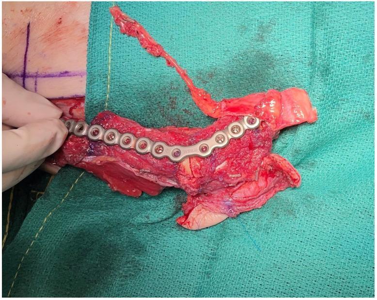
This clinical photograph displays an intraoperative view of a complex mandibular reconstruction construct on a surgical drape. The image shows a composite free flap consisting of a free fibula flow-through flap coupled with a medial femoral trochlea (MFT) flap, intended for mandibular ramus and condylar reconstruction. A silver-colored, contoured titanium reconstruction plate is secured to the bony segments of the fibula using multiple fixation screws. The construct features integrated soft tissue, including a vascularized cartilage component from the MFT flap, which is visible at the distal end of the assembly. A long, thin vascular pedicle extends superiorly from the tissue mass, prepared for anastomosis. This educational image illustrates the use of CAD-CAM surgical planning and multi-component free tissue transfer to restore both the structural integrity of the posterior mandible and the functional joint surface of the condyle.
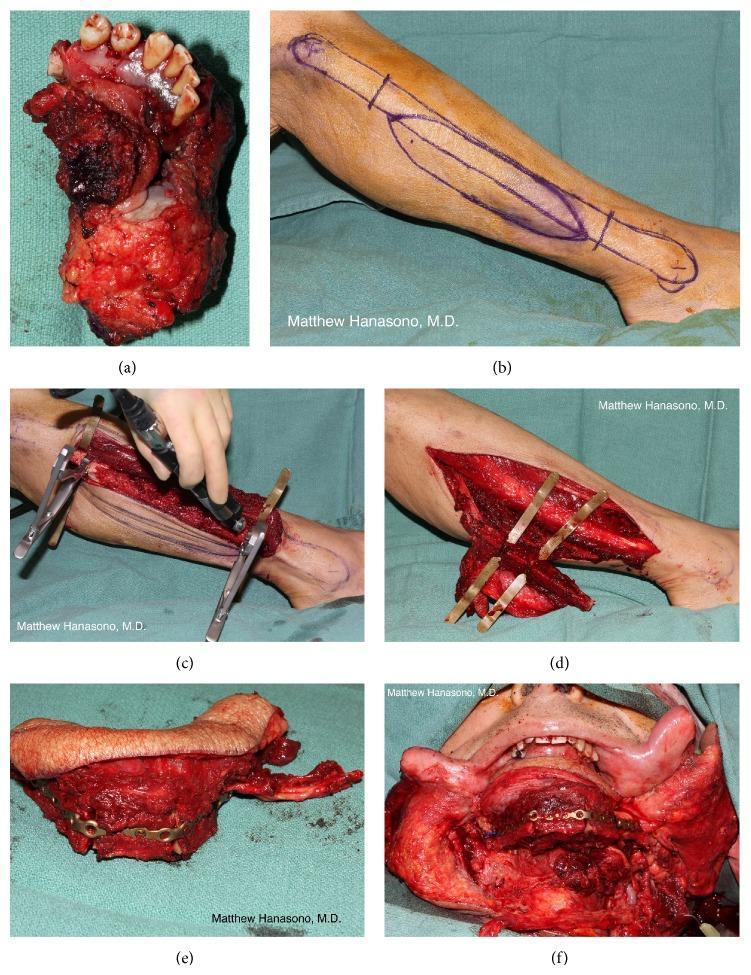
A six-panel clinical photograph series (a-f) documenting a composite anterior mandibular reconstruction using a fibula osteocutaneous free flap. (a) Displays the surgical specimen: a resected segment of the anterior mandible and floor of mouth containing primary tumor and teeth. (b) Shows preoperative skin markings on the lateral lower leg for the planned fibula flap skin paddle. (c) Illustrates the surgical elevation of the fibula with the bone and associated vascular pedicle, utilizing retractors for exposure. (d) Demonstrates the execution of wedge osteotomies on the vascularized fibula to contour the bone. (e) Shows the completed fibula osteocutaneous flap, where multiple bone segments are rigidly fixed with a contoured titanium reconstruction plate and screws to mimic the mandibular arch. (f) Displays the final flap inset into the oral cavity, showing the restored mandibular projection and soft tissue closure. This sequence highlights the complex reconstruction of functional and aesthetic continuity following oncologic resection in head and neck surgery.
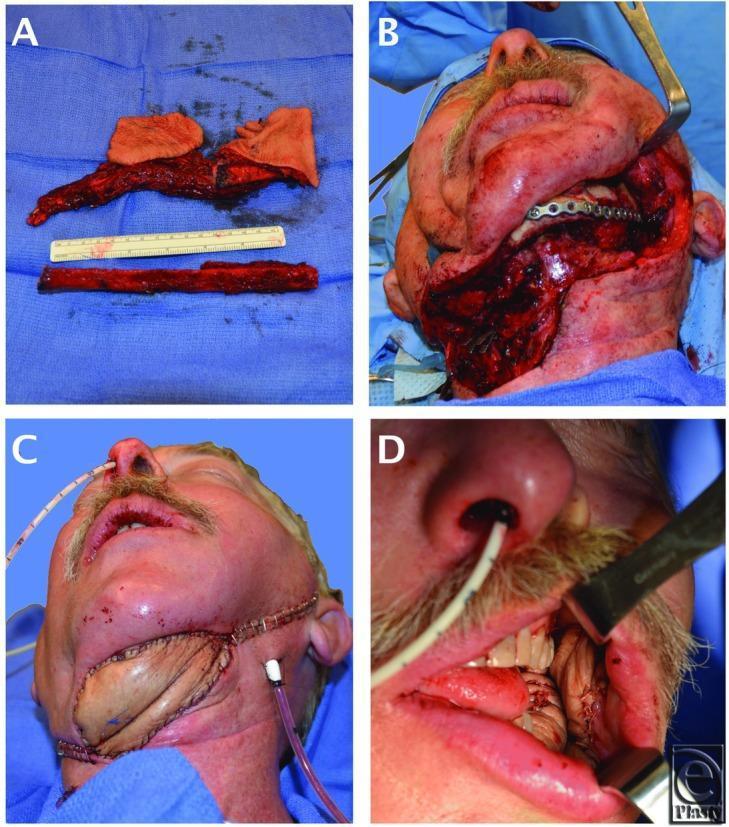
This four-panel clinical series documents a complex head and neck reconstruction using a double-skin paddle fibula free flap. Panel (a) displays the harvested osteocutaneous flap on a surgical drape next to a ruler; it features a segmented fibula bone and two distinct skin islands (double skin paddle) designed to provide independent coverage for internal and external defects. Panel (b) shows the recipient site: a large composite defect of the lateral mandible and submental region, with a metallic reconstruction plate stabilizing the mandibular osteotomy. Panel (c) demonstrates the first skin paddle inset to reconstruct the external submental cutaneous defect, secured with surgical staples. Panel (d) provides an intraoral view showing the second skin paddle inset to restore the mucosal lining of the oral cavity. This case illustrates a modified fibula free flap technique for 'Type 4' composite through-and-through mandibular defects, requiring both structural bone support and dual-surface soft tissue reconstruction.
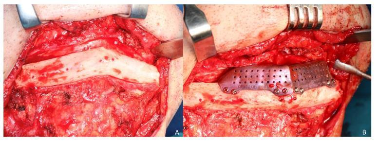
Clinical photograph of an intraoperative maxillofacial reconstruction procedure using a fibula free flap. Figure A shows the surgical exposure of a two-segment fibula flap via a cervical approach. The bone appears pale white with visible surface punctate bleeding, indicating healthy vascularization, and is surrounded by dissected cervical soft tissue and muscle. Figure B demonstrates the placement of a secondary cortico-cancellous iliac crest graft on top of the fibula segments to increase mandibular vertical height. The graft is secured using a custom CAD/CAM titanium mesh, which features a perforated design with 1.5 mm pores to facilitate tissue ingrowth and vascularization. The mesh is fixed to the underlying bone segments with several small diameter (1.5 mm) osteosynthesis screws. This image illustrates complex reconstructive surgery for mandibular defects, specifically addressing vertical discrepancy after primary fibula flap reconstruction.
Mandibular Reconstruction - Techniques in Detail
Introduction
Common Causes of Mandibular Defects
- Oral cavity malignancy (most common)
- Benign tumors (ameloblastoma, odontogenic keratocyst)
- Trauma
- Infection (osteomyelitis)
- Osteoradionecrosis (ORN)
Goals of Mandibular Reconstruction
- Maintain oral competency
- Maintain occlusal relationships of remaining teeth
- Maintain temporomandibular joint (TMJ) alignment and architecture
- Maintain contour of the lower third of the face
- Allow for dental rehabilitation
Classification of Mandibular Defects
- H - Hemi-mandible (posterior segment including condyle)
- C - Central/anterior segment (from canine to canine, crossing the midline)
- L - Lateral segment (body, angle, ramus without condyle)
Principles of Reconstruction Planning
- Size and location of the defect - anterior vs. lateral/posterior
- Pathology - benign vs. malignant; likely need for radiation
- Patient's overall medical status and surgical risk
- Preoperative oral function and goals
- Dentition - edentulous vs. dentulous patients
- Need for adjuvant therapy (radiation compromises non-vascularized grafts)
Small-to-moderate lateral defects (<5 cm) can sometimes be managed with soft tissue alone or plate + soft tissue, especially in edentulous patients. Larger defects, anterior defects, and defects in dentulous patients require bony reconstruction.
Techniques of Mandibular Reconstruction
1. Soft Tissue Reconstruction Only (No Bone)
- Either a pedicled flap or free soft tissue flap fills the defect
- No bony reconstruction is attempted
- The remaining mandible drifts toward the deficient side causing malocclusion, facial asymmetry, and TMJ deviation
- Pectoralis major myocutaneous flap (pedicled) - workhorse for lateral defects
- Radial forearm free flap - thin, pliable soft tissue
- Anterolateral thigh (ALT) flap - large surface area, good for bulky defects
2. Reconstruction Plate Alone (No Bone Graft)
- A rigid titanium reconstruction plate is contoured to span the bony defect
- No bone graft is used
- Must be covered with adequate soft tissue (local, pedicled, or free flap) to prevent plate exposure
- Maintains mandibular contour and prevents deviation
- Shorter operative time
- Avoids donor site morbidity
- Plate fracture (especially in dentulous patients under masticatory stress)
- Plate exposure (infection, radiation, inadequate soft tissue coverage)
- Hardware loosening
- Not a permanent solution for long defects - not suitable for anterior arches
3. Non-Vascularized (Free) Bone Grafts
- Autograft - iliac crest, rib (most reliable)
- Allograft (cadaveric bone)
- Recombinant human BMP (rhBMP) combined with titanium mesh crib
- Bone chips (crushed autograft or allograft) are packed into a titanium mesh crib
- Acts as a scaffold for new bone ingrowth
- Requires intact soft tissue envelope for vascularization
4. Vascularized Free Tissue Transfer (Free Flap) - THE GOLD STANDARD
- Heal in irradiated and contaminated fields
- Provide bulk for soft tissue reconstruction simultaneously
- Allow dental rehabilitation with osseointegrated implants
- Low bone resorption compared to non-vascularized grafts
A. Fibula Free Flap (Most Common - Preferred)
- Supplied by the peroneal artery
- Pedicle length: 6-8 cm
- Bone length: 16-20 cm (up to 25 cm in Cummings)
- Bicortical, thick bone with a short (~1.5 cm) height
- Skin paddle: reliable, thin, pliable with large surface area
- Longest bone length - allows total/subtotal mandibular reconstruction
- Can be osteotomized multiple times (wedge osteotomies) to recreate the mandibular arch curve
- Thin, pliable skin paddle
- Long pedicle
- Easy harvest in supine position (simultaneous two-team approach)
- Excellent osseointegration potential for dental implants
- Suitable for pediatric reconstruction
- Short bone height (~1.5 cm) - may be insufficient for dental implants without augmentation
- Requires vascular assessment of leg circulation (Allen's test equivalent for leg - Doppler assessment)
- Skin paddle orientation limited relative to bone
- Total/subtotal mandibular reconstruction
- Bone-only defects (ORN)
- Atrophic mandible
- Pediatric reconstruction
- Secondary reconstruction
B. Iliac Crest Free Flap (Deep Circumflex Iliac Artery - DCIA Flap)
- Supplied by the deep circumflex iliac artery (DCIA)
- Pedicle length: 6-8 cm
- Bone length: 10-15 cm
- Bone characteristics: tall/thick, abundant cancellous bone - excellent osseointegration (best for dental implants)
- Tallest bone height - closest to native mandibular height
- Best osseointegration potential for dental implants (rated +++ in Scott-Brown's)
- Robust, thick bone stock
- Donor site complications: post-operative pain, gait disturbance, risk of hernia (abdominal wall weakness)
- Pedicle is short
- Limited bone length
- Associated soft tissue is thick and limited - difficult to use for intraoral lining
- Difficult positioning (two-team approach requires repositioning)
C. Scapular / Subscapular System Free Flap
- Based on the subscapular artery system (circumflex scapular artery or thoracodorsal artery)
- Bone: lateral border or tip of scapula
- Lateral border: 10-12 cm, thin and short
- Scapula tip: ~9 cm, ideal shape for angle defects
- Pedicle length: 4-8 cm
- Multiple independent soft tissue islands (subscapular system): skin from thoracodorsal territory + parascapular/scapular skin + latissimus dorsi muscle - all on a single pedicle
- Ideal for complex composite defects involving internal lining + external skin
- Latissimus dorsi can be included for large volume defects
- Minimal functional donor site morbidity
- Thin, short bone - generally NOT adequate to support dental implants (poor osseointegration, rated ++ in Scott-Brown's)
- Requires prone or lateral decubitus positioning (simultaneous two-team surgery not possible in most cases)
- Limited bone length
D. Radial Forearm Osteocutaneous Free Flap
- Based on the radial artery
- Bone: up to 10 cm from the radius (maximum)
- Pedicle length: 8-10 cm (longest pedicle)
- Bone: very thin, blood supply tenuous
- Long pedicle - excellent reach to neck vessels
- Very thin, pliable skin - best for intraoral lining
- Technically straightforward harvest
- Bone stock is too thin and fragile - high risk of donor site radius fracture
- Poor osseointegration potential (rated + in Scott-Brown's)
- Limited bone length (max 10 cm)
- Blood supply to bone is tenuous (endosteal supply rather than periosteal)
5. Temporomandibular Joint (TMJ) Reconstruction
| Method | Description |
|---|---|
| Fibula end in condylar fossa | Round end of fibula placed against temporal bone; heals by scarring; adequate mouth opening |
| Free condylar transfer | Resected condyle (if oncologically safe) plated as free graft onto fibula flap |
| Rib cartilage graft | Rib cartilage secured to fibula tip as load-bearing surface |
| Mechanical prosthetic joint | Metallic condylar prosthesis (head + fossa components) |
| Custom manufactured condylar plate | CAD-manufactured condylar component integrated into reconstruction plate |
Pedicled Regional Flaps for Soft Tissue Coverage
| Flap | Blood Supply | Use |
|---|---|---|
| Pectoralis major myocutaneous | Pectoral branch of thoracoacromial artery | Workhorse for lateral/posterior defects; large volume |
| Submental island flap | Submental artery | Anterior floor of mouth; good color match |
| Temporalis muscle flap | Deep temporal artery | Intraoral coverage |
| Platysma flap | Submental and subplatysmal plexus | Small lateral oral defects |
Comparison Table: Osseocutaneous Free Flaps
| Flap | Bone Length (cm) | Pedicle Length (cm) | Donor Complications | Osseointegration | Skin Reliability |
|---|---|---|---|---|---|
| Fibula | 16-20 | 6-8 | +++ | ++ | ++ |
| Scapula | 8-12 | 4-8 | ++ | ++ | +++ |
| Iliac crest | 10-14 | 6-8 | + | +++ | + |
| Radius | 8-12 | 8-10 | + | + | +++ |
Advanced Technologies in Mandibular Reconstruction
Virtual Surgical Planning (VSP) / Surgical Design and Simulation (SDS)
- 3D-printed cutting guides - placed on the mandible and fibula intraoperatively for precise, pre-planned osteotomies
- Prebent custom titanium plates - manufactured to match the planned reconstruction exactly
- Implant drilling guides - for immediate osseointegrated implant placement
- External fixators and surgical templates - to maintain spatial relationships
- Better 3D understanding of mandibular pathology
- More accurate reconstruction
- Reduced flap ischemia and operative time
- Improved potential for dental implantation and occlusal reconstruction
- Enhanced surgical training and education
- Cost
- Uncertainty in planning resection margins preoperatively in oncologic cases (tumor growth between planning and surgery date may make cutting guides irrelevant)
- Time lag between planning and surgery
Occlusion and Dental Rehabilitation
Bone-Driven Reconstruction (Traditional)
- Lower border of mandible used as template for reconstruction
- Relies on freehand intraoperative techniques
- Limitation: alveolar height may not align with tooth-bearing process
Delayed Osseointegrated Implants
- Reconstruction first, implants placed later (after adjuvant therapy and healing)
- Advantage: selects disease-free, motivated patients for implants
- Major disadvantage: very long delay (reported up to 73 months for complete rehabilitation)
Immediate Osseointegrated Implants (with VSP)
- Implants placed at time of reconstruction using pre-planned drilling guides
- Reduces overall treatment time significantly
- Requires excellent planning (VSP/SDS)
Occlusion-Driven Reconstruction
- Plans reconstruction from the occlusal plane downward rather than from the lower border upward
- Ensures the bone is positioned to optimally support dental implants and prosthetics
- Enabled by VSP technologies
Summary: Choosing the Right Technique
| Clinical Scenario | Preferred Technique |
|---|---|
| Small lateral defect, edentulous, poor surgical risk | Soft tissue flap only (pectoralis or free flap) |
| Short lateral defect, high surgical risk, no radiation | Reconstruction plate + soft tissue coverage |
| Benign tumor, no planned radiation | Non-vascularized bone graft + titanium mesh |
| Any significant defect, malignancy, radiation planned | Vascularized free flap (fibula preferred) |
| Total/subtotal mandibular reconstruction | Fibula free flap |
| Short defect, dental implants priority | Iliac crest DCIA flap |
| Complex composite defect (through-and-through) | Scapular system flap |
| Secondary reconstruction or revision | Fibula free flap |
| High precision dental rehabilitation | VSP/SDS-guided fibula + osseointegrated implants |
Key Points to Remember (Exam Focus)
- Gold standard = Vascularized free tissue transfer with rigid fixation
- Most common free flap = Fibula (longest bone, multiple osteotomies possible, reliable skin paddle)
- Best osseointegration = Iliac crest (tallest bone, thick cancellous stock)
- Best soft tissue options = Scapular system (multiple independent tissue islands)
- Non-vascularized grafts fail in irradiated fields
- Reconstruction plates alone are best for short posterior defects in edentulous patients
- Anterior (C-segment) defects are the most functionally devastating - must have bony reconstruction
- VSP/SDS reduces ischemia time, improves accuracy, and facilitates dental rehabilitation
- Andy Gump deformity = loss of anterior mandibular arch - bilateral muscle pull causes retrodisplacement of tongue, threatening airway