
Polyuria and its causes
Reading File
Now let me search the medical library for polyuria content in parallel.
Finding Sources
Finding Sources
Searching PubMed
polyuria causes diagnosis management
Good results. Now let me read the most comprehensive sections from Harrison's and Goldman-Cecil in parallel.
Reading File
Reading File
Reading File
Let me read the Goldman-Cecil table of causes and fetch the Harrison's diagnostic flowchart image.
Reading File
Reading File
Loading Image
Excellent - I now have comprehensive information. Let me also get the Goldman-Cecil table of polyuria causes and the Brenner & Rector content on osmotic diuresis.
Finding Sources
Finding Sources
I now have a comprehensive set of information from multiple authoritative textbooks. Let me compile the full answer.
Polyuria and Its Causes
Definition
Polyuria is conventionally defined as a urine output exceeding 3 L/day in adults (some sources use >2.5 L/day). It must be distinguished from urinary frequency, where small volumes are voided repeatedly but total daily output remains normal. A 24-hour urine collection is often needed to confirm true polyuria. - Harrison's Principles of Internal Medicine 22E, p. 387
Brenner & Rector offer a physiology-based definition: polyuria is present when urine flow is higher than expected for the prevailing rate of effective osmole excretion and the state of vasopressin (AVP) action, even if the volume does not exceed 2.5 L/day. - Brenner and Rector's The Kidney, p. 1024
Diagnostic Flowchart
The first step in evaluating polyuria is measuring urine osmolality, which divides all cases into two major categories:
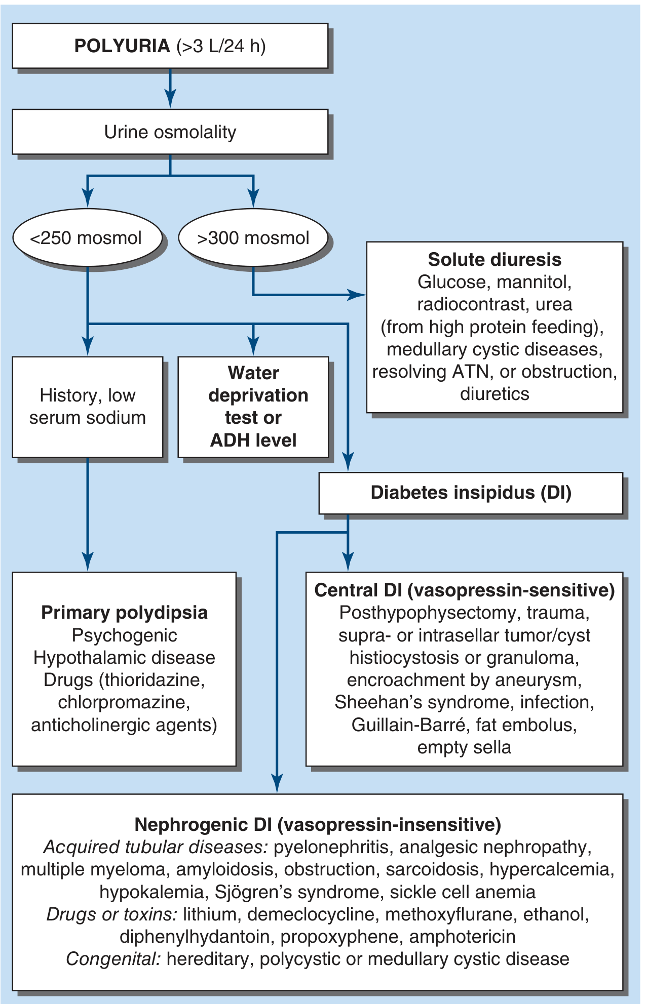
| Urine Osmolality | Category |
|---|---|
| < 250 mosmol/L | Water diuresis |
| > 300 mosmol/L | Solute (osmotic) diuresis |
Category 1: Water Diuresis (Urine Osmolality < 250 mosmol/L)
Water diuresis results from impaired AVP secretion or renal resistance to AVP. There are three sub-categories:
A. Primary Polydipsia (Psychogenic / Dipsogenic)
Excessive water intake overwhelms normal renal diluting mechanisms. AVP is appropriately suppressed due to normal-to-low plasma osmolality. Urine can be maximally dilute at ~50 mosmol/L.
Causes:
- Psychiatric disorders (e.g., schizophrenia, anxiety)
- Hypothalamic disease affecting the thirst center
- Drugs that stimulate thirst or dry the mouth: thioridazine, chlorpromazine, anticholinergic agents
B. Central Diabetes Insipidus (CDI) - Vasopressin-Sensitive
Inadequate secretion of AVP from the posterior pituitary. Urine volumes can reach up to 20 L/day. Responds to exogenous desmopressin (DDAVP).
Causes: - Goldman-Cecil Medicine, p. 1205; Robbins Pathologic Basis of Disease
- Idiopathic (most common - often autoimmune destruction of AVP neurons)
- Post-hypophysectomy or surgical trauma
- Head trauma
- Suprasellar/intrasellar tumors or cysts (craniopharyngioma, germinoma)
- Histiocytosis X (Langerhans cell histiocytosis)
- Granulomas (sarcoidosis, tuberculosis)
- Encroachment by aneurysm
- Sheehan's syndrome (postpartum pituitary necrosis)
- Infection (meningitis, encephalitis)
- Guillain-Barre syndrome
- Fat embolus
- Empty sella syndrome
- Inflammatory hypothalamic disease
C. Nephrogenic Diabetes Insipidus (NDI) - Vasopressin-Insensitive
The kidney fails to respond to circulating AVP. AVP cannot insert AQP2 water channels into the luminal membrane of collecting duct principal cells. Does NOT respond to DDAVP.
Causes: - Harrison's p. 387; Goldman-Cecil p. 1205
Acquired tubular diseases:
- Pyelonephritis
- Analgesic nephropathy
- Multiple myeloma
- Amyloidosis
- Urinary tract obstruction / post-obstructive diuresis
- Sarcoidosis
- Hypercalcemia (impairs AQP2 insertion)
- Hypokalemia
- Sjögren's syndrome
- Sickle cell disease / sickle cell anemia
- Chronic kidney disease
Drugs and toxins:
- Lithium (most common drug cause - used in bipolar disorder; blocks AQP2 channel insertion)
- Demeclocycline (tetracycline)
- Methoxyflurane
- Ethanol
- Diphenylhydantoin (phenytoin)
- Propoxyphene
- Amphotericin B
Congenital (genetic):
- Mutation in vasopressin V2 receptor gene (X-linked, most common hereditary form)
- Mutation in aquaporin-2 (AQP2) water channel gene (autosomal recessive)
- Hereditary polycystic or medullary cystic disease
Category 2: Solute (Osmotic) Diuresis (Urine Osmolality > 300 mosmol/L)
Excess poorly-reabsorbed solutes in the tubular lumen prevent water reabsorption. The medullary concentration gradient is also washed out, and urine output can be massive.
Causes: - Harrison's p. 387; Goldman-Cecil p. 1205-1206
| Solute | Clinical Setting |
|---|---|
| Glucose | Uncontrolled diabetes mellitus (most common cause of solute diuresis) |
| Urea | High-protein enteral/parenteral nutrition; post-obstructive diuresis; resolving acute tubular necrosis (ATN) |
| Mannitol | Iatrogenic (IV mannitol for cerebral edema) |
| Radiocontrast | Post-imaging with contrast agents |
| Sodium | Salt-wasting nephropathy (Bartter's syndrome, tubulointerstitial disease, resolving ATN) |
| Sodium | Cystic renal diseases |
Since urine sodium is less than plasma sodium in glucose-driven diuresis, more water than sodium is lost, resulting in hypernatremia and serum hypertonicity.
Other Important Causes
- Chronic renal failure: As nephron mass is reduced, each surviving nephron handles an increased solute load and cannot concentrate urine. Polyuria and nocturia are early signs of CKD. - Goldman-Cecil Medicine, p. 1206
- Tubulointerstitial kidney diseases: Direct medullary tubular damage reduces NaCl and water reabsorption (e.g., sickle cell nephropathy, analgesic nephropathy).
- Gestational diabetes insipidus: Pregnancy is associated with increased vasopressinase activity (from the placenta), accelerating AVP degradation. Resolves after delivery. - Creasy & Resnik's Maternal-Fetal Medicine
- Hypercalcemia / Hypokalemia: Both cause NDI by interfering with AVP-mediated water reabsorption in the collecting duct.
Summary Table
| Category | Urine Osmolality | Key Causes |
|---|---|---|
| Primary polydipsia | < 100 mosmol/L | Psychiatric, hypothalamic disease, drugs |
| Central DI | 50-200 mosmol/L | Trauma, tumors, surgery, infection, idiopathic |
| Nephrogenic DI | 50-200 mosmol/L | Lithium, hypercalcemia, hypokalemia, genetic mutations |
| Solute diuresis | > 300 mosmol/L | Diabetes mellitus, mannitol, urea, contrast |
Diagnostic Workup Summary
- 24-hour urine collection - confirm volume > 3 L/day
- Urine osmolality - differentiates water vs. solute diuresis
- Plasma glucose, calcium, potassium, urea - screen for solute causes
- Water deprivation test + exogenous DDAVP - distinguishes central DI, nephrogenic DI, and primary polydipsia
- Plasma AVP (or copeptin) - elevated in NDI (AVP present but ineffective); low/absent in central DI
- Hypertonic saline-stimulated copeptin - can substitute for water deprivation testing where available
- Harrison's Principles of Internal Medicine 22E, p. 387-388; Goldman-Cecil Medicine, p. 1205-1206; Brenner and Rector's The Kidney, p. 1024
This is a shared conversation. Sign in to Orris to start your own chat.