
Classify gall stones. Discuss the etiopathogenesis. Clinical features, investigation and treatment of acute calculous cholecystits.Describe etiology, clinical features, investigations, differential diagnosis and management of Gallstones***** ➢ Discuss obstructive jaundice with investigations and management***
Reading File
Now I'll run parallel searches across the medical library:
Finding Sources
Finding Sources
Reading File
Reading File
Good - substantial content returned. Let me read more of both files to get the full clinical picture:
Reading File
Reading File
Reading File
Reading File
Reading File
Finding Sources
Reading File
Reading File
Reading File
Reading File
Reading File
Reading File
I now have comprehensive content from the textbooks. Let me fetch a couple of key images and get the obstructive jaundice management content:
Reading File
Loading Image
I now have comprehensive content from multiple authoritative textbooks. Let me compile the full, detailed answer.---
GALLSTONES (CHOLELITHIASIS) AND ACUTE CALCULOUS CHOLECYSTITIS
PART 1: CLASSIFICATION OF GALLSTONES
Gallstones affect 10-20% of adult populations in high-resource countries. More than 95% of biliary tract disease is attributable to gallstones. In the USA/Europe, over 80% are cholesterol or mixed stones; in Asia, 80% are pigment stones.
Three Main Types:
| Type | Composition | Key Associations |
|---|---|---|
| Cholesterol stones | >50% crystalline cholesterol monohydrate | Obesity, female sex, OCP, pregnancy, rapid weight loss, ileal disease |
| Black pigment stones | Bilirubin polymer + calcium phosphate + calcium bicarbonate; <30% cholesterol | Haemolysis (hereditary spherocytosis, sickle cell disease), cirrhosis, advancing age |
| Brown pigment stones | Calcium bilirubinate + calcium palmitate + calcium stearate + cholesterol | Bile stasis, infection (E. coli, Clonorchis sinensis, Ascaris lumbricoides), bile duct foreign bodies |
Mixed stones contain 50-99% pure cholesterol with an admixture of calcium salts, bile acids, bile pigments and phospholipids.
PART 2: ETIOPATHOGENESIS OF GALLSTONES
Cholesterol Stone Formation - Three Steps:
Step 1 - Supersaturation of bile with cholesterol:
- Cholesterol is insoluble in water and secreted from the canalicular membrane in phospholipid vesicles
- When bile is supersaturated with cholesterol and/or bile acid concentrations are low, unstable unilamellar phospholipid vesicles form
- Factors increasing cholesterol secretion: obesity, high-calorie diet, oral contraceptives, oestrogens (increase hepatic lipoprotein receptors and stimulate hepatic cholesterol synthesis)
- Factors depleting bile acid pool: ileal disease (Crohn's), ileal resection, cholestyramine
Step 2 - Nucleation of cholesterol crystals:
- Cholesterol monohydrate crystals nucleate from multilamellar vesicles
- Nucleating factors include mucus, glycoproteins, and infection
Step 3 - Gallbladder hypomotility/stasis:
- Abnormal gallbladder emptying aids aggregation of nucleated crystals
- This is why removing stones without removing the gallbladder leads to recurrence
Factors in gallstone formation:
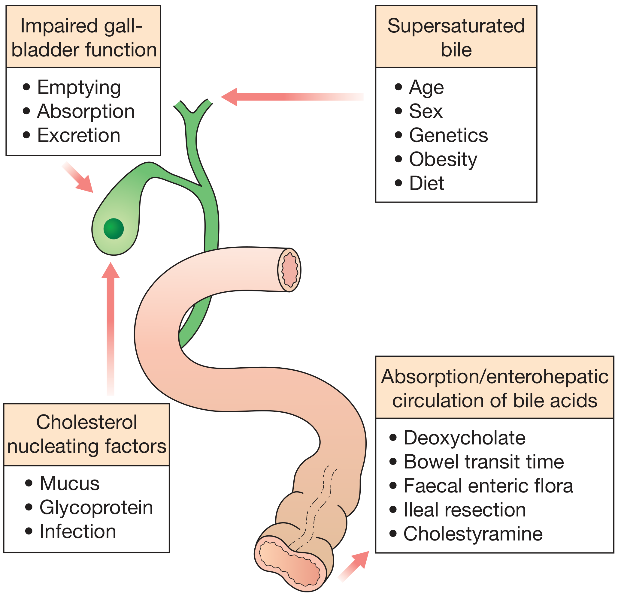
Risk Factors (The "5 F's" mnemonic expanded):
- Fat - obesity, metabolic syndrome, hyperlipidaemia
- Female - higher prevalence due to oestrogen exposure
- Fertile - pregnancy increases biliary cholesterol secretion
- Forty - increasing age; predominantly middle to older age
- Fair - Northern Europeans, North/South Americans, Native Americans (Pima, Hopi, Navajo tribes up to 75%)
- Additional: oral contraceptives, rapid weight reduction, gallbladder stasis, inborn bile acid metabolism disorders, total parenteral nutrition, vagotomy
Pigment Stone Pathogenesis:
- Black stones: Unconjugated bilirubin increases due to haemolysis → precipitates as bilirubin polymer in concentrated supersaturated bile
- Brown stones: Bacterial β-glucuronidase (from E. coli, Bacteroides) deconjugates bilirubin glucuronide → insoluble unconjugated bilirubinate precipitates in infected stagnant bile in ducts
PART 3: CLINICAL FEATURES OF GALLSTONES
Asymptomatic ("silent") gallstones - >80% of cases:
- Prophylactic cholecystectomy generally NOT indicated
- 20-year follow-up: only 18% develop biliary pain; mean yearly probability is 2% in first 5 years, declining to 0.5% by years 10-20
Symptomatic gallstones:
- Right upper quadrant (RUQ) or epigastric pain, radiating to the back or right shoulder tip
- Pain is typically dull, continuous, and severe (not truly "colicky") - lasts minutes to hours
- Associated nausea, vomiting, anorexia
- Pain often starts at night (wakes the patient)
- Relieved when stone slips back into the gallbladder body
- Dyspepsia, flatulence, fatty food intolerance (less specific symptoms)
Complications - clinical presentations:
- Acute cholecystitis: fever, positive Murphy's sign, RUQ mass (omental wall-off)
- Empyema of gallbladder: high fever, rigors, severely ill patient
- Gangrene and perforation: peritonitis
- Choledocholithiasis: obstructive jaundice (dark urine, pale stools, pruritus)
- Cholangitis: Charcot's triad (RUQ pain + fever/rigors + jaundice); Reynolds' pentad adds shock + confusion
- Pancreatitis: epigastric pain radiating to the back, elevated amylase/lipase
Differential Diagnosis of biliary colic:
- Peptic ulcer disease / gastritis
- Acute pancreatitis
- Hepatitis (acute)
- Right-sided renal colic
- Myocardial infarction (inferior MI can mimic biliary pain)
- Irritable bowel syndrome
- Hiatus hernia / GORD
- Appendicitis (atypical)
PART 4: INVESTIGATIONS FOR GALLSTONES
Bloods:
- FBC - leucocytosis in cholecystitis (WBC >18,000/mm³ indicates moderate severity by Tokyo Guidelines)
- LFTs - elevated ALP, GGT, conjugated bilirubin if CBD involvement; transaminases elevated in hepatitis/pancreatitis
- Serum amylase/lipase - if pancreatitis suspected
- CRP - inflammatory marker to assess severity
- PT/INR - may be prolonged if jaundiced (Vitamin K deficiency)
- Serum albumin - marker of nutritional status
Imaging:
| Investigation | Role | Sensitivity |
|---|---|---|
| Ultrasound (1st line) | Detects gallstones, GB wall thickening (>4mm), pericholecystic fluid, acoustic shadowing, CBD diameter | ~95% for GB stones |
| MRCP | Best non-invasive test for CBD stones, strictures, choledocholithiasis | ~95% overall for biliary obstruction |
| CT abdomen | Excludes complications (perforation, abscess), detects CBD stones (less sensitive than MRCP), staging of malignancy | High for masses >2cm |
| HIDA scan (hepatobiliary iminodiacetic acid) | Functional assessment; non-filling = cystic duct obstruction (acute cholecystitis) when US equivocal | ~95% sensitivity for acute cholecystitis |
| ERCP | Therapeutic gold standard for CBD stones - sphincterotomy, stone extraction, stenting | Also diagnostic for strictures |
| EUS (endoscopic ultrasound) | Excellent for distal CBD, ampullary region tumors, FNA for tissue; sensitivity 84-91% for CBD stones | Better than MRCP for small stones |
Ultrasound criteria for acute cholecystitis:
- Sonographic Murphy's sign
- Gallbladder wall thickening >4mm
- Pericholecystic fluid
- Distended gallbladder with gallstones
PART 5: ACUTE CALCULOUS CHOLECYSTITIS
Pathogenesis:
Acute calculous cholecystitis accounts for 90% of all acute cholecystitis cases. The sequence is:
- Stone impaction in the cystic duct or gallbladder neck → outflow obstruction
- Chemical inflammation - mucosal phospholipases hydrolyze luminal lecithins to toxic lysolecithins → disrupts the protective glycoprotein mucus layer → bile salts exert direct detergent action on mucosa
- Prostaglandin release from distended gallbladder wall → mucosal and mural inflammation
- Distension and raised intraluminal pressure → compromised mucosal blood flow
- Bacterial superinfection (later phase) - bacteria from: E. coli, Klebsiella, Enterococcus, Bacteroides, Clostridium
- Progression → gangrenous cholecystitis → perforation → empyema/peritonitis
Clinical Features:
- Onset: Progressive RUQ or epigastric pain lasting >6 hours (distinguishes it from biliary colic)
- Fever: Low-grade initially; high fever suggests gangrenous cholecystitis or empyema
- Nausea and vomiting
- Anorexia, tachycardia, sweating
Physical signs:
- Murphy's sign (pathognomonic) - RUQ tenderness exacerbated during inspiration when palpating the right subcostal region; the patient catches their breath
- RUQ tenderness and guarding - tenderness, voluntary guarding
- Palpable mass - inflamed gallbladder walled off by omentum
- Jaundice (in ~20%) - due to CBD compression by periductal inflammation (Mirizzi syndrome) or concurrent choledocholithiasis
Complications:
- Empyema of the gallbladder - pus filling the lumen; high fever, rigors, very tender mass
- Gangrenous cholecystitis - wall necrosis; gas-forming organisms → emphysematous cholecystitis
- Perforation - generalised biliary peritonitis or localised pericholecystic abscess
- Fistula - cholecystoduodenal, cholecystocolic, or cholecystogastric
- Gallstone ileus - large stone erodes into duodenum, causes small bowel obstruction at ileocaecal valve
- Mirizzi syndrome - stone in cystic duct compresses common hepatic duct
Tokyo Guidelines 2018 - Severity Grading:
Grade I (Mild): Acute cholecystitis in a healthy patient with no organ dysfunction; mild inflammatory changes in the gallbladder
Grade II (Moderate): Any one of:
- Elevated WBC >18,000/mm³
- Palpable tender mass in RUQ
- Duration of symptoms >72 hours
- Marked local inflammation (gangrenous/emphysematous cholecystitis, pericholecystic abscess)
Grade III (Severe): Organ dysfunction in any one of:
- Cardiovascular: hypotension requiring dopamine ≥5 μg/kg/min or any epinephrine
- Neurological: decreased level of consciousness
- Respiratory: PaO₂/FiO₂ ratio <300
- Renal: oliguria; creatinine >2.0 mg/dL
- Hepatic: PT-INR >1.5
- Haematological: platelets <100,000/mm³
Management of Acute Calculous Cholecystitis:
Initial (non-operative) treatment:
- Nil by mouth + IV fluid resuscitation until pain resolves
- Analgesia - NSAIDs (diclofenac IM/IV reduces intraluminal pressure via prostaglandin inhibition), opioids (pethidine preferred over morphine to avoid Oddi spasm)
- IV Antibiotics - as cystic duct is blocked, serum concentration matters more than biliary concentration:
- Mild-moderate: cefazolin, cefuroxime, cefoxitin, or ciprofloxacin
- Severe: piperacillin-tazobactam or 3rd-generation cephalosporin + metronidazole
- Add anaerobic cover if gangrenous/emphysematous cholecystitis suspected
- Monitoring: temperature, pulse, WBC, CRP
Investigations to confirm:
- USG to confirm (first-line)
- If jaundiced: MRCP to exclude choledocholithiasis
- If complicated: CT scan
Surgical Management - Timing:
| Timing | Recommendation | Evidence |
|---|---|---|
| Early cholecystectomy (within 72h-7 days) | Preferred; safe if experienced surgeon available | Shorter total hospital stay (9.6 vs 17.8 days combined RCT data) |
| Delayed/interval cholecystectomy (6 weeks later) | If early surgery not feasible or Grade II with no deterioration | 26% failure requiring emergency surgery before 6 weeks |
| Emergency surgery | Grade III; imminent perforation; diagnostic uncertainty | Urgent |
- Laparoscopic cholecystectomy is the gold standard (higher conversion rate in acute vs. elective but still preferred)
- Tokyo Guidelines algorithm: Grade I → early laparoscopic cholecystectomy; Grade II → early LC if experienced surgeon available, otherwise conservative + delayed LC; Grade III → ICU stabilisation first, then LC when stable or cholecystostomy as bridge
- Percutaneous cholecystostomy - for Grade III patients unfit for surgery; tube drainage of gallbladder as a temporising measure
OBSTRUCTIVE JAUNDICE
DEFINITION
Obstructive (post-hepatic/cholestatic) jaundice is the clinical manifestation of impaired flow of conjugated bilirubin through the biliary tree into the duodenum. Total serum bilirubin is elevated, predominantly the direct (conjugated) fraction (>50% of total).
ETIOLOGY / CAUSES
Malignant (Painless, Progressive):
- Carcinoma of the head of pancreas (most common malignant cause)
- Cholangiocarcinoma (Klatskin tumour at hilum)
- Carcinoma of the gallbladder
- Ampullary carcinoma
- Metastatic disease compressing the biliary system
Benign (often Painful, Fluctuating):
- Choledocholithiasis (most common cause overall - painful, fluctuating)
- Primary or secondary sclerosing cholangitis
- Benign biliary strictures (post-operative, post-inflammatory)
- Choledochal cyst
- Parasites (Clonorchis sinensis, Ascaris)
- Haemobilia
- Pancreatic pseudocysts
- Chronic pancreatitis (pseudotumoral)
- Duodenal diverticulum / ampullary scarring
Congenital:
- Biliary atresia
- Caroli's disease
Key clinical pointer: Painful jaundice → likely stones; Painless progressive jaundice → likely malignancy (Courvoisier's law: a palpable non-tender gallbladder in the presence of jaundice is unlikely to be due to stones)
CLINICAL FEATURES
Symptoms:
- Jaundice - yellow discolouration of skin, sclerae, mucous membranes
- Dark urine (conjugated bilirubin - water soluble, excreted in urine → "Coca-Cola urine")
- Pale/clay-coloured stools (absence of stercobilinogen)
- Pruritus - due to bile salt deposition in skin; often severe
- Fatigue
- Abdominal pain - RUQ colicky pain in stones; epigastric pain in pancreatic cancer
- Weight loss, anorexia - suggests malignancy
- Fever and rigors - suggests cholangitis (Charcot's triad: pain + fever + jaundice)
Signs:
- Icteric sclerae, jaundiced skin
- Scratch marks (pruritus)
- Palpable, non-tender gallbladder (Courvoisier's sign) → malignant obstruction
- Hepatomegaly
- Splenomegaly in portal hypertension
- Cachexia, Virchow's node (left supraclavicular lymph node) - in GI malignancy
- Hepatic flap/asterixis if hepatic failure developing
DIFFERENTIAL DIAGNOSIS
| Type | Examples | Bilirubin | ALP | AST/ALT | Urine | Stools |
|---|---|---|---|---|---|---|
| Pre-hepatic | Haemolysis, spherocytosis | Indirect ↑↑ | Normal | Normal | Normal (no bili) | Normal/dark |
| Hepatic | Viral hepatitis, cirrhosis, drugs | Both ↑ | Mildly ↑ | Markedly ↑↑ | Bilirubin+ | Variable |
| Post-hepatic (Obstructive) | Stones, malignancy | Direct ↑↑ | Markedly ↑↑↑ | Mildly ↑ | Dark (bilirubin+) | Pale |
INVESTIGATIONS
Blood Tests:
| Test | Finding in Obstructive Jaundice |
|---|---|
| Total bilirubin | Elevated; repeat every 3-4 days to monitor trend |
| Direct (conjugated) bilirubin | >50% of total; markedly elevated |
| Serum alkaline phosphatase (ALP) | 3-4 fold elevation in 75% of cholestasis cases |
| GGT / 5'-nucleotidase | Elevated (confirms hepatobiliary source vs bone) |
| AST/ALT (transaminases) | Mildly elevated; very high levels (>1000 IU/L) suggest hepatocellular cause |
| Serum albumin | Low in chronic disease / malnutrition |
| PT/INR | Prolonged (Vitamin K malabsorption); corrects with IV Vitamin K in post-hepatic jaundice (unlike hepatic jaundice) - this response differentiates the two |
| FBC | Anaemia of malignancy; leucocytosis in cholangitis |
| Serum amylase/lipase | Elevated in pancreatic pathology |
| CA 19-9, CEA | Tumour markers for pancreatic and biliary malignancy |
| Alpha-fetoprotein | Hepatocellular carcinoma |
| Hepatitis serology (HBsAg, anti-HCV, HAV IgM) | Exclude viral hepatitis |
| ANA, AMA (anti-mitochondrial Ab) | Primary biliary cirrhosis (AMA+); autoimmune hepatitis |
Imaging Investigations:
-
Ultrasound (US) - FIRST LINE:
- Detects biliary duct dilation; establishes level of obstruction in ~67% of cases
- Sensitivity for CBD stones only 21-63% (poor for distal stones)
- Shows liver parenchyma, gallbladder, hepatic/pancreatic masses
-
MRCP (Magnetic Resonance Cholangiopancreatography):
- Non-invasive test of choice for biliary anatomy
- Sensitivity 95% for biliary obstruction; best for CBD stones
- Differentiates strictures but distinguishing benign vs malignant can be challenging
- No therapeutic capability
-
CT abdomen/pelvis:
- Excellent for pancreatic tumours >2cm, staging of malignancy
- Identifies vascular involvement, lymphadenopathy, metastases
- Less sensitive than MRCP for CBD stones
-
EUS (Endoscopic Ultrasound):
- Best for distal CBD, ampullary region, pancreatic head
- Enables fine-needle aspiration (FNA): sensitivity 84-91%, specificity 71-100%
- Can identify vascular invasion and lymph node metastases
-
ERCP (Endoscopic Retrograde Cholangiopancreatography):
- Primarily therapeutic (not just diagnostic)
- Cannulation successful in >90% cases
- Enables: sphincterotomy, stone extraction, balloon sweep, stent placement, brush cytology
- Complications: pancreatitis, cholangitis, bleeding, perforation
-
Percutaneous Transhepatic Cholangiography (PTC):
- Used when ERCP fails or inaccessible anatomy
- Allows external biliary drainage, internal stenting
-
Liver biopsy:
- Reserved for suspected parenchymal disease
- Contraindicated if PT prolonged and unresponsive to Vitamin K, or platelets <50,000/mm³
MANAGEMENT OF OBSTRUCTIVE JAUNDICE
General Preoperative Preparation:
-
Correction of coagulation:
- IV Vitamin K₁ 20 mg → rapid normalisation of PT within 12-24 hours (if liver synthetic function intact)
- Good response = post-hepatic obstruction (favourable surgical risk)
- Poor response = hepatocellular disease
-
Renal protection:
- Jaundiced patients at high risk of hepatorenal syndrome
- Aggressive IV fluid hydration pre- and post-operatively
- Monitor urine output; consider mannitol infusion
- Avoid nephrotoxic drugs
-
Nutritional support - correct malnutrition, hypoalbuminaemia
-
Antibiotics - treat concurrent cholangitis with broad-spectrum cover
Surgical mortality in jaundiced patients: ~20%; recognised risk factors:
- Old age
- Malnutrition
- Ongoing biliary infection
- Malignancy
- High serum urea (renal impairment)
Definitive Management Based on Cause:
A. Choledocholithiasis (CBD stones):
- ERCP + endoscopic sphincterotomy + stone extraction (first-line)
- Basket or balloon retrieval of stones
- Lithotripsy for large stones
- Followed by laparoscopic cholecystectomy for the gallbladder
B. Malignant Obstruction (Pancreatic/Bile Duct Cancer):
Resectable disease (~20% of periampullary cancers):
- Pancreaticoduodenectomy (Whipple's procedure) for periampullary/pancreatic head tumours
- Preoperative biliary drainage: controversial - not proven to reduce operative mortality; however, needed if neoadjuvant chemotherapy is planned
Non-resectable disease:
- Biliary decompression/palliation:
- ERCP + stenting (first-line for distal obstruction)
- Plastic stents: cheap but occlude every 3-4 months (biofilm formation); patency ~43 days
- Self-expanding metallic stents (SEMS): wider bore, patency ~125 days; preferred for >6 months survival
- Covered SEMS (cSEMS): prevent tumour ingrowth, removable; meta-analysis shows fewer adverse events vs uncovered SEMS
- Percutaneous transhepatic drainage (when ERCP fails)
- Surgical bypass (hepaticojejunostomy or choledochojejunostomy) if endoscopic approach not possible
- ERCP + stenting (first-line for distal obstruction)
- Only 25-30% of liver volume needs to be drained for jaundice resolution
- Right hepatic duct drains 50-55% of liver
- Left hepatic duct drains 30-35%
- Caudate lobe drains 10%
C. Benign Biliary Strictures (Bismuth Classification):
| Type | Level | Treatment |
|---|---|---|
| I | Low CHD stump >2 cm | ERCP dilation + plastic stents |
| II | Proximal CHD stump <2 cm | ERCP or surgical repair |
| III | Hilar; confluence preserved | Hepaticojejunostomy |
| IV | Hilar; confluence lost | Complex hepaticojejunostomy |
| V | Aberrant right sectoral duct | Surgical |
- Endoscopic treatment: serial large-bore plastic stents exchanged every 3-4 months; success 74-90%, recurrence up to 30% at 2 years
- Surgical hepaticojejunostomy (Roux-en-Y) for failed endoscopic treatment
D. Sclerosing Cholangitis:
- Ursodeoxycholic acid (medically)
- Dominant strictures: ERCP dilation + stenting
- Liver transplantation for end-stage disease
Summary Algorithm for Obstructive Jaundice:
Jaundice suspected
      ↓
Blood tests (LFTs, bilirubin fractions, ALP, transaminases, PT)
      ↓
Ultrasound abdomen (1st line)
      ↓
Biliary dilation?
   YES                          NO
    ↓                            ↓
MRCP/CT/EUS              Consider hepatic/pre-hepatic cause
    ↓
Cause identified?
   Stones → ERCP + extraction    Malignancy → Resectable? → Whipple's
                                             → Non-resectable → ERCP stent / PTC
   Benign stricture → ERCP or surgical bypass
Sources:
- Bailey and Love's Short Practice of Surgery, 28th Edition - Chapters on Gallstones, Acute Cholecystitis
- Robbins, Cotran & Kumar Pathologic Basis of Disease - Gallbladder pathology
- Sleisenger and Fordtran's Gastrointestinal and Liver Disease - Acute Cholecystitis, Obstructive Jaundice
- Current Surgical Therapy, 14th Edition - Obstructive Jaundice evaluation and endoscopic management
- Pye's Surgical Handicraft, 22nd Edition - Management of obstructive jaundice, preoperative preparation
This is a shared conversation. Sign in to Orris to start your own chat.