
Heartrate in valsalva maneuver
During the Valsalva maneuver, heart rate follows a characteristic 4-phase pattern: it initially rises slightly (phase I), then increases more during straining (phase II) due to reduced venous return and compensatory tachycardia, then briefly drops sharply (phase III), and finally shows a reflex bradycardia (phase IV) as blood pressure overshoots and the baroreceptor reflex kicks in.
Searching sources...
Reading File
Finding Sources
Finding Sources
Reading File
Reading File
Reading File
Let me also fetch the diagram image from Braunwald's and check the Adams & Victor neurology content for the Valsalva ratio:
Reading File
Reading File
Loading Image
Here is a complete, textbook-grounded answer on heart rate changes during the Valsalva maneuver.
Heart Rate During the Valsalva Maneuver
The Valsalva maneuver is a forced expiration against a closed glottis (or into a closed system at ~40 mmHg for ~10-15 seconds). It produces a characteristic, 4-phase sequence of blood pressure and heart rate changes driven by baroreceptor reflexes.
The 4 Phases
| Phase | What Happens to BP | Heart Rate | Mechanism |
|---|---|---|---|
| I (onset of straining) | Brief rise | Slight transient slowing | Increased intrathoracic pressure transmitted to aorta raises BP momentarily; baroreceptors cause brief vagal slowing |
| II (continued straining) | Falls (reduced venous return → reduced CO) | Rises (tachycardia) | Reduced BP + pulse pressure inhibits baroreceptors → sympathetic activation + parasympathetic withdrawal → tachycardia + peripheral vasoconstriction |
| III (release of straining) | Sharp brief drop | Brief further rise or transient drop | Sudden drop in intrathoracic pressure transiently pools blood in the pulmonary vasculature |
| IV (after release) | Overshoots above baseline | Reflex bradycardia | Venous return and CO are restored into a still-vasoconstricted circulation → BP overshoots → baroreceptors stimulated → parasympathetic predominance → bradycardia and return to normal |
Key Mechanisms
Phase II tachycardia:
"The fall in blood pressure is sensed and sympathetic stimulation and parasympathetic withdrawal brings the blood pressure back up toward baseline and the heart rate elevates."
- Braunwald's Heart Disease, p. 1264
Phase IV bradycardia:
"The blood pressure therefore rises above normal, and this stimulates the baroreceptors, causing bradycardia and a drop in pressure to normal levels."
- Ganong's Review of Medical Physiology, p. 592
"With the release of intrathoracic pressure, the venous return, stroke volume, and blood pressure rise to higher-than-normal levels; reflex parasympathetic influence then predominates and a bradycardia results."
- Adams and Victor's Principles of Neurology, p. 554
Marey's Law: Pulse rate moves in a reciprocal direction to arterial blood pressure - this is the foundational principle governing all phases. (Barash, Cullen, and Stoelting's Clinical Anesthesia, p. 1068)
Normal Blood Pressure Response (with pulse rate as reciprocal)
The image below shows the normal arterial pressure trace (Panel A) vs. an abnormal response (Panel B, e.g. in autonomic failure):
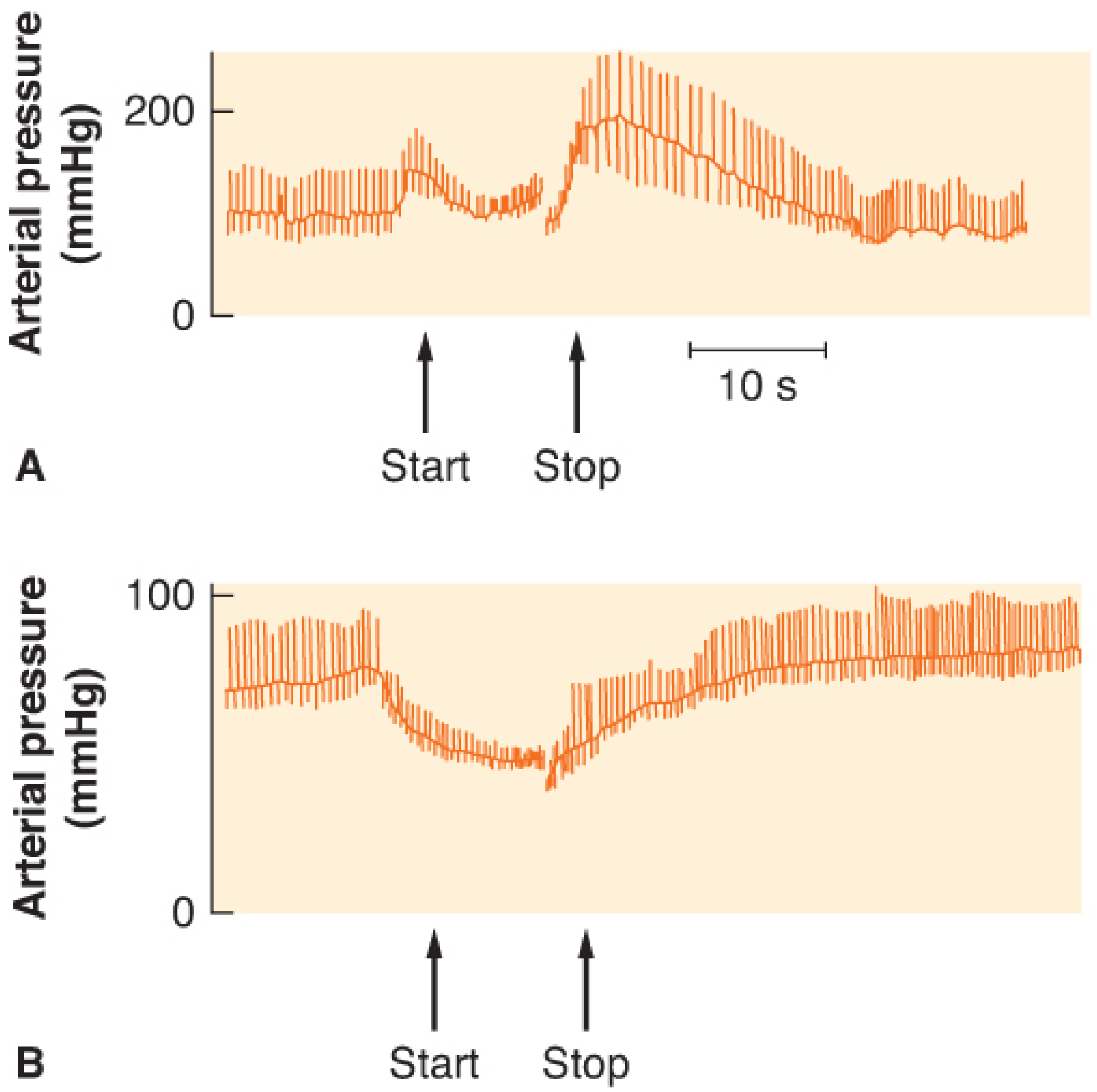
In Panel A (normal): BP rises at onset, falls during straining, then overshoots after release - heart rate mirrors the inverse of this curve.
In Panel B (autonomic failure): BP falls continuously during straining with no overshoot - heart rate fails to compensate.
Valsalva Ratio (Clinical Use)
The Valsalva ratio = maximum heart rate during the maneuver ÷ lowest heart rate within 30 seconds after that peak.
- Normal ratio: ≥ 1.5 (varies slightly by age)
- Used to test autonomic integrity - specifically parasympathetic (vagal) function
- Failure of HR to rise during straining (Phase II) → sympathetic dysfunction
- Failure of HR to slow during BP overshoot (Phase IV) → parasympathetic (vagal) dysfunction
(Adams and Victor's Principles of Neurology, p. 554)
Abnormal Response Pattern
In autonomic failure (e.g. Shy-Drager syndrome, diabetic autonomic neuropathy):
- The fall in BP during straining is not corrected (no sympathetic compensation)
- There is no BP overshoot on release
- Therefore no Phase IV bradycardia occurs
- Heart rate changes are absent or blunted throughout
(Ganong's Review of Medical Physiology, p. 592; Braunwald's Heart Disease, p. 1264)
Summary
| Direction | Phase | Driver |
|---|---|---|
| HR increases | Phase II (straining) | Baroreceptor-mediated sympathetic activation + vagal withdrawal |
| HR decreases (reflex bradycardia) | Phase IV (after release) | BP overshoot activates baroreceptors → vagal predominance |
How to interpret abnormal Valsalva responses in autonomic testing
Abnormal Valsalva responses point to dysfunction in either the sympathetic or parasympathetic limb of the autonomic reflex arc. In brief: failure of HR to rise during straining = sympathetic failure; failure of HR to slow (no bradycardia) after release = parasympathetic failure; absent BP overshoot = adrenergic failure; and a "square wave" response = elevated venous pressure (e.g. heart failure).
Searching sources...
Finding Sources
Finding Sources
Finding Sources
Finding Sources
Reading File
Reading File
Finding Sources
Finding Sources
Finding Sources
Finding Sources
Reading File
Reading File
Searching the Web
Valsalva maneuver autonomic testing interpretation abnormal responses adrenergic failure square wave
Reading File
Reading Web Page
https://paulogentil.com/pdf/The%20Valsalva%20manoeuvre%20-%2…
Reading File
Now I have everything needed for a comprehensive, well-structured answer.
Interpreting Abnormal Valsalva Responses in Autonomic Testing
The Valsalva maneuver probes two distinct arms of the autonomic baroreflex:
| Parameter | Autonomic Limb Tested |
|---|---|
| Heart rate response / Valsalva ratio | Parasympathetic (vagal) - Phase II tachycardia & Phase IV bradycardia |
| BP recovery time + adrenergic index | Sympathetic (adrenergic) - Phase II BP recovery & Phase IV overshoot |
The Two Core Scores
1. Valsalva Ratio (VR)
= Maximum HR during maneuver ÷ Lowest HR within 30 s after that peak
- Normal: ≥ 1.5 in young adults; decreases with age (≥ 1.2 by age 40, approaches 1.04 beyond age 60)
- Measures the parasympathetic arm of the baroreflex
- Diminished in diabetes, α-synuclein disorders (Parkinson's, MSA, DLB), and with anticholinergic medications
2. Adrenergic Index / BP Recovery
- The magnitude of the BP drop during Phase II, BP recovery time, and BP overshoot in Phase IV
- Measures the sympathetic (adrenergic) arm of the baroreflex
(Harrison's Principles of Internal Medicine 22E, p. 451)
Pattern Recognition: 4 Abnormal Response Types
Type 1 - Adrenergic (Sympathetic) Failure
BP pattern: Large, uncorrected hypotension in early Phase II, no late Phase II recovery, no Phase IV overshoot.
"Large hypotension in early Phase II and lack of pressure recovery and pressure overshoot in late Phase II and Phase IV can be observed in patients with orthostatic hypotension (α-adrenergic failure). Dramatic drops in BP in Phase II in subjects with adrenergic dysfunction may even lead to syncope."
- Physiology and Clinical Examples of the Valsalva Manoeuvre (Sandroni et al. 1985 cited)
HR implication: HR may still rise in Phase II (if baroreceptor afferents and vagal pathways are intact) but there is no Phase IV bradycardia because there is no BP overshoot to trigger it.
"Dysfunction of the SNS is implicated if exaggerated and prolonged hypotension develops during the forced expiration phase (≥50% fall from resting MAP). In addition, the overshoot at the end of the Valsalva maneuver is absent."
- Barash, Cullen, and Stoelting's Clinical Anesthesia, p. 1068
Type 2 - Vagal (Parasympathetic) Failure
HR pattern: HR fails to slow appropriately during the Phase IV BP overshoot. Valsalva ratio is reduced or absent.
"Failure of the heart rate to increase during the positive intrathoracic pressure phase of the Valsalva maneuver points to sympathetic dysfunction, and failure of the rate to slow during the period of blood pressure overshoot points to a parasympathetic disturbance."
- Adams and Victor's Principles of Neurology 12th Ed., p. 554
"Dysfunction of the PNS can be assumed if the HR does not respond appropriately to the blood pressure changes."
- Barash, Cullen, and Stoelting's Clinical Anesthesia, p. 1068
Type 3 - Complete Autonomic Failure (combined sympathetic + parasympathetic)
Pattern: The BP falls continuously throughout Phase II and never recovers. No BP overshoot on release. No Phase IV bradycardia. HR changes are absent throughout.
"In patients with autonomic failure, the fall in blood pressure is not aborted during the last few seconds of increased intrathoracic pressure, and there is no overshoot of blood pressure when the breath is released."
- Adams and Victor's Principles of Neurology 12th Ed., p. 554
"In patients whose sympathetic nervous system is not functional, heart rate changes still occur because the baroreceptors and the vagi are intact. However, in patients with autonomic insufficiency, a syndrome in which autonomic function is widely disrupted, the heart rate changes are absent."
- Ganong's Review of Medical Physiology, p. 592
Seen in: MSA, pure autonomic failure (PAF), severe diabetic autonomic neuropathy.
Type 4 - "Square Wave" Response (Elevated Venous Pressure / Cardiac Failure)
Pattern: No Phase II BP fall - instead, BP is maintained or even rises throughout straining ("flat-top" pressure trace). No Phase II tachycardia. No Phase IV bradycardia. This is because venous return is NOT reduced by straining.
Mechanism: When left ventricular filling pressure is already elevated (heart failure, constrictive pericarditis, mitral stenosis, atrial septal defect), the increased intrathoracic pressure does not reduce venous return significantly - the elevated pulmonary blood volume acts as a buffer. There is no fall in CO, so no baroreceptor-mediated HR response occurs.
(Physiology and Clinical Examples of the Valsalva Manoeuvre; Levin 1966, Zema et al. 1980)
Summary Interpretation Table
| Finding | What It Indicates |
|---|---|
| Low Valsalva ratio (low HR rise in Ph II, no bradycardia in Ph IV) | Parasympathetic (vagal) failure |
| No Phase II BP recovery + no Phase IV overshoot | Sympathetic (adrenergic) failure |
| Severe Phase II hypotension ± syncope | Severe α-adrenergic failure |
| No HR or BP response at all | Complete autonomic failure |
| "Square wave" - no Phase II BP drop | Elevated venous pressure / cardiac failure |
| Adrenergic index low but VR normal | Isolated sympathetic involvement, vagal intact |
Confounders to Account For
- Age: VR normally decreases with age - always compare to age-matched norms. Values approaching 1.04 can be normal over age 60.
- Medications: Anticholinergics blunt VR; antihypertensives, alpha- and beta-blockers directly affect both arms of the baroreflex.
- Technique: Suboptimal straining pressure (<40 mmHg) or duration (<15 s) may prevent late Phase II from forming, yielding a false-positive abnormal result.
- Position: Supine position increases the incidence of abnormal BP responses; standardize position between tests.
(Adams and Victor's Principles of Neurology, p. 554; The Dysautonomia Project - Valsalva)
Clinical Conditions Causing Abnormal Responses
| Condition | Pattern |
|---|---|
| Diabetic autonomic neuropathy | Low VR (early parasympathetic involvement), later adrenergic failure |
| MSA / Parkinson's with autonomic failure | Both VR and adrenergic index abnormal |
| Pure autonomic failure (PAF) | Severe adrenergic failure, no overshoot |
| Autoimmune autonomic ganglionopathy | Variable, depending on ganglionic AChR antibody titer |
| Heart failure / constrictive pericarditis | Square wave response |
| Spinal cord injury (high cervical) | Absent adrenergic response below lesion |
| Primary hyperaldosteronism | Absent HR changes and BP overshoot (returns to normal after tumor removal) |
Significance of valsalva maneuver
The Valsalva maneuver has wide clinical significance: it tests autonomic integrity, helps terminate SVT, differentiates cardiac murmurs, equalizes middle ear pressure, and occurs physiologically during coughing, defecation, and childbirth. Abnormal responses can diagnose autonomic neuropathy, heart failure, and baroreflex dysfunction.
Searching sources...
Finding Sources
Finding Sources
Reading File
Reading File
Reading File
Finding Sources
Finding Sources
Finding Sources
Reading File
Reading File
Finding Sources
Reading File
Reading File
Finding Sources
I now have rich content from multiple textbooks covering all the major domains of Valsalva significance. Let me compile the comprehensive answer.
Clinical Significance of the Valsalva Maneuver
The Valsalva maneuver (forced expiration against a closed glottis) holds significance across multiple medical disciplines - from bedside cardiology to neurology, urology, ophthalmology, and anesthesia.
1. Autonomic Function Testing
The maneuver is the cornerstone bedside test of baroreceptor reflex integrity, probing both arms of the autonomic nervous system simultaneously.
"The integrity of the baroreceptor reflex can be tested with the Valsalva maneuver, which is expiring against a closed glottis as during coughing, defecation, or heavy lifting."
-
Costanzo Physiology 7th Edition, p. 172
-
Valsalva Ratio (max HR during maneuver ÷ lowest HR in 30 s after) = parasympathetic (vagal) function
-
Adrenergic Index (magnitude of BP fall + overshoot) = sympathetic (adrenergic) function
Diminished in: diabetes, α-synuclein disorders (MSA, Parkinson's, DLB), anticholinergic drug use
(Harrison's Principles of Internal Medicine 22E)
2. Termination of SVT (Supraventricular Tachycardia)
The Valsalva maneuver is a first-line vagal maneuver for terminating AV node-dependent tachycardias.
"During wide-QRS tachycardias with a 1:1 relationship between the P waves and QRS complexes, vagal influence can terminate or slow a supraventricular tachycardia (SVT) that depends on the AV node for perpetuation."
- Braunwald's Heart Disease, p. Physical Examination section
"Mean changes in bradycardia are greatest with the Valsalva maneuver... As the maneuver is sustained, vagal tone is increased, thereby leading to a compensatory decrease in SA and AV conduction."
- Roberts and Hedges' Clinical Procedures in Emergency Medicine
SVTs terminated by Valsalva: AVNRT, AVRT, sinus node reentry, adenosine-sensitive AT, idiopathic RVOT tachycardia.
Technique (modified/REVERT method): Patient supine, blows to 40 mmHg for 10-20 s, then passive leg raise immediately after release - enhances venous return and augments the vagal response.
Also useful diagnostically: In wide-complex tachycardia, if vagal maneuver causes AV dissociation, this confirms VT rather than SVT with aberrancy.
3. Dynamic Cardiac Auscultation - Murmur Differentiation
The Valsalva maneuver is a standard bedside tool for differentiating cardiac murmurs by transiently reducing preload (venous return) during the strain phase.
"The majority of murmurs decrease in intensity during the strain phase of the maneuver. Two notable exceptions are the murmurs associated with MVP and HOCM, both of which become louder during the Valsalva maneuver."
- Harrison's Principles of Internal Medicine 22E, p. 332
| Condition | Effect of Valsalva (strain phase) | Mechanism |
|---|---|---|
| HOCM | Murmur increases | Reduced preload → smaller LV cavity → worse LVOT obstruction |
| MVP | Click moves earlier; murmur longer and louder | Smaller LV → earlier leaflet prolapse |
| Aortic stenosis | Murmur decreases | Reduced preload → less flow across valve |
| MR, VSD, AR | Murmur decreases | Reduced preload and filling |
| Most other murmurs | Decrease | Reduced venous return/cardiac output |
"The left ventricular outflow tract obstruction of HOCM is dynamic... Preload is decreased during the straining phase of the Valsalva maneuver, which leads to an increase in the degree of outflow obstruction with an associated increase in the intensity of the murmur."
- Frameworks for Internal Medicine
4. Physiological Occurrences (Normal Life)
The Valsalva maneuver occurs involuntarily in several everyday activities:
- Defecation / straining - most common physiological Valsalva
- Heavy lifting / weight training
- Coughing and sneezing
- Childbirth (second stage of labor)
- Playing wind instruments
- Ear pressure equalization (e.g., during air travel or diving)
This is why patients with heart failure, severe aortic stenosis, or autonomic failure can experience dizziness, presyncope, or syncope during these activities.
5. Diagnosis of Heart Failure ("Square Wave" Response)
In patients with elevated left ventricular filling pressures (heart failure, constrictive pericarditis, mitral stenosis), the normal Phase II BP fall does not occur - instead, BP is maintained ("square wave" or flat-top pattern) because the large pulmonary blood volume buffers the effect of reduced venous return.
This square wave response is a bedside sign of elevated left heart pressures and was historically used to diagnose heart failure at the bedside (Sharpey-Schafer, 1955).
6. Urological Applications
Valsalva voiding - straining to raise intravesical pressure to overcome bladder outlet resistance - is used in selected patients with:
- Areflexic/neurogenic bladder with low outlet resistance
- T11-L2 spinal cord injury with sympathetic denervation
- Detrusor underactivity (DUA) with incompetent sphincter
"'Voiding' by the Credé or Valsalva maneuver is generally discouraged because it is non-physiologic... The best chance for success is in the patient with an areflexic bladder and some degree of outlet denervation."
- Campbell Walsh Wein Urology
Contraindicated in: vesicoureteral reflux, neurogenic bladder with poor detrusor compliance (risk of upper tract deterioration).
7. Stress Incontinence Provocation
A Valsalva (cough/sneeze/laugh/strain) increases intra-abdominal pressure and is used clinically to provoke and demonstrate stress urinary incontinence - patients leak urine when urethral sphincter resistance is insufficient to overcome the pressure rise.
8. Ophthalmology - Valsalva Retinopathy
Sudden, intense Valsalva (e.g., heavy lifting, vomiting, vigorous coughing) can cause a sudden rise in intraocular/venous pressure, rupturing superficial retinal capillaries and producing Valsalva retinopathy - typically pre-retinal (subhyaloid) hemorrhage at the macula, causing acute, often severe visual loss in an otherwise healthy young person.
9. Anesthesia Significance
The Valsalva maneuver identifies patients at risk for autonomic instability during anesthesia, particularly when patients are on drugs depleting catecholamines (e.g., reserpine). It is also relevant to:
- Managing elevated ICP (straining increases intracranial pressure - should be avoided)
- Predicting hemodynamic instability under general anesthesia
- Assessing baroreflex sensitivity preoperatively
(Barash, Cullen and Stoelting's Clinical Anesthesia)
Summary Table
| Domain | Significance |
|---|---|
| Autonomic testing | Tests both sympathetic (BP response) and parasympathetic (HR/Valsalva ratio) baroreflex integrity |
| Cardiology - Arrhythmia | Terminates AV node-dependent SVT; distinguishes SVT from VT |
| Cardiology - Murmurs | Increases HOCM and MVP murmurs; decreases most others |
| Cardiology - Heart failure | Square wave response indicates elevated LV filling pressure |
| Urology | Valsalva voiding in selected neurogenic bladder patients; provokes stress incontinence |
| Ophthalmology | Valsalva retinopathy (pre-retinal hemorrhage) |
| Anesthesia | Predicts ANS instability; ICP concerns |
| Physiology | Occurs during defecation, coughing, lifting, childbirth, ear pressure equalization |
Baroreceptor reflexes
Baroreceptors are stretch-sensitive mechanoreceptors in the carotid sinus and aortic arch that detect changes in arterial wall tension (a proxy for blood pressure) and trigger rapid autonomic reflexes to restore BP toward normal. Rising BP increases baroreceptor firing → inhibits sympathetic outflow + increases vagal tone → vasodilation + bradycardia. Falling BP does the opposite.
Searching sources...
Finding Sources
Finding Sources
Reading File
Reading File
Reading File
Reading File
Reading File
Reading File
Reading File
Reading File
Reading File
Loading Image
Loading Image
Loading Image
Reading File
Reading File
Finding Sources
Loading Image
Loading Image
Excellent - I have rich, comprehensive content from multiple major physiology textbooks plus four highly relevant diagrams. Here is the complete answer.
Baroreceptor Reflexes
Overview
The baroreceptor reflex is the primary fast, neurally mediated mechanism for moment-to-moment blood pressure control. It works by sensing arterial wall stretch and driving compensatory changes in autonomic outflow within seconds.
"The baroreceptor mechanisms are fast, neurally mediated reflexes that attempt to keep arterial pressure constant via changes in the output of the sympathetic and parasympathetic nervous systems to the heart and blood vessels."
- Costanzo Physiology 7th Ed., p. 169
1. Anatomy of Baroreceptors
Baroreceptors are spray-type mechanoreceptors (stretch receptors) located in:
- Carotid sinus - a small dilation of the internal carotid artery just above the carotid bifurcation
- Aortic arch - in the adventitia of the aortic wall
They are also found in smaller numbers in almost every large thoracic and neck artery.
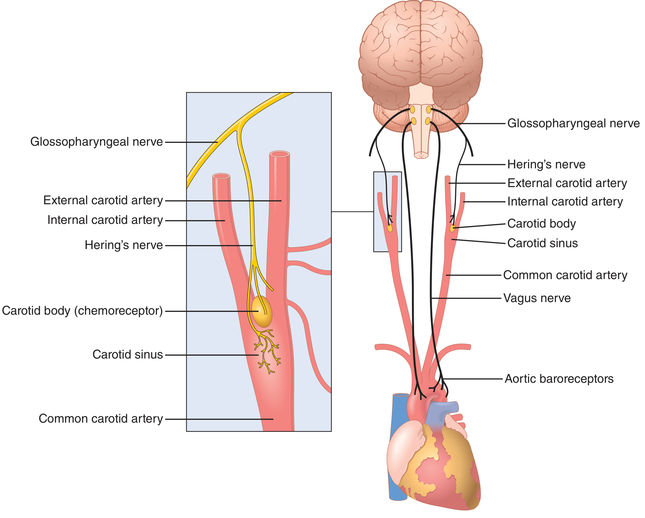
Afferent pathways:
- Carotid sinus baroreceptors → Hering's nerve → Glossopharyngeal nerve (CN IX) → Nucleus tractus solitarius (NTS) of medulla
- Aortic arch baroreceptors → Aortic depressor nerve → Vagus nerve (CN X) → NTS of medulla
"Baroreceptors are spray-type nerve endings that lie in the walls of the arteries and are stimulated when stretched... extremely abundant in the carotid sinus and the aortic arch."
- Guyton and Hall Textbook of Medical Physiology
Key difference between the two receptors:
- Carotid sinus baroreceptors: respond to both increases AND decreases in arterial pressure; more sensitive; threshold ~50 mmHg; saturates at ~200 mmHg
- Aortic arch baroreceptors: primarily responsive to increases in arterial pressure; operate at pressure levels ~30 mmHg higher than carotid receptors; continue responding at pressures where carotid receptors have saturated
(Costanzo Physiology, p. 170; Medical Physiology)
2. Firing Characteristics
The firing rate of the carotid sinus nerve follows a sigmoidal (S-shaped) curve with pressure - sensitivity is maximum in the normal operating range (~100 mmHg MAP), exactly where it is most needed.
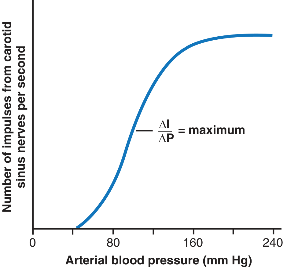
Key properties:
- Threshold: ~50 mmHg (no activity below this)
- Maximum firing: ~200 mmHg (throughout the cardiac cycle)
- Most sensitive to: rapid changes in pressure (rate of change), not just absolute pressure
- Pulsatile pressure sensitivity: a fall in pulse pressure without change in mean pressure still reduces firing and triggers a pressor response
- Firing increases during systole and decreases during diastole (phasic pattern)
"The strongest stimulus for the baroreceptors is a rapid change in arterial pressure!"
- Costanzo Physiology, p. 170
"The baroreceptors respond rapidly... the rate of impulse firing increases in the fraction of a second during each systole and decreases again during diastole. Furthermore, the baroreceptors respond much more to a rapidly changing pressure than to a stationary pressure."
- Guyton and Hall Textbook of Medical Physiology
3. Central Processing - The Medullary Circuit
The NTS is the first synapse. From there, a well-defined 4-neuron brainstem circuit controls both sympathetic and parasympathetic output:
Neurotransmitters and relay nuclei:
| Step | Structure | Neurotransmitter | Effect |
|---|---|---|---|
| 1 | NTS (first relay) | Glutamate | Activates CVLM |
| 2 | CVLM (caudal ventrolateral medulla) | GABA | Inhibits RVLM |
| 3 | RVLM (rostral ventrolateral medulla) | Glutamate (suppressed) | Sympathetic outflow decreases |
| 4 | NTS → nucleus ambiguus / dorsal motor nucleus | Excitatory | Vagal (parasympathetic) outflow increases |
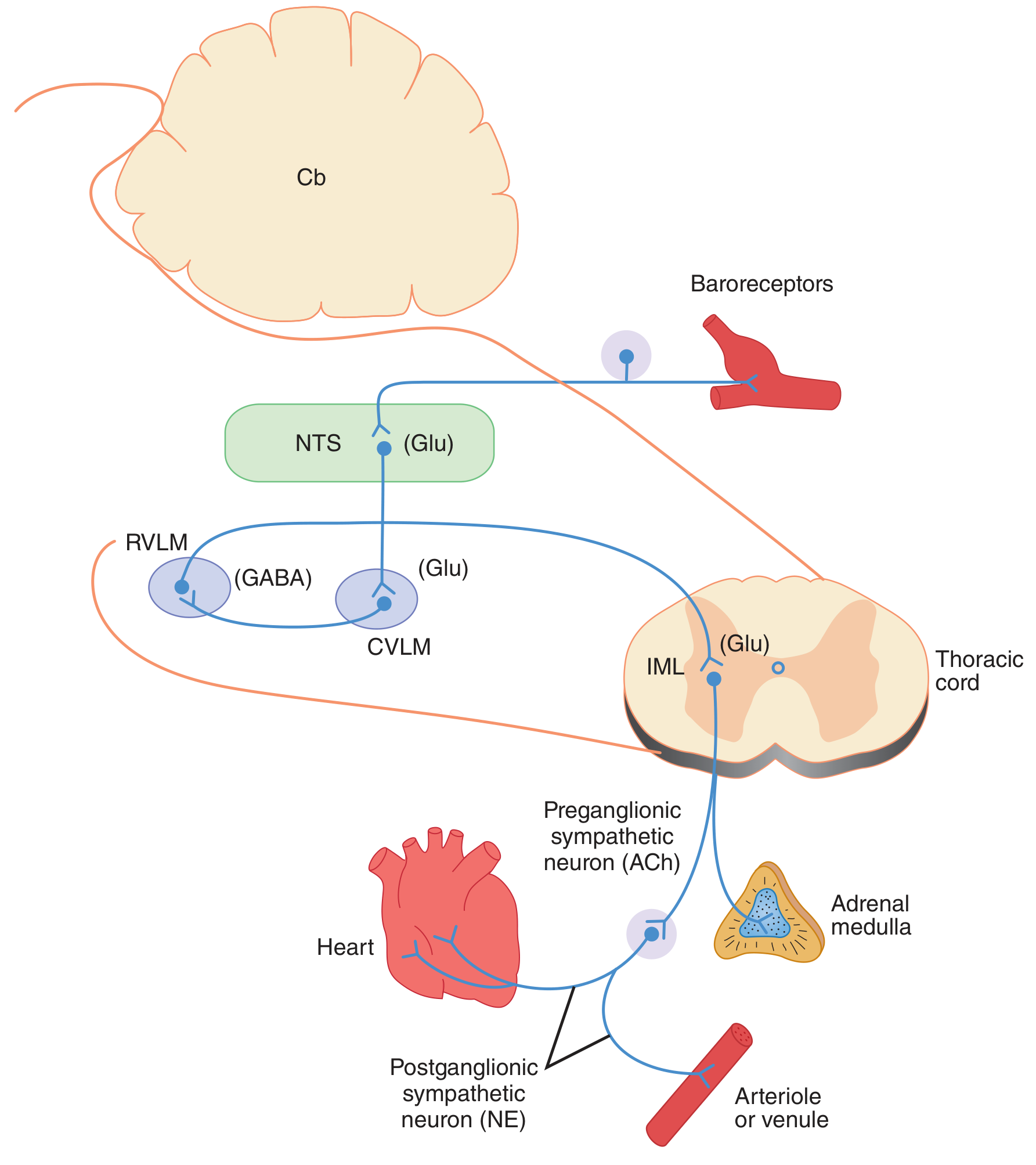
"NTS neurons excite neurons in the CVLM. These CVLM neurons release GABA into the RVLM to reduce the firing rate of these [sympathetic] neurons. Excitatory projections also extend from the NTS to the vagal motor neurons. Thus, increased baroreceptor discharge inhibits the tonic discharge of sympathetic nerves and excites the vagal innervation of the heart. These neural changes produce vasodilation, venodilation, hypotension, bradycardia, and a decrease in cardiac output."
- Ganong's Review of Medical Physiology, p. 589
Brainstem cardiovascular centers (tonically active):
- Vasoconstrictor center (C1/RVLM): upper medulla + lower pons → sympathetic → arteriolar and venous constriction
- Cardiac accelerator center: sympathetic → increases SA node firing, AV conduction velocity, and contractility
- Cardiac decelerator center: parasympathetic (vagus) → decreases SA node firing rate
4. Integrated Reflex Arc
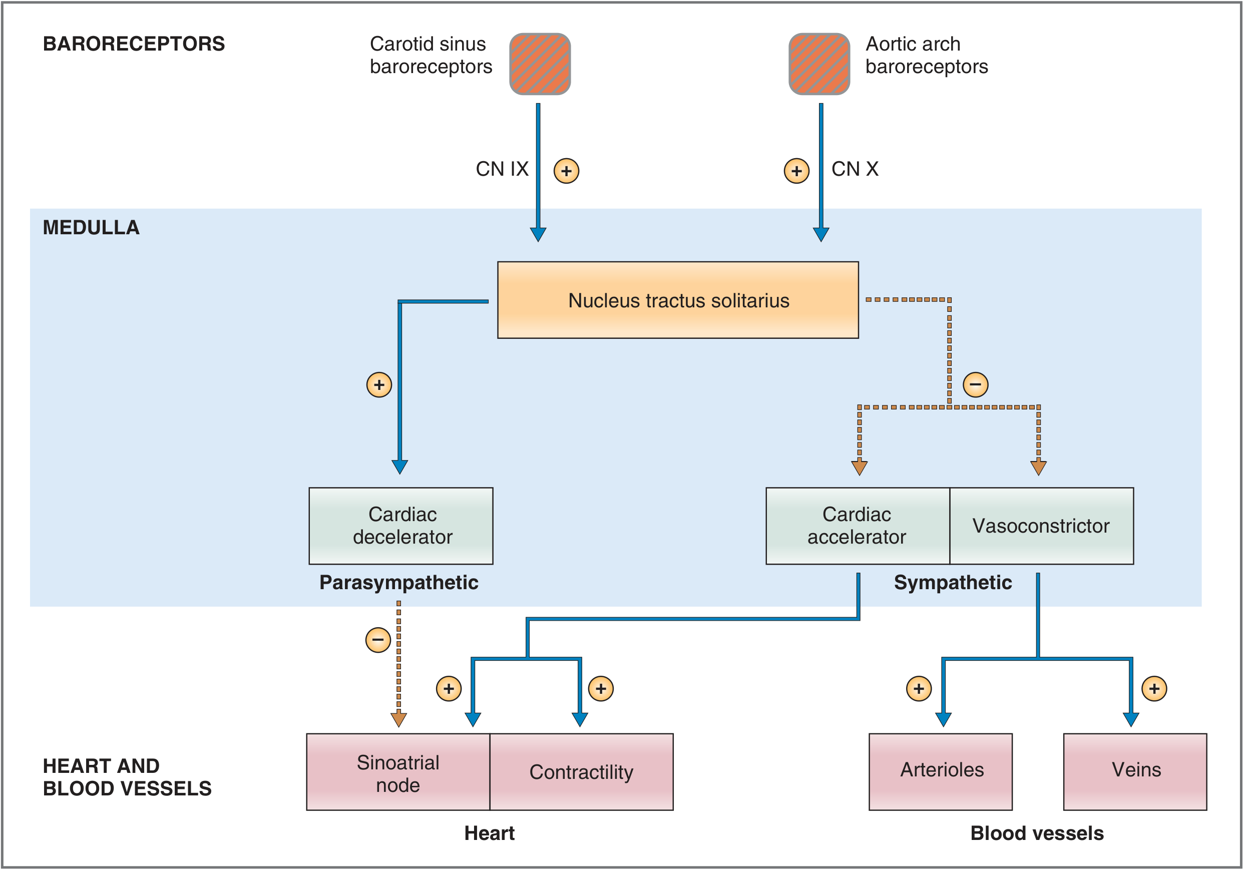
Response to INCREASED arterial pressure (e.g., acute hypertension):
- ↑ BP → ↑ stretch → ↑ baroreceptor firing rate (CN IX, CN X → NTS)
- NTS activates CVLM → CVLM GABAergically inhibits RVLM → ↓ sympathetic outflow
- NTS also excites vagal motor nuclei → ↑ parasympathetic outflow
- Net effects:
- ↓ Heart rate (vagus on SA node)
- ↓ Contractility (↓ sympathetic to myocardium)
- Arteriolar vasodilation → ↓ TPR
- Venodilation → ↑ unstressed volume → ↓ venous return
- ↓ Cardiac output + ↓ TPR → BP falls back toward normal
Response to DECREASED arterial pressure (e.g., hemorrhage, standing up):
- ↓ BP → ↓ stretch → ↓ baroreceptor firing
- Less CVLM activity → less GABA inhibition of RVLM → ↑ sympathetic outflow
- Less NTS output to vagal nuclei → ↓ parasympathetic outflow
- Net effects:
- ↑ Heart rate (β1 on SA node)
- ↑ Contractility (β1 on myocardium)
- ↑ TPR (arteriolar vasoconstriction via α1)
- Venoconstriction → ↓ unstressed volume → ↑ venous return
- ↑ Cardiac output + ↑ TPR → BP rises toward normal
(Costanzo Physiology, pp. 170-171; Guyton and Hall)
5. Responses in Specific Situations
Response to Hemorrhage
Decreased blood volume → ↓ stressed volume → ↓ PA → baroreceptor reflex activated:
| Parameter | Response |
|---|---|
| Carotid sinus nerve firing | ↓ |
| Heart rate | ↑ |
| Contractility | ↑ |
| Cardiac output | ↑ |
| TPR | ↑ (arteriolar vasoconstriction) |
| Venous unstressed volume | ↓ (venoconstriction increases venous return) |
| Renin, Ang II, Aldosterone | ↑ (slower hormonal reinforcement) |
(Costanzo Physiology, Table 4.9)
Response to Standing (Orthostatic stress)
Blood pools in leg veins → ↓ venous return → ↓ CO → ↓ PA → baroreceptor reflex drives tachycardia + arteriolar and venous constriction to maintain cerebral perfusion.
"Immediately on standing, arterial pressure in the head and upper body tends to fall, and marked reduction could cause loss of consciousness. However, the falling pressure at the baroreceptors elicits an immediate reflex, resulting in strong sympathetic discharge throughout the body."
- Guyton and Hall
6. "Pressure Buffer" Function
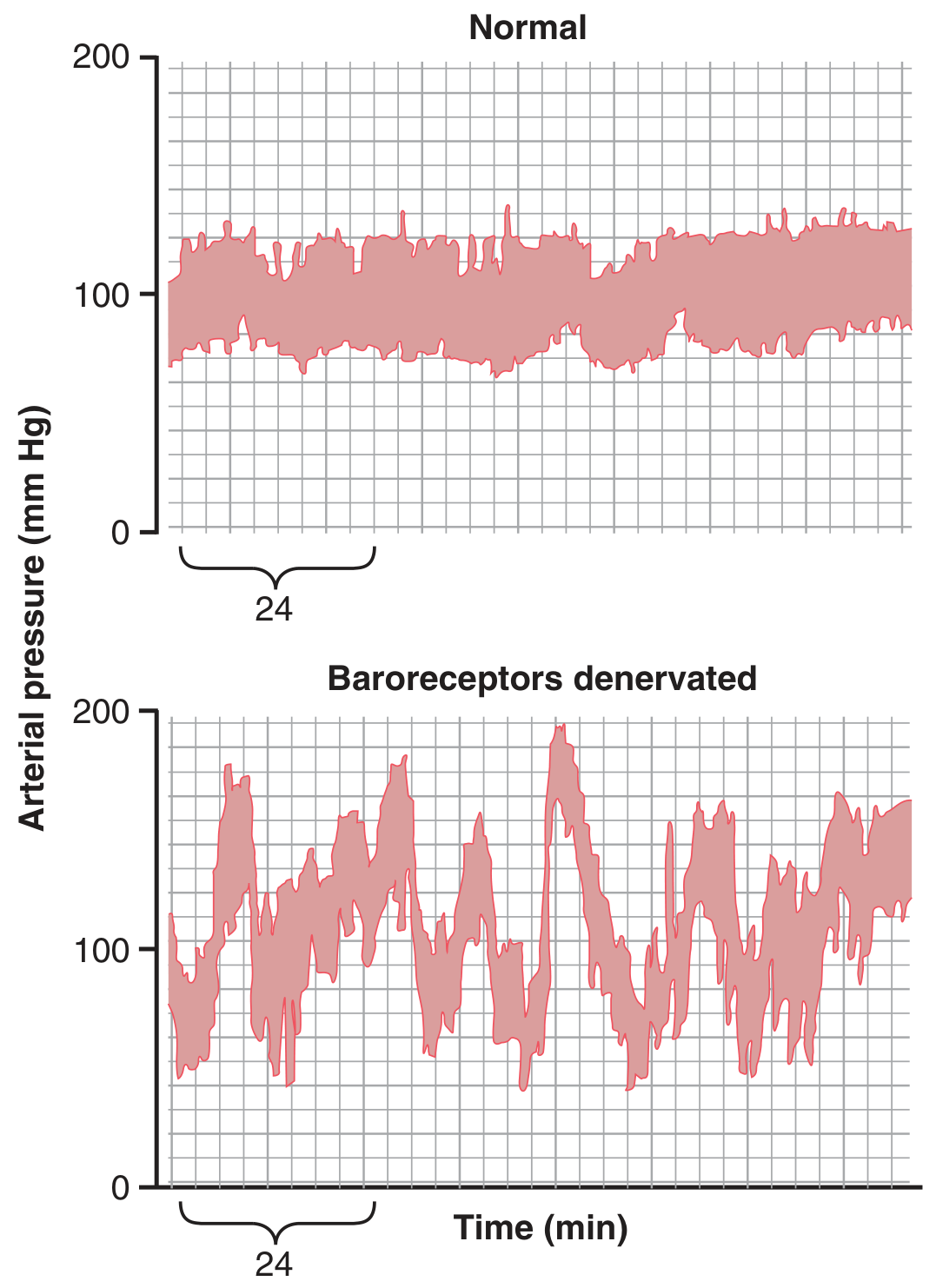
The baroreceptors serve as a minute-to-minute blood pressure buffer, dampening variability from everyday activities (eating, defecation, emotional stress, posture changes).
"A primary purpose of the arterial baroreceptor system is to reduce the minute-by-minute variation in arterial pressure to about one-third of what would occur if the baroreceptor system were not present."
- Guyton and Hall
Experimental evidence: Dogs with baroreceptor denervation showed BP swings from 50 to >160 mmHg during daily activities, compared to the normal narrow range of 85-115 mmHg.
7. Baroreceptor Resetting
With sustained hypertension, baroreceptors gradually reset to accept the higher pressure as normal (over 1-2 days). This is why:
- Chronic hypertension is maintained rather than corrected by the baroreceptor reflex
- The mechanism involves decreased mechanoreceptor sensitivity, not resetting of central CNS vasomotor centers
"In chronic hypertension, the baroreceptors do not 'see' the elevated blood pressure as abnormal. In such cases, the hypertension will be maintained, rather than corrected, by the baroreceptor reflex."
- Costanzo Physiology, p. 170
However, baroreceptors may still contribute to long-term regulation by modulating renal sympathetic nerve activity - increased firing promotes sodium and water excretion, gradually reducing blood volume and BP. Chronic carotid sinus nerve stimulation (baroreflex activation therapy) exploits this to lower BP by 15-20 mmHg in refractory hypertension.
(Guyton and Hall, p. 229)
8. Low-Pressure (Cardiopulmonary) Baroreceptors
Beyond the arterial baroreceptors, low-pressure receptors exist in:
- Right atrium and great veins (Bainbridge reflex)
- Pulmonary arteries
- Atria (volume/stretch sensors)
These detect changes in venous filling/preload rather than arterial pressure. They respond opposite to arterial baroreceptors:
- ↑ Atrial stretch → ↑ HR (Bainbridge reflex)
- ↓ Venous pressure → ↓ HR
"Venous baroreceptors sample preload by stretch of the atrium. Arterial baroreceptors survey resistance, or afterload, as reflected in the mean arterial pressure. Afterload and preload produce opposite effects on CO; thus the venous and arterial baroreceptors produce opposing effects after a similar stretch stimulus."
- Barash, Cullen and Stoelting's Clinical Anesthesia
9. Clinical Relevance Summary
| Situation | Baroreceptor Role |
|---|---|
| Orthostatic hypotension | Reflex tachycardia + vasoconstriction on standing; failure = dysautonomia |
| Hemorrhagic shock | Tachycardia + vasoconstriction preserve BP; failure = catastrophic hypotension |
| Carotid sinus hypersensitivity | Exaggerated reflex bradycardia/asystole with minimal carotid pressure |
| Chronic hypertension | Baroreceptors reset to new set point - hypertension perpetuated |
| Baroreflex activation therapy | Chronic electrical carotid sinus stimulation - lowers BP 15-20 mmHg in drug-resistant hypertension |
| Heart failure | Impaired baroreflex sensitization → chronic sympathetic excess → disease progression |
| Valsalva testing | Phase II tachycardia (baroreceptor-mediated) + Phase IV bradycardia (overshoot detection) |
| Carotid sinus massage | Induces reflex bradycardia/AV block - terminates SVT or confirms carotid sinus hypersensitivity |
This is a shared conversation. Sign in to Orris to start your own chat.