
Mbbs 1 st year biochemistry professional examination answer type Answer this question of the topic (Phenyl ketonoriya, gluconeogenesis, uriya circle Give me detailed answers
Reading File
Finding Sources
Finding Sources
Finding Sources
Reading File
Reading File
Finding Sources
Reading File
Reading File
Finding Sources
Reading File
Reading File
Finding Sources
Reading File
Finding Sources
Reading File
Reading File
Finding Sources
Loading Image
Loading Image
Loading Image
Excellent! I have all the content and diagrams needed. Now I'll compose the full, detailed MBBS 1st year professional examination answers for all three topics.
MBBS 1st Year Biochemistry: Detailed Professional Examination Answers
TOPIC 1: PHENYLKETONURIA (PKU)
Definition
Phenylketonuria (PKU) is an autosomal recessive inborn error of amino acid metabolism caused by a deficiency of the enzyme phenylalanine hydroxylase (PAH), which normally converts phenylalanine to tyrosine.
- Incidence: ~1 in 10,000-15,000 live births
- First described as a cause of intellectual disability in 1934 by Asbjorn Folling
Biochemical Basis (Enzyme Defect)
Under normal conditions:
Phenylalanine + O2 + Tetrahydrobiopterin (BH4) → Tyrosine + Dihydrobiopterin (BH2) + H2O (catalyzed by Phenylalanine hydroxylase, a mixed-function oxygenase)
In PKU, this reaction is blocked. Phenylalanine accumulates and is shunted into alternative catabolic pathways, producing:
| Metabolite | Formed by | Clinical significance |
|---|---|---|
| Phenylpyruvic acid | Transamination | Excreted in urine ("phenylketone") |
| Phenylacetate | Oxidative decarboxylation | Musty/mousy odor of urine |
| Phenyllactate | Reduction | Detected in urine |
| Phenylacetylglutamine | Conjugation | Excreted in urine |
The name "phenylketonuria" refers to the characteristic excretion of phenylpyruvic acid (a phenylketone) in the urine.
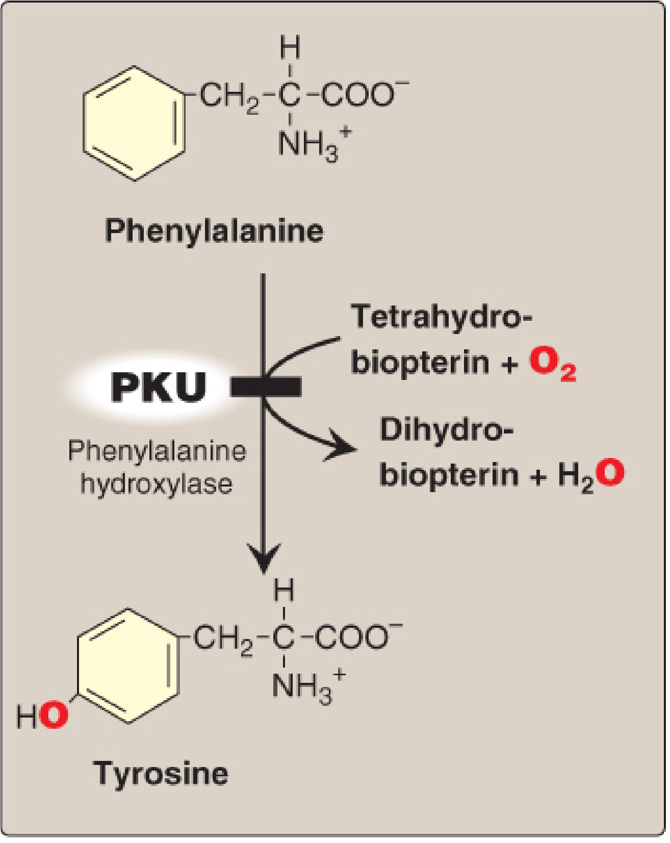
Figure: In PKU, the enzyme phenylalanine hydroxylase is blocked, preventing conversion of phenylalanine to tyrosine. BH4 (tetrahydrobiopterin) is required as a cofactor.
Types of Hyperphenylalaninemia
| Type | Defect |
|---|---|
| Classical PKU (Type I) | Phenylalanine hydroxylase (PAH) deficiency |
| Type II | Dihydrobiopterin reductase (DHPR) deficiency |
| Type III | Dihydrobiopterin reductase deficiency (allelic to II) |
| Types IV & V | Defects in BH4 biosynthesis |
In BH4-deficiency variants (Types II-V), simply restricting phenylalanine in the diet is insufficient because BH4 is also required for:
- Tyrosine hydroxylase - synthesis of catecholamines (dopamine, norepinephrine)
- Tryptophan hydroxylase - synthesis of serotonin
So these patients also need BH4 supplementation + L-DOPA + 5-hydroxytryptophan.
Pathogenesis of Brain Damage
- Hyperphenylalaninemia - phenylalanine competes with large neutral amino acids (LNAA) for transport across the blood-brain barrier
- Decreased entry of other amino acids (tyrosine, tryptophan) into brain
- Impaired synthesis of neurotransmitters (dopamine, serotonin, norepinephrine)
- Disrupted myelination and protein synthesis
- Phenylalanine is directly toxic at high concentrations - inhibits brain enzymes
Clinical Features
Affected infants appear normal at birth (maternal enzyme protects in utero). Manifestations begin within weeks to months after birth:
A. Neurological:
- Severe intellectual disability (IQ < 50 in untreated cases)
- Seizures (often infantile spasms)
- Hyperreflexia, tremors
- Microcephaly
- Behavioral problems - hyperactivity, aggression
B. Pigmentation:
- Fair skin, light hair, blue eyes (due to decreased melanin synthesis - tyrosine is the precursor of melanin)
- Eczema-like skin rash
C. Musty/Mousy body odor - due to phenylacetate accumulation
D. Maternal PKU:
Women with PKU who stop dietary treatment during pregnancy have high phenylalanine levels. Their children (even heterozygotes) suffer transplacental phenylalanine toxicity:
- Congenital heart defects
- Microcephaly
- Intellectual disability
- IUGR
Diagnosis
Newborn Screening (Guthrie Test / Tandem Mass Spectrometry):
- Blood is taken from a heel prick after 48-72 hours of feeding
- Detects elevated blood phenylalanine
- All states/countries mandate this screen
- Guthrie test: Bacterial inhibition assay using Bacillus subtilis (phenylalanine reverses inhibition by beta-2-thienylalanine)
- Tandem mass spectrometry (modern standard) - more reliable and can screen for >50 disorders simultaneously
Other tests:
- Urinary FeCl3 test - urine turns green in PKU (older, less reliable - now largely replaced)
- Plasma amino acid analysis
- DNA-based prenatal diagnosis (for known familial mutations)
Blood phenylalanine levels:
- Normal: < 2 mg/dL (120 μmol/L)
- PKU: > 20 mg/dL (1200 μmol/L)
- Mild hyperphenylalaninemia: 2-20 mg/dL
Treatment
-
Low-phenylalanine diet - Phenylalanine is an essential amino acid, so it cannot be completely eliminated. The diet provides just enough phenylalanine for growth.
- Special formula (Lofenalac, PKU-specific amino acid mixtures)
- Avoid high-protein foods (meat, eggs, dairy, nuts, beans)
- Avoid aspartame (artificial sweetener that contains phenylalanine)
- Must supplement with tyrosine (becomes essential in PKU)
-
BH4 (Sapropterin, Kuvan) - for BH4-responsive PKU (~25-50% of patients respond)
-
Pegvaliase (enzyme substitution therapy) - pegylated phenylalanine ammonia lyase; FDA approved for adults
-
Gene therapy - under clinical trials
-
Dietary treatment must begin within the first 2-3 weeks of life to prevent irreversible brain damage. Treatment should ideally continue lifelong.
TOPIC 2: GLUCONEOGENESIS
Definition
Gluconeogenesis is the metabolic pathway by which glucose is synthesized from non-carbohydrate precursors. It is essentially the reverse of glycolysis except at three key irreversible steps.
Physiological Significance
The brain, red blood cells, renal medulla, lens, cornea, and testes are obligate glucose consumers - they cannot survive without a steady glucose supply. After 2-3 hours of fasting, liver glycogen stores begin to deplete, and gluconeogenesis becomes the primary means of maintaining blood glucose.
Site
- Primary site: Liver (cytoplasm and mitochondria)
- Secondary site: Kidney cortex (important during prolonged starvation - ~40% of glucose production)
- Does NOT occur in muscle (lacks glucose-6-phosphatase)
Precursors (Substrates) of Gluconeogenesis
| Precursor | Source | Entry Point |
|---|---|---|
| Lactate | Anaerobic glycolysis in RBCs, muscle, exercising skeletal muscle | Pyruvate (via lactate dehydrogenase) |
| Pyruvate | Amino acid catabolism, glycolysis | Oxaloacetate (via pyruvate carboxylase) |
| Alanine | Muscle protein (glucose-alanine cycle) | Pyruvate (via transamination) |
| Glutamine | Gut, kidney, other tissues | TCA cycle intermediates |
| Glycerol | Lipolysis of triacylglycerols (adipose tissue) | Glycerol-3-phosphate → DHAP |
| Propionate | Odd-chain fatty acid oxidation | Succinyl-CoA → OAA |
| Glucogenic amino acids | Protein catabolism | Various TCA intermediates |
Note: Acetyl CoA (from even-chain fatty acid oxidation and ketone bodies) cannot serve as a net gluconeogenic precursor in mammals, because the two carbons it contributes to the TCA cycle are lost as CO2.
Key Reactions: The Three Bypass Steps
Gluconeogenesis is largely the reverse of glycolysis, but three steps of glycolysis are irreversible and require bypass enzymes:
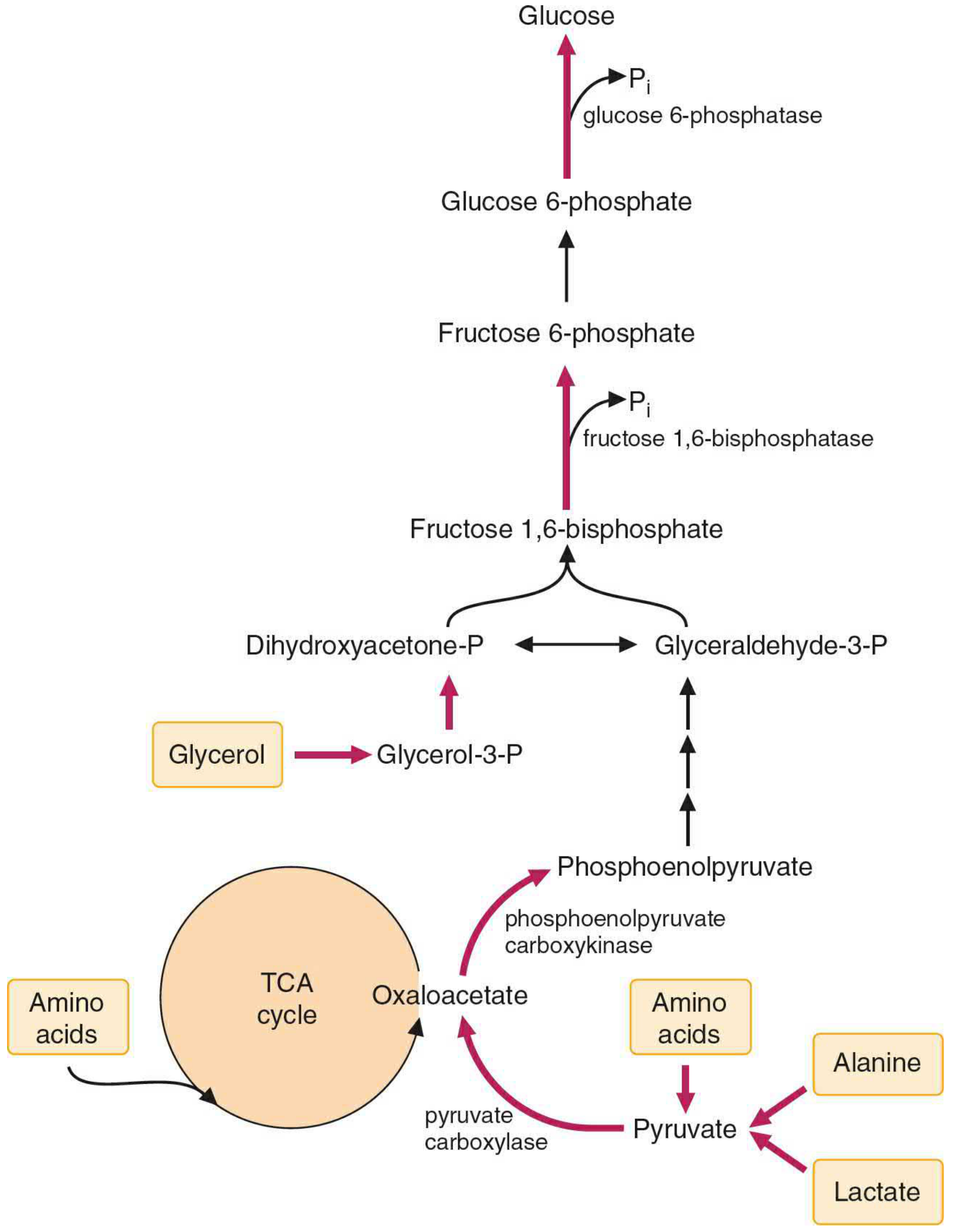
Figure 28.3 - Key reactions of gluconeogenesis. Red arrows indicate the three steps that differ from glycolysis. Precursors include amino acids (especially alanine), lactate, and glycerol. (Basic Medical Biochemistry, 6e)
Bypass 1: Pyruvate → Phosphoenolpyruvate (PEP)
This is the most complex bypass. It requires two enzymes and involves the mitochondria:
Step 1a:
Pyruvate + CO2 + ATP → Oxaloacetate (OAA) + ADP + Pi Enzyme: Pyruvate carboxylase (mitochondria) Cofactor: Biotin (CO2 carrier), allosterically activated by Acetyl CoA
Step 1b:
OAA + GTP → Phosphoenolpyruvate (PEP) + CO2 + GDP Enzyme: Phosphoenolpyruvate carboxykinase (PEPCK) (cytosol/mitochondria)
Why two steps? Because pyruvate kinase (glycolytic enzyme) is strongly exergonic and irreversible. The bypass uses two ATP equivalents to overcome this energy barrier.
Bypass 2: Fructose 1,6-bisphosphate → Fructose 6-phosphate
Fructose 1,6-bisphosphate + H2O → Fructose 6-phosphate + Pi Enzyme: Fructose 1,6-bisphosphatase (FBPase-1) (cytosol)
This bypasses phosphofructokinase-1 (PFK-1) of glycolysis. FBPase-1 is inhibited by AMP and fructose 2,6-bisphosphate.
Bypass 3: Glucose 6-phosphate → Glucose
Glucose 6-phosphate + H2O → Glucose + Pi Enzyme: Glucose 6-phosphatase (ER membrane of liver and kidney)
This bypasses hexokinase/glucokinase of glycolysis. Muscle lacks this enzyme, which is why muscle cannot release free glucose into the blood.
Energy Cost of Gluconeogenesis
For synthesis of 1 glucose from 2 pyruvate molecules:
- 6 ATP equivalents are consumed (2 ATP + 2 GTP + 2 NADH)
- Compare to glycolysis which generates 2 ATP per glucose
Regulation of Gluconeogenesis
Allosteric Regulation:
| Factor | Effect on Gluconeogenesis |
|---|---|
| AMP | Inhibits (activates glycolysis instead) |
| ATP | Activates |
| Acetyl CoA | Activates pyruvate carboxylase |
| Fructose 2,6-bisphosphate | Inhibits FBPase-1 |
| ADP | Inhibits PEPCK |
Hormonal Regulation:
| Hormone | Effect | Mechanism |
|---|---|---|
| Glucagon (fasting) | Activates | Increases cAMP, activates PEPCK gene transcription, decreases fructose 2,6-bisphosphate |
| Cortisol | Activates | Induces PEPCK, glucose-6-phosphatase |
| Epinephrine | Activates | Similar to glucagon |
| Insulin | Inhibits | Decreases cAMP, decreases PEPCK transcription |
Substrate (Feed-forward) Regulation:
When more precursors are available (e.g., high protein diet, prolonged fasting with protein catabolism), gluconeogenesis accelerates.
Cori Cycle (Lactic Acid Cycle)
During intense exercise, muscles produce large amounts of lactate via anaerobic glycolysis. This lactate is transported to the liver, where it is reconverted to glucose by gluconeogenesis and sent back to the muscles. This is the Cori cycle. It transfers the metabolic burden from muscle to liver.
Glucose-Alanine Cycle
Muscle releases alanine (formed by transamination of pyruvate with glutamate). Liver takes up alanine, deaminates it to pyruvate, and uses pyruvate for gluconeogenesis. The nitrogen is excreted as urea.
Clinical Relevance
- Metformin (diabetes drug) inhibits gluconeogenesis (by inhibiting mitochondrial Complex I, reducing NADH availability)
- Von Gierke disease (Type Ia GSD): Glucose-6-phosphatase deficiency - both gluconeogenesis and glycogenolysis are impaired, causing severe fasting hypoglycemia
- DKA (Diabetic Ketoacidosis): Gluconeogenesis is markedly elevated due to high glucagon:insulin ratio
TOPIC 3: UREA CYCLE (Ornithine Cycle / Krebs-Henseleit Cycle)
Introduction
The urea cycle was first proposed in 1932 by Hans Krebs and Kurt Henseleit (hence also called the Krebs-Henseleit cycle). It is the primary mechanism for disposal of ammonia (NH3/NH4+), which is toxic - especially to the brain and CNS.
- Normal blood ammonia: 30-60 μmol/L
- Urea is the primary nitrogenous excretory product in humans (ureotelism)
Why is Ammonia Toxic?
- NH4+ enters neurons and depletes alpha-ketoglutarate (by forming glutamate), reducing TCA cycle activity and ATP production
- This impairs neuronal energy metabolism, causing hepatic encephalopathy (asterixis, confusion, coma)
- GABA levels also increase in liver failure, adding to CNS depression
Location
- Liver (hepatocytes) - primary site
- First two steps: mitochondrial matrix
- Remaining steps: cytosol
Nitrogen Sources
Urea contains two nitrogen atoms:
- One from NH4+ (directly from amino acid catabolism via glutamate dehydrogenase)
- One from aspartate (via transamination from oxaloacetate)
Steps of the Urea Cycle
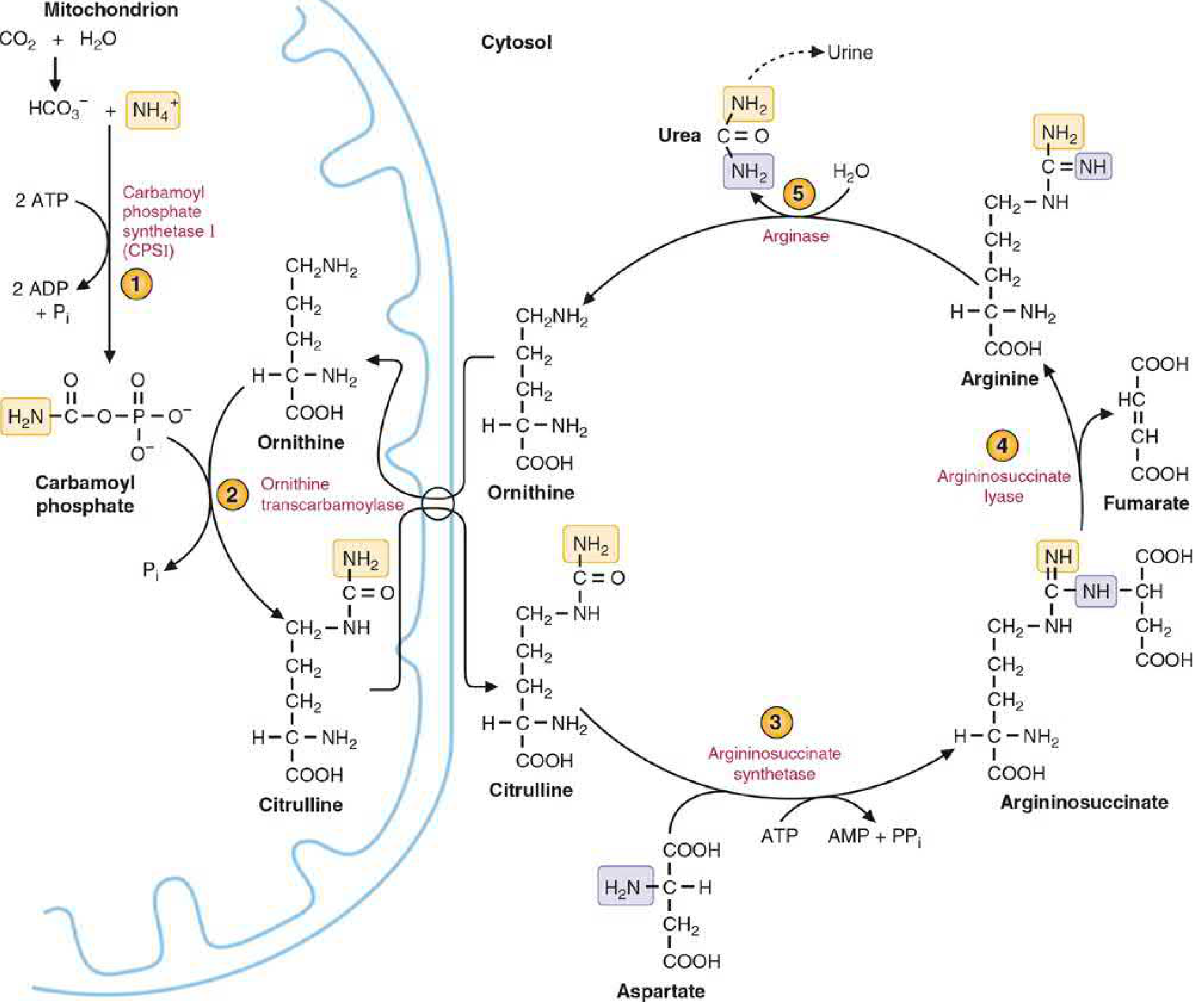
Figure 36.10 - The Urea Cycle. Steps 1-2 occur in mitochondria; steps 3-5 in cytosol. (Basic Medical Biochemistry, 6e)
Step 1 (Mitochondria): Formation of Carbamoyl Phosphate
NH4+ + HCO3- + 2 ATP → Carbamoyl phosphate + 2 ADP + Pi Enzyme: Carbamoyl Phosphate Synthetase I (CPS-I) Location: Mitochondrial matrix Activator: N-Acetylglutamate (NAG) - allosteric
Note: CPS-II (cytosolic) uses glutamine as nitrogen source and is involved in pyrimidine biosynthesis.
Step 2 (Mitochondria): Carbamoyl Phosphate + Ornithine → Citrulline
Carbamoyl phosphate + Ornithine → Citrulline + Pi Enzyme: Ornithine Transcarbamoylase (OTC) Location: Mitochondrial inner membrane
Citrulline is transported from mitochondria to cytosol (in exchange for ornithine).
Step 3 (Cytosol): Citrulline + Aspartate → Argininosuccinate
Citrulline + Aspartate + ATP → Argininosuccinate + AMP + PPi Enzyme: Argininosuccinate Synthetase The second nitrogen (from aspartate) is incorporated here
Step 4 (Cytosol): Argininosuccinate → Arginine + Fumarate
Argininosuccinate → Arginine + Fumarate Enzyme: Argininosuccinate Lyase (Argininosuccinase)
Fumarate enters the TCA cycle (via cytoplasmic fumarase → malate → OAA), linking the urea cycle with energy metabolism. This connection is called the "urea cycle-TCA cycle bicycle."
Step 5 (Cytosol): Arginine → Ornithine + Urea
Arginine + H2O → Ornithine + Urea Enzyme: Arginase
Ornithine is regenerated and transported back into mitochondria to begin the next cycle. Urea is the final product and is excreted in urine.
Summary Table of Urea Cycle Steps
| Step | Reaction | Enzyme | Location |
|---|---|---|---|
| 1 | NH4+ + HCO3- → Carbamoyl phosphate | CPS-I | Mitochondria |
| 2 | Carbamoyl-P + Ornithine → Citrulline | OTC | Mitochondria |
| 3 | Citrulline + Aspartate → Argininosuccinate | Argininosuccinate synthetase | Cytosol |
| 4 | Argininosuccinate → Arginine + Fumarate | Argininosuccinate lyase | Cytosol |
| 5 | Arginine → Ornithine + Urea | Arginase | Cytosol |
Energy Cost
Each turn of the urea cycle consumes 4 ATP equivalents:
- Step 1: 2 ATP used
- Step 3: 1 ATP → AMP + PPi (equivalent to 2 ATP hydrolysis; PPi is hydrolyzed by pyrophosphatase)
Regulation of the Urea Cycle
1. Allosteric regulation (short-term):
- N-Acetylglutamate (NAG) is the obligatory allosteric activator of CPS-I
- NAG is synthesized from acetyl CoA + glutamate by N-Acetylglutamate Synthase (NAGS)
- NAGS is activated by arginine (feed-forward: as more protein is eaten, more arginine forms, stimulating NAG, which activates CPS-I)
- So high dietary protein → high arginine → high NAG → high CPS-I activity → more urea made
2. Substrate availability (feed-forward):
- Higher ammonia production (high protein diet, fasting, catabolic states) drives the cycle faster
3. Enzyme induction (long-term):
- High-protein diet and prolonged fasting induce synthesis of all five urea cycle enzymes
Urea Cycle Disorders (Hyperammonemias)
| Enzyme Deficiency | Disorder | Characteristic Feature |
|---|---|---|
| CPS-I | CPS-I deficiency | Severe hyperammonemia, no orotic aciduria |
| OTC | OTC deficiency (most common) | X-linked; orotic aciduria (excess carbamoyl-P floods pyrimidine pathway) |
| Argininosuccinate synthetase | Citrullinemia (Type I) | Elevated citrulline in blood and urine |
| Argininosuccinate lyase | Argininosuccinic aciduria | Elevated argininosuccinate in blood and urine; brittle hair (trichorrhexis nodosa) |
| Arginase | Argininemia | Elevated arginine; spastic diplegia |
| NAGS | NAGS deficiency | Responds to N-carbamyl glutamate treatment |
Key Differentiator: OTC deficiency is the only urea cycle disorder associated with orotic aciduria (elevated orotic acid in urine) because the excess carbamoyl phosphate is diverted into the pyrimidine synthesis pathway.
Clinical Features of Hyperammonemia
- Neonatal presentation: poor feeding, vomiting, lethargy, seizures, coma
- Older children: intellectual disability, episodic vomiting, avoidance of high-protein foods (self-selection)
- In adults: episodes triggered by high-protein loads, infections, stress, valproate use
Treatment of Urea Cycle Disorders
- Low-protein diet - reduce nitrogen load
- Essential amino acid supplements - maintain growth
- Sodium benzoate - conjugates glycine → hippurate (excreted, removes nitrogen without urea)
- Sodium phenylacetate/phenylbutyrate - conjugates glutamine → phenylacetylglutamine (excreted)
- N-carbamyl glutamate (carglumic acid) - for NAGS deficiency; activates CPS-I
- Liver transplant - definitive treatment (provides all urea cycle enzymes)
- Hemodialysis - for acute hyperammonemic crisis
Quick Comparison Summary
| Feature | PKU | Gluconeogenesis | Urea Cycle |
|---|---|---|---|
| Type | Inborn error of metabolism | Metabolic pathway | Metabolic pathway |
| Key enzyme | Phenylalanine hydroxylase | Pyruvate carboxylase, PEPCK, FBPase-1, G6Pase | CPS-I, OTC, Arginase etc. |
| Primary organ | Liver | Liver (mainly) | Liver |
| Inheritance | Autosomal recessive | N/A | Enzyme deficiency: AR (except OTC = X-linked) |
| Key metabolite accumulated/produced | Phenylalanine, phenylpyruvate | Glucose | Urea |
| Clinical consequence of failure | Intellectual disability | Hypoglycemia | Hyperammonemia, encephalopathy |
| Key test | Blood Phe (Guthrie/Tandem MS) | Blood glucose, lactate | Plasma ammonia, amino acids, orotic acid |
Sources: Lippincott's Illustrated Reviews: Biochemistry, 8th ed.; Basic Medical Biochemistry: A Clinical Approach, 6e; Robbins & Kumar Basic Pathology; Harper's Illustrated Biochemistry, 32nd ed.; Park's Textbook of Preventive and Social Medicine.
This is a shared conversation. Sign in to Orris to start your own chat.