
Aha guidlines for CPCR in details take a reference from standard text books of anaesthesia
AHA 2020 2024 guidelines CPR CPCR cardiopulmonary resuscitation key updates
AHA adult cardiac arrest algorithm CPR ACLS 2020
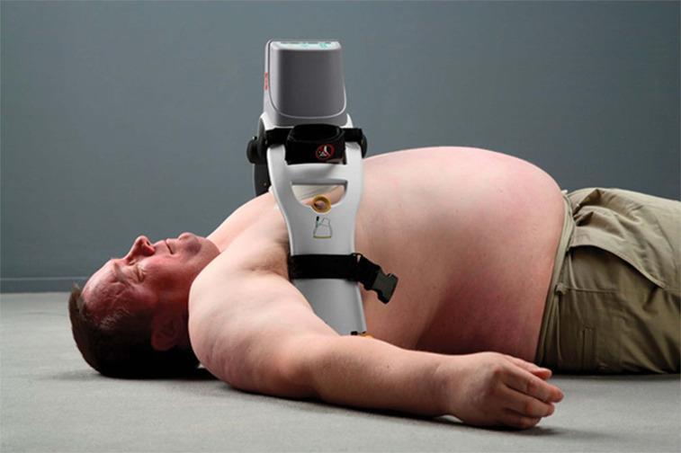
A clinical photograph demonstrating the application of a LUCAS 3 mechanical chest compression system on an adult male in a supine position. The image serves as a procedural demonstration of Active Compression-Decompression Cardiopulmonary Resuscitation (ACD-CPR). The device is positioned centrally over the sternum, featuring a white support arch that spans the torso and is secured with black stabilization straps. The grey upper housing contains the motor and control interface, while a vertical piston with a circular suction cup/pad is visible in contact with the lower third of the sternum to deliver consistent, high-quality chest compressions. The patient is bare-chested to show proper anatomical placement of the device between the axillary lines. This image is used for training medical professionals in emergency medicine and advanced cardiac life support (ACLS) to illustrate mechanical adjuncts that reduce provider fatigue and maintain perfusion during cardiac arrest.
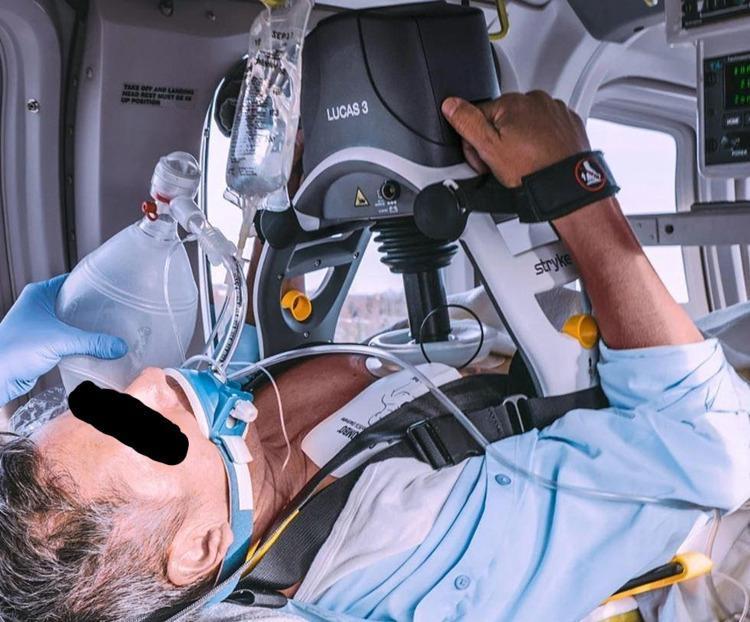
This clinical photograph captures a male patient undergoing Advanced Cardiac Life Support (ACLS) in an intensive care or transport setting, featuring the implementation of mechanical cardiopulmonary resuscitation (CPR). The primary focus is the LUCAS 3 Chest Compression System, an automated device positioned over the patient's sternum. The system's upper unit is mounted on a rigid backplate via a support frame, with the suction cup plunger centered on the chest to deliver consistent compressions. Concurrent interventions include airway management via a bag-valve-mask (BVM) ventilation setup and a bag of intravenous fluids suspended overhead. Visible monitoring equipment includes ECG leads and adhesive pads on the patient's chest. The image illustrates the integration of automated technology in managing cardiac arrest, demonstrating the positioning of mechanical compression devices alongside manual ventilation and pharmacological access. This visual is intended for healthcare professionals to understand the practical application of automated CPR systems in critical care scenarios.
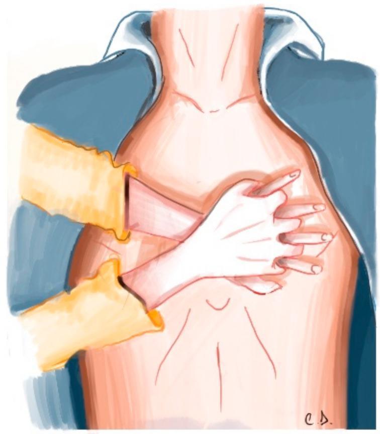
This medical illustration depicts the correct anatomical hand placement for high-quality Cardiopulmonary Resuscitation (CPR) in an adult. The view is an anterior-superior perspective of the patient's torso. The rescuer's hands are shown interlocked, with the heel of one hand placed firmly on the lower half of the patient's sternum, centered between the nipples. The second hand is positioned directly on top of the first, with fingers interlaced to ensure pressure is localized to the sternum rather than the ribs. The patient is shown with an open shirt (blue collar and yellow sleeves) to emphasize the importance of exposing the chest during emergency cardiac care. Anatomical landmarks include the neck, clavicular area, and the midline of the thorax, representing the path for external chest compressions (ECC). This graphic serves as a pedagogical tool for Basic Life Support (BLS) and Advanced Cardiac Life Support (ACLS) training, illustrating the initial mechanical step in the Out-of-Hospital Chain of Survival.
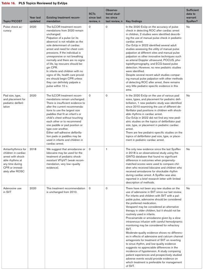
Table 16. PLS Topics Reviewed by EvUps <table><thead><tr><th>Topic/PICOST</th><th>Year last updated</th><th>Existing treatment recommendation</th><th>RCTs since last review, n</th><th>Observational studies since last review, n</th><th>Key findings</th><th>Sufficient data to warrant SysRev?</th></tr></thead><tbody><tr><td>Pulse check accuracy</td><td>2020</td><td>The ILCOR treatment recommendations from 2020 remain unchanged: Palpation of a pulse (or its absence) is not reliable as the sole determinant of cardiac arrest and need for chest compressions. If the individual is unresponsive or not breathing normally and there are no signs of life, lay rescuers should begin CPR. In infants and children with no signs of life, health care providers should begin CPR unless they can definitely palpate a pulse within 10 s.</td><td>0</td><td>0</td><td>In the 2020 EvUp on the accuracy of pulse check in determining ROC after cardiac arrest in children, 2 studies were identified describing the use of manual pulse check in pediatric cardiac arrest. Our EvUp in 2022 identified several adult studies assessing the utility of manual pulse palpation at different sites and manual pulse palpation vs other innovative techniques such as the use of Doppler ultrasound, POCUS, photoplethysmography, and ECG-based pulse detection. However, no new pediatric studies were identified. Despite several recent adult studies comparing manual pulse palpation with other methods of detecting ROC after arrest, there remains very little pediatric-specific evidence in this area.</td><td>No</td></tr><tr><td>Pad size, type, and placement for pediatric defibrillation</td><td>2020</td><td>The ILCOR treatment recommendations remain unchanged: There is insufficient evidence to alter the current recommendations to use the largest size paddles that fit an infant's or child's chest without touching each other or to recommend one paddle or pad position or type over another. Either self-adhesive defibrillation pads or paddles may be used in infants and children in cardiac arrest.</td><td>0</td><td>0</td><td>In the 2020 EvUp on the use of various pad sizes, types, and placement for pediatric defibrillation, 1 new pediatric study was identified since 2010 examining the use of different defibrillator pad positions in children with shockable rhythms in cardiac arrest. Our EvUp in 2022 did not find any new pediatric studies on the topics of defibrillator pad size, type, or placement in pediatric cardiac arrest. There are few pediatric-specific studies on the topics of defibrillator pad size, type, or placement in pediatric cardiac arrest.</td><td>No</td></tr><tr><td>Antiarrhythmics for children in cardiac arrest with shockable rhythms at any time during CPR or immediately after ROSC</td><td>2018</td><td>We suggest that amiodarone or lidocaine may be used for the treatment of pediatric shock-resistant VF/pVT (weak recommendation, very low–quality evidence).</td><td>0</td><td>1</td><td>The only new evidence since the last SysRev in 2018 is an observational study using the GWTG database that found no significant difference in outcomes when propensity-matched scores were used to compare children who received lidocaine and children who received amiodarone for shockable rhythm during cardiac arrest. A SysRev was also reported in a brief research letter with limited description of methods.</td><td>No</td></tr><tr><td>Adenosine use in SVT</td><td>2020</td><td>This treatment recommendation is unchanged from 2010. For infants and children with SVT with a palpable pulse, adenosine should be considered the preferred medication. Verapamil may be considered an alternative therapy in older children, but it should not be routinely used in infants. Procainamide or amiodarone given by a slow intravenous infusion with careful hemodynamic monitoring may be considered for refractory SVT. Moderate-quality evidence shows no differences in effects of adenosine and calcium channel antagonists for treatment of SVT reverting to sinus rhythm, and low-quality evidence suggests no appreciable differences in the incidence of hypotension. A study comparing patient experiences and prospectively studied adverse events would provide evidence on which treatment is preferable for management of SVT.</td><td>0</td><td>0</td><td>There have not been any new studies on the use of adenosine in SVT since our last review. For infants and children with SVT with a palpable pulse, adenosine should be considered the preferred medication. Verapamil may be considered an alternative therapy in older children, but it should not be routinely used in infants. Procainamide or amiodarone given by a slow intravenous infusion with careful hemodynamic monitoring may be considered for refractory SVT. Moderate-quality evidence shows no differences in effects of adenosine and calcium channel antagonists for treatment of SVT reverting to sinus rhythm, and low-quality evidence suggests no appreciable differences in the incidence of hypotension. A study comparing patient experiences and prospectively studied adverse events would provide evidence on which treatment is preferable for management of SVT.</td><td>No</td></tr></tbody></table>
AHA Guidelines for CPCR (Cardiopulmonary-Cerebral Resuscitation) - Detailed Review
1. DEFINITIONS AND OVERVIEW
- Maintain oxygen and blood supply to vital organs
- Restore spontaneous circulation (ROSC)
- Minimize post-resuscitation organ injury
- Improve neurologic outcome and survival
2. PATHOPHYSIOLOGY OF SUDDEN CARDIAC ARREST (SCA)
- After SCA, forward systemic arterial blood flow continues until the pressure gradient between the aorta and right heart reaches equilibrium (approximately 5 minutes)
- Cardiac output during CPR with effective, uninterrupted chest compression is at best 25-30% of normal spontaneous circulation
- Blood flow (not oxygen content) is the limiting factor for oxygen delivery during CPR
- Rescue breaths are therefore less important than initiating effective chest compressions immediately
- Thoracic pump theory: compression raises intrathoracic pressure, driving blood from thorax to periphery; venous valves prevent retrograde flow
- Cardiac pump theory: direct compression of the heart between sternum and vertebral column ejects blood from ventricles
- Overzealous ventilation raises intrathoracic pressure, impairs venous return, and reduces ROSC chances
3. THE CHAIN OF SURVIVAL
- Recognition of cardiac arrest and activation of emergency response
- Early high-quality CPR
- Rapid defibrillation
- Advanced resuscitation by EMS and emergency teams
- Post-cardiac arrest care
- Recovery (including rehabilitation)
4. BASIC LIFE SUPPORT (BLS)
Recognition
- Check for unresponsiveness
- Check for absence of breathing or abnormal (agonal) breathing - agonal gasps are common in cardiac arrest and do NOT indicate true respiratory effort
- Lay rescuers: do NOT check pulse - just start CPR
- Health care providers: check pulse for no more than 10 seconds
High-Quality Chest Compressions
| Parameter | Target |
|---|---|
| Rate | 100-120/min |
| Depth (adults) | ≥5 cm (2 inches); not >6 cm |
| Chest recoil | Complete (hands off chest between compressions) |
| Interruptions | Minimize; pre-shock pause <10 seconds |
| Compression fraction | >60% |
- Position: patient supine on firm surface; rescuer directly over the patient
- Hand position: heel of one hand on lower half of sternum, second hand on top, fingers interlaced
- Change compressor every 2 minutes or sooner if fatigued
Compression:Ventilation Ratio
- 30:2 for adults (single or two rescuers) until advanced airway placed
- Once advanced airway in place: continuous compressions at 100-120/min + 1 breath every 6 seconds (10 breaths/min)
- Tidal volume: 500-600 mL; 1 second per breath; visible chest rise
Early Defibrillation
- Cardiac arrest with VF: survival declines 7-10% for every minute without defibrillation
- AEDs should be deployed as early as possible in both in-hospital and public settings
5. THE ADULT CARDIAC ARREST ALGORITHM (2020 AHA)
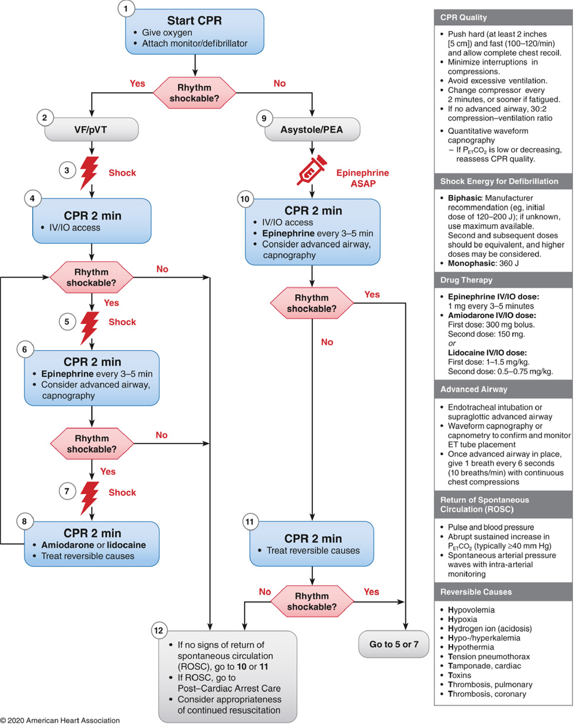
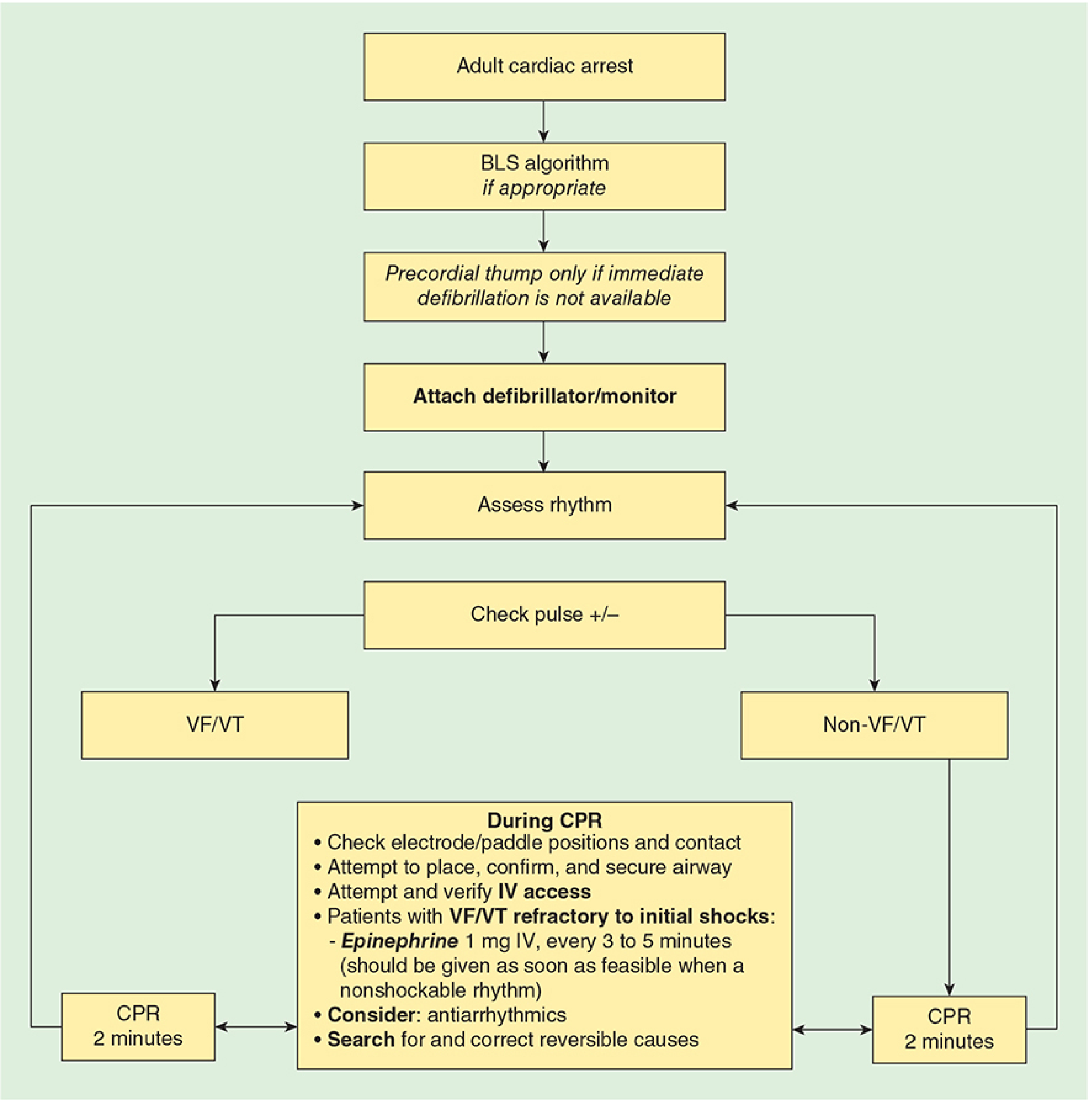
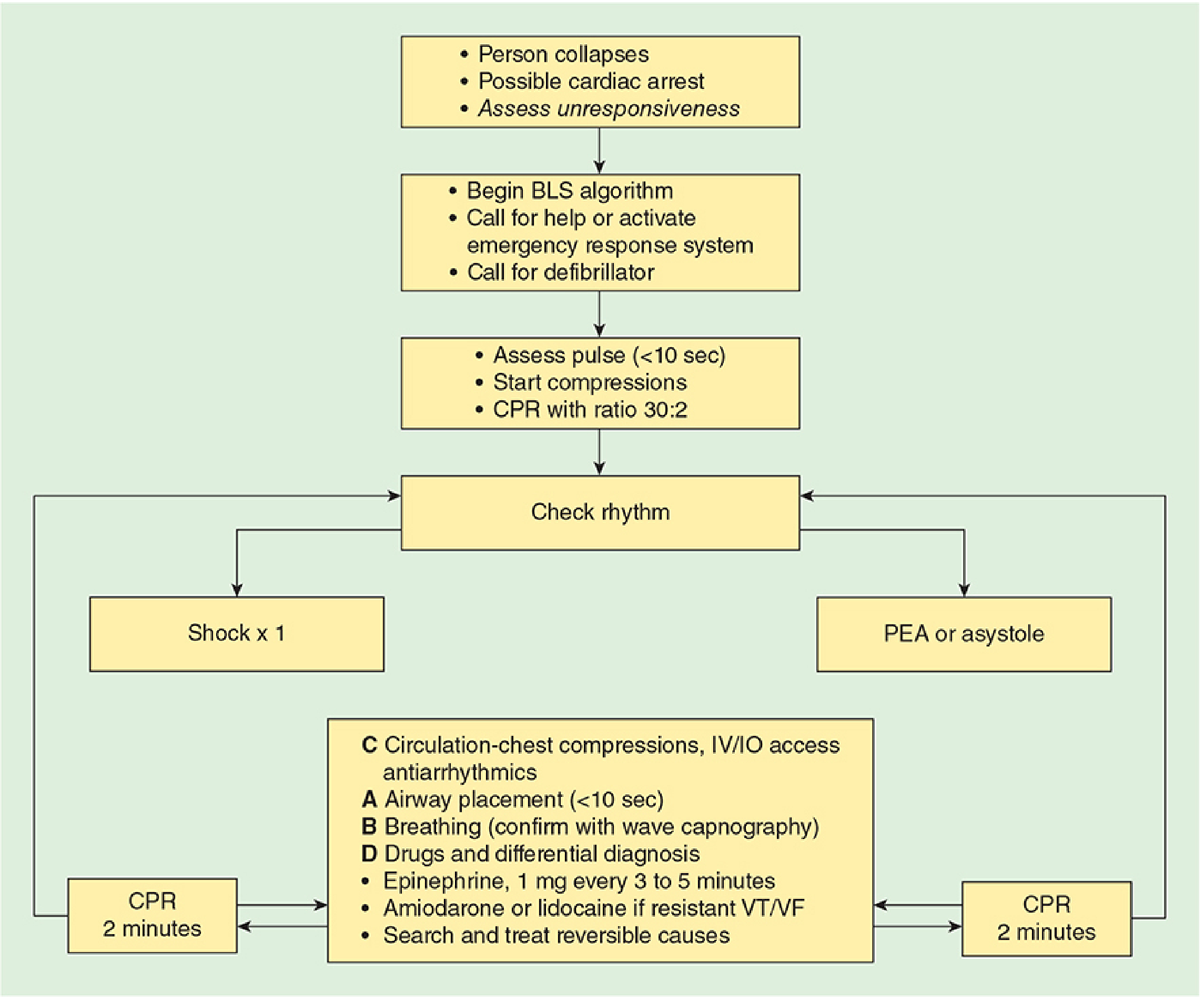
Algorithm Summary (CABD Sequence):
- Give oxygen, attach monitor/defibrillator
- Shock (Step 3)
- CPR 2 min + IV/IO access (Step 4)
- If VF/pVT persists: Shock again (Step 5)
- CPR 2 min + Epinephrine every 3-5 min + consider advanced airway (Step 6)
- If VF/pVT persists: Shock (Step 7)
- CPR 2 min + Amiodarone or Lidocaine + treat reversible causes (Step 8)
- Epinephrine ASAP (Step 9)
- CPR 2 min + IV/IO + Epi every 3-5 min + advanced airway (Step 10)
- CPR 2 min + treat reversible causes (Step 11)
- If ROSC: Post-Cardiac Arrest Care
- If no ROSC: continue algorithm
6. AIRWAY MANAGEMENT IN CARDIAC ARREST
Sequence
- Initial: Bag-mask ventilation with head tilt-chin lift or jaw thrust (recommended for initial airway control in most circumstances)
- Advanced airway: ETT or supraglottic airway (SGA) device
- Both bag-mask and advanced airway are acceptable; choice depends on provider skill
- No evidence of superior outcomes with ETT vs. SGA
- Failed airway: Transtracheal jet ventilation or cricothyrotomy (not recommended in children <10 years)
Endotracheal Intubation Rules
- Do not interrupt compressions >10 seconds for any intubation attempt
- Intubation may occur during CPR or during the pulse check
- Confirm placement: continuous waveform capnography is the most reliable method
- False negatives (no CO2 despite tracheal intubation): severe PE, low cardiac output, severe obstructive lung disease
- After advanced airway: 1 breath every 6 seconds with continuous compressions
Oxygen
- Provide 100% (maximum) FiO2 during CPR - oxygen delivery is flow-limited, not content-limited during CPR
- Do NOT extrapolate hyperoxia concerns from post-arrest care to active CPR
Ventilation Rate
- 6 breaths/min after advanced airway placement
- Faster rates impede cardiac output by raising intrathoracic pressure
- VD/VT increases during CPR - minute ventilation may need to increase 50-100% once circulation is restored
7. DEFIBRILLATION
Energy Recommendations
| Device | Energy |
|---|---|
| Biphasic (truncated exponential) | 150-200 J |
| Biphasic (rectilinear) | 120 J |
| Biphasic (unknown type) | 200 J |
| Monophasic | 360 J |
| Second/subsequent biphasic | Same or higher dose |
| AEDs | Energy pre-selected |
Key Principles
- Single shock strategy - immediately resume compressions after each shock
- Pre-shock pause should be <10 seconds
- Early defibrillation is the most important determinant of survival in VF arrest
- Biphasic shocks have 85-98% first-shock success rate
- For implanted pacemakers/AICDs: place pads ≥10 cm from device; anteroposterior pad placement preferred to avoid energy shunting
Precordial Thump
- Only if immediate defibrillation is NOT available and arrest is witnessed
- Not a substitute for defibrillation
8. PHARMACOTHERAPY IN CARDIAC ARREST
Epinephrine
- Dose: 1 mg IV/IO every 3-5 minutes
- Mechanism: Alpha-adrenergic effects increase coronary perfusion pressure (CPP) and cerebral perfusion pressure during CPR
- For non-shockable rhythms (PEA/Asystole): administer ASAP - early administration improves ROSC, survival to discharge, and neurologically intact survival
- For shockable rhythms: give after first shock fails
- High-dose epinephrine NOT recommended for routine use (exceptions: beta-blocker/calcium channel blocker overdose)
- Atropine is no longer recommended for asystole or PEA
Antiarrhythmics (for shock-refractory VF/pVT)
| Drug | First Dose | Second Dose |
|---|---|---|
| Amiodarone | 300 mg IV/IO bolus | 150 mg |
| Lidocaine | 1-1.5 mg/kg IV/IO | 0.5-0.75 mg/kg |
- Amiodarone or lidocaine may be used; amiodarone may be superior in patients with implanted devices
Vasopressin
- Can be given via ETT (2-2.5x IV dose, diluted in 5-10 mL NS or distilled water)
- No longer routinely combined with epinephrine in the latest guidelines
Sodium Bicarbonate
- NOT recommended for routine cardiac arrest
- Can be given via ETT
- Because CO2 (not bicarbonate) crosses cell membranes and the blood-brain barrier, arterial hypercapnia causes intracellular tissue acidosis - paradoxical worsening with bicarb
ETT Drug Delivery (when IV/IO not available)
- Drugs that CAN be given via ETT: Lidocaine, Epinephrine, Atropine, Naloxone, Vasopressin (mnemonic: LEAN-V)
- Drugs that CANNOT be given via ETT: Sodium bicarbonate
- Dose: 2-2.5 times the IV dose, diluted in 5-10 mL NS or distilled water
Vascular Access Priority
- Pre-existing central line (ideal)
- Peripheral IV (antecubital or external jugular) - flush with 20 mL bolus + elevate limb
- Intraosseous (IO) - proximal tibia (2-3 cm below tibial tuberosity at 45° away from epiphysis), distal femur
- Can deliver all drugs, crystalloids, colloids, blood
- Flow rates >100 mL/h (gravity); higher with 300 mmHg pressure
9. RECOGNIZING AND TREATING REVERSIBLE CAUSES: THE "H's and T's"
| H's | T's |
|---|---|
| Hypovolemia | Tension pneumothorax |
| Hypoxia | Tamponade (cardiac) |
| Hydrogen ion (acidosis) | Toxins |
| Hypo-/Hyperkalemia | Thrombosis (pulmonary embolism) |
| Hypothermia | Thrombosis (coronary) |
- Acute massive pulmonary embolism
- Air embolism
- Drug overdose (beta-blockers, calcium channel blockers, digoxin, tricyclic antidepressants)
10. NON-SHOCKABLE RHYTHMS
Asystole
- Complete absence of electrical activity - extremely poor prognosis
- Differentiate from: agonal bradycardia and fine VF (look in multiple leads)
- Treatment: effective chest compressions + oxygen + IV epinephrine
- Atropine no longer recommended
Pulseless Electrical Activity (PEA)
- Organized electrical activity WITHOUT a palpable pulse
- Priority: identify and correct reversible causes (Hs and Ts)
- Prompt chest compressions + 1 mg epinephrine while searching for cause
- Common in trauma: hypovolemia, tamponade, tension pneumothorax
- Intraoperative arrest: also consider PE, air embolism, drug effects, electrolyte derangements
11. MONITORING CPR QUALITY
| Parameter | Target |
|---|---|
| Compression rate | 100-120/min |
| Compression depth | ≥5 cm |
| Complete recoil | Yes |
| PETCO2 | >10 mmHg; ideally >20 mmHg at 5-10 min post-intubation |
| Coronary perfusion pressure | >15-20 mmHg |
- PETCO2 is the most practical real-time proxy for CPR quality and ROSC
- An abrupt sustained increase in PETCO2 (≥40 mmHg) signals ROSC
- If PETCO2 is low or falling, reassess CPR quality
12. RETURN OF SPONTANEOUS CIRCULATION (ROSC)
- Return of palpable pulse and blood pressure
- Abrupt sustained increase in PETCO2 (typically ≥40 mmHg)
- Spontaneous arterial pressure waves on intra-arterial monitoring
13. POST-CARDIAC ARREST CARE
Immediate Priorities
- Avoid hyperoxia - titrate FiO2 to target SpO2 94-99%
- Avoid hypotension - target MAP ≥65 mmHg
- Avoid hypoglycemia and hyperglycemia
- 12-lead ECG - if STEMI, urgent coronary angiography/PCI
Targeted Temperature Management (TTM)
- Indicated for comatose survivors of cardiac arrest
- Target: 32-36°C for 24 hours, then controlled rewarming
- Prevents secondary neuronal injury from excitotoxicity and reperfusion injury
- 2019-2021 evidence has refined guidance: avoid fever (>37.5°C) is mandatory; the specific cool temperature target (33°C vs 36°C) remains debated
Neurological Prognostication
- Should not be performed earlier than 72 hours post-arrest
- Multimodal assessment: clinical exam, EEG, SSEP, brain imaging, biomarkers (NSE)
14. SPECIAL CIRCUMSTANCES
Perioperative/In-Hospital Cardiac Arrest
- AHA guidelines alone are insufficient for OR arrest - OR arrest has unique features
- Common causes in OR: respiratory arrest (hypoxemia), anesthetic overdose, vasovagal, anaphylaxis, PE, air embolism, local anesthetic systemic toxicity (LAST)
- Maintain situation awareness; anesthesiologist must recognize different pathophysiology from community arrest
Patients with LVADs
- Ensure LVAD connected, battery charged, audible hum present
- Peripheral pulses may be absent even in non-arrest (LVAD provides non-pulsatile flow)
- Follow standard ACLS algorithms for cardiac arrest
- Standard CPR may cause device displacement - use caution
Patients with Pacemakers/AICDs
- Standard CPR is safe in pacemaker/AICD patients
- If AICD shocks are impeding CPR: apply magnet over device corner to disable
- External defibrillation: place pads ≥10 cm from pulse generator
- Anteroposterior pad configuration preferred
- Use lowest effective energy
- After ROSC: cardiologist/electrophysiologist should interrogate device
Pediatric CPR
- Compression:ventilation ratio: 15:2 for two healthcare providers; 30:2 for single rescuer
- Check pulse at brachial (infants) or femoral/carotid artery
- IO access: proximal tibia 2-3 cm below tibial tuberosity (needle at 45° away from epiphysis)
- Use actual body weight to calculate initial drug doses
- Epinephrine: 0.01 mg/kg IV/IO (max 1 mg)
- Amiodarone or lidocaine for shock-refractory VF/pVT (both acceptable)
15. QUALITY IMPROVEMENT IN CPR
- Standardized quality control of CPR practice is mandatory
- A continuous quality improvement model may be more important to improved outcomes than any new intervention
- Real-time feedback devices during CPR are recommended
- ACLS training of in-hospital responders is associated with significantly improved 1-year survival (IHCA)
Summary Table: Key AHA 2020 CPR Parameters
| Component | Recommendation |
|---|---|
| Compression rate | 100-120/min |
| Depth (adult) | ≥5 cm, ≤6 cm |
| Compression:ventilation | 30:2 (no advanced airway); 10 breaths/min (with advanced airway) |
| Tidal volume | 500-600 mL |
| Oxygen (during CPR) | 100% FiO2 |
| Pulse check time | <10 seconds |
| Pre-shock pause | <10 seconds |
| Defibrillation (biphasic) | 120-200 J |
| Epinephrine | 1 mg IV/IO q3-5 min |
| Amiodarone (1st dose) | 300 mg IV/IO bolus |
| Lidocaine (1st dose) | 1-1.5 mg/kg IV/IO |
| Compressor change | Every 2 minutes |
- Miller's Anesthesia, 10th Edition - Chapter 82 (Advanced Cardiac Life Support), p. 12186-12207
- Morgan & Mikhail's Clinical Anesthesiology, 7th Edition - Chapter 55 (Cardiopulmonary Resuscitation), p. 2377-2400
- Barash, Cullen & Stoelting's Clinical Anesthesia, 9th Edition - Chapter on Resuscitation and Special Circumstances, p. 4929-4930
- 2020 AHA Guidelines for CPR and ECC - Circulation, 2020;142(16 Suppl 2)