DKA Secondary spontaneous pneumothorax in COPD Methanol intoxication Closed abdominal trauma emergency topics make osce station cases

Reading File
Now let me search all four topics in parallel from the medical library:
Finding Sources
Finding Sources
Finding Sources
Finding Sources
Good. Now let me read the most relevant sections in parallel for each topic:
Reading File
Reading File
Reading File
Reading File
Excellent - I now have rich textbook content. Let me read deeper into the DKA chapter and continue gathering content for the OSCE stations:
Finding Sources
Reading File
Reading File
Reading File
Now let me get the DKA chapter from Rosen's and more SSP/abdominal trauma content:
Reading File
Reading File
Reading File
Now let me read the DKA management section more deeply and get SSP management:
Reading File
Reading File
I now have comprehensive content from all four topics. Let me compile the four OSCE station cases.Here are the four fully developed OSCE station cases:

OSCE Emergency Medicine Station Cases


STATION 1: Diabetic Ketoacidosis (DKA)

Candidate Instructions

You have 10 minutes. A 24-year-old male with known Type 1 Diabetes Mellitus presents to the ED with a 2-day history of vomiting, abdominal pain, and increasing drowsiness. He ran out of insulin 3 days ago. Take a focused history, perform a relevant examination, interpret the investigations provided, and outline your management plan.

Patient Scenario (Actor Brief)

  • Name: Ahmed, 24 years old, T1DM x 6 years
  • Missed insulin for 3 days
  • Nausea, vomiting x 2 days, diffuse abdominal pain
  • Polyuria, polydipsia
  • Drowsy but responsive
  • Fruity breath odor

Examination Findings

  • GCS: 13/15 (E3V4M6)
  • BP: 90/60 mmHg | HR: 124 bpm | RR: 32 breaths/min (deep, sighing - Kussmaul breathing) | Temp: 37.8°C | SpO2: 98%
  • Mucous membranes: dry; skin turgor reduced
  • Abdomen: diffuse tenderness, no peritonism
  • Breath: acetone/fruity odor

Investigations Provided

ParameterResultReference
Glucose28.4 mmol/L3.9-5.6
Na+131 mEq/L135-145
K+5.8 mEq/L3.5-5.0
Cl-98 mEq/L98-106
Bicarbonate8 mEq/L22-26
Urea12 mmol/L2.5-6.4
Creatinine140 µmol/L62-106
Arterial pH7.187.35-7.45
pCO218 mmHg35-45
Anion Gap25 mEq/L8-12
Urinalysis+++ ketones, +++ glucose
WBC16 x 10^9/L4-11
ECGTented T waves

Marking Scheme

History (2 marks)
  • Identifies insulin omission as precipitant
  • Asks about symptoms duration, vomiting frequency, fluid intake
Examination (2 marks)
  • Recognizes Kussmaul breathing and explains significance (compensation for metabolic acidosis)
  • Assesses hydration status and level of consciousness
Interpretation (3 marks)
  • Diagnoses DKA: hyperglycemia (>11 mmol/L) + ketosis + acidosis (pH <7.3, bicarb <15)
  • Calculates corrected Na: Na + 1.6 x [(glucose - 5.6)/5.6] = ~138 (true hypernatremia masked)
  • Recognizes high anion gap metabolic acidosis (AG = 131 - 8 - 98 = 25)
  • Notes ECG: peaked T-waves consistent with hyperkalemia; states true body K+ is depleted and will drop sharply with insulin
Management (3 marks)
  • IV fluids: 1 L NS over 1st hour (adult without shock); 2 L in first 1-3 hours, then switch to 0.45% NaCl
  • Potassium: K+ currently 5.8 - do NOT give K+ yet; reassess once <5.5 mEq/L and urine output confirmed
  • Insulin: IV regular insulin infusion 0.1 unit/kg/hr; hold if K+ <3.3 until repleted
  • Add glucose to IV fluid (dextrose) when blood glucose falls to <14 mmol/L to prevent hypoglycemia while continuing insulin
  • Monitoring: hourly glucose, electrolytes q2h, continuous cardiac monitoring
  • Identify and treat precipitant (missed insulin, possible infection - WBC elevated)
Critical Safety Points (bonus)
  • States NOT to give bicarbonate (not recommended unless pH <6.9)
  • Recognizes K+ must be replaced before insulin if K+ <3.3 mEq/L

Examiner Notes

DKA is a syndrome of insulin deficiency + glucagon excess producing hyperglycemia, dehydration, and ketoacidosis. Average fluid deficit in severe DKA is 70-120 mL/kg. The initial K+ is often falsely elevated due to acidosis; total body K+ is always depleted. Potassium falls precipitously with insulin and fluid therapy. The ADA recommends: if K+ <3.3 hold insulin; if K+ 3.3-5.5 supplement with insulin; if K+ >5.5 hold supplementation. - Rosen's Emergency Medicine, Chapter 115


STATION 2: Secondary Spontaneous Pneumothorax in COPD

Candidate Instructions

You have 10 minutes. A 67-year-old male with severe COPD (ex-smoker, 45 pack-years) presents to the ED with sudden-onset worsening breathlessness and right-sided chest pain that started 1 hour ago. Examine, interpret investigations, and manage.

Patient Scenario (Actor Brief)

  • Name: Robert, 67 years old, known severe COPD on home nebulizers
  • Sudden deterioration in dyspnea - much worse than his usual COPD breathlessness
  • Right-sided pleuritic chest pain
  • No fever, no recent travel, no leg swelling

Examination Findings

  • GCS: 14/15 (slightly confused)
  • BP: 108/70 mmHg | HR: 118 bpm | RR: 30/min | SpO2: 82% on room air | Temp: 37.2°C
  • Trachea: deviated to the LEFT
  • Right hemithorax: absent breath sounds, hyper-resonant to percussion, reduced chest expansion
  • Left hemithorax: reduced breath sounds bilaterally (baseline COPD)
  • JVP: mildly elevated
  • No lower limb edema

CXR Report Provided

"Right-sided pneumothorax with complete collapse of right lung. Mediastinal shift to the left. No rib fractures."

Investigations

ParameterResult
ABG (room air)pH 7.28, pO2 48, pCO2 62, HCO3 28 (type 2 respiratory failure)
ECGSinus tachycardia
Spirometry (on file)FEV1/FVC 0.48, FEV1 34% predicted (severe obstruction)

Marking Scheme

Recognition (2 marks)
  • Identifies secondary spontaneous pneumothorax (SSP) - not primary, because of underlying COPD
  • Recognizes clinical features of tension pneumothorax (tracheal deviation, hypotension, hypoxia, tachycardia)
Immediate Management - Tension Pneumothorax Protocol (3 marks)
  • DOES NOT wait for CXR if clinical tension PTX - immediate needle decompression
  • Needle decompression: 2nd intercostal space, midclavicular line, right side (or 4th/5th ICS, anterior axillary line)
  • Followed immediately by tube thoracostomy (chest drain) - NOT simple aspiration
  • States: SSP in COPD always requires chest drain (tube thoracostomy); simple needle aspiration is only appropriate for primary spontaneous pneumothorax
Chest Drain Specifics (2 marks)
  • Large bore chest drain (20-28 Fr) for SSP - smaller tubes fail more often due to viscous secretions
  • Drain inserted: 4th/5th ICS, mid-axillary line (the "safe triangle")
  • Water-seal drainage system, not suction initially
Oxygen (1 mark)
  • Controlled low-flow oxygen (target SpO2 88-92%) - NOT high-flow oxygen (risk of CO2 retention in COPD)
Additional Management (2 marks)
  • Urgent surgical referral / thoracic surgery consult
  • Continue treatment of underlying COPD (bronchodilators, steroids)
  • Discuss definitive management: pleurodesis/VATS to prevent recurrence (high recurrence rate in COPD)
  • ICU/HDU admission given severity

Examiner Notes

Secondary spontaneous pneumothorax occurs most commonly with COPD in the US due to rupture of blebs weakened by chronic inflammation. SSP is poorly tolerated because there is NO reserve lung function. Tube thoracostomy is the standard of care for SSP - less invasive approaches have high failure rates. Tension pneumothorax requires immediate needle decompression without waiting for imaging. - Rosen's Emergency Medicine, Chapter 63


STATION 3: Methanol Intoxication

Candidate Instructions

You have 10 minutes. A 38-year-old male is brought to the ED by his flatmate after being found confused at home. He has been drinking heavily over the past 2 days. He complains that his vision is "blurry like a snowstorm." Manage this patient.

Patient Scenario (Actor Brief)

  • Name: Ivan, 38-year-old male, history of alcohol use disorder
  • Has been consuming a mixture of beverages, possibly industrial alcohol (his flatmate found an unlabeled bottle)
  • Complains of visual disturbance ("everything looks white and hazy")
  • Abdominal pain, vomiting
  • Progressively confused over last 3 hours

Examination Findings

  • GCS: 12/15 (E3V3M6)
  • BP: 100/65 mmHg | HR: 112 bpm | RR: 28/min | SpO2: 95%
  • Eyes: sluggish pupillary response bilaterally, optic disc hyperemia
  • Abdomen: generalized tenderness
  • No alcohol odor on breath (this is a key clue - methanol is relatively non-inebriating compared to ethanol)

Investigations

ParameterResultReference
Arterial pH7.127.35-7.45
Bicarbonate6 mEq/L22-26
Anion Gap32 mEq/L8-12
Serum Na+138 mEq/L
Measured osmolality360 mOsm/kg275-295
Calculated osmolality308 mOsm/kg
Osmolar Gap52 mOsm/kg<10
Lactate4.8 mmol/L<2
Methanol level85 mg/dL(confirms diagnosis)
Ethanol levelUndetectable
Formate levelElevated

Marking Scheme

Diagnosis (3 marks)
  • Recognizes the clinical triad: visual disturbance + high anion gap metabolic acidosis + high osmolar gap
  • Calculates osmolar gap: Measured osm - Calculated osm = 360 - 308 = 52 (markedly elevated, normal <10)
  • Calculated osmolality formula: 2[Na] + glucose/18 + BUN/2.8 = 2(138) + glucose + BUN
  • Differentiates from ethanol intoxication: no ethanol odor, visual symptoms, no improvement over time
  • States the "latency period" of 1-72 hours where patient may appear to improve as methanol is metabolized to formic acid
Pathophysiology Key Points (1 mark)
  • Methanol → (alcohol dehydrogenase) → formaldehyde → formic acid → mitochondrial cytochrome oxidase inhibition → metabolic acidosis + optic nerve toxicity
Immediate Management (3 marks)
  • Fomepizole (4-methylpyrazole): alcohol dehydrogenase inhibitor - FIRST LINE
    • Loading dose: 15 mg/kg IV
    • Maintenance: 10 mg/kg IV every 12 hours x 48 hours, then 15 mg/kg every 12 hours
  • Hemodialysis - urgent indication given: pH <7.3, methanol level >50 mg/dL, visual symptoms, severe acidosis
    • HD removes both methanol AND formate, corrects acidosis
    • Adjust fomepizole dosing during HD (give every 4 hours during HD session)
  • Folinic acid (leucovorin) 1 mg/kg IV every 4-6 hours - enhances formate metabolism
  • Sodium bicarbonate - IV sodium bicarb to correct severe acidosis (pH <7.3) - reduces formic acid diffusion across cell membranes and decreases ocular toxicity
  • IV fluids, supportive care, airway management if GCS falls
Monitoring (1 mark)
  • Serial methanol levels, serial ABG, electrolytes, ophthalmologic assessment
  • Endpoint of HD/fomepizole: methanol <20 mg/dL + normal acid-base status
Alternatives if Fomepizole Unavailable (1 mark)
  • Ethanol: maintain serum ethanol 100-150 mg/dL (ADH has 10x higher affinity for ethanol vs. methanol); however more complex, more adverse effects, not preferred

Examiner Notes

Methanol's metabolite formic acid binds iron in mitochondrial cytochrome oxidase, inhibiting oxidative metabolism in a "circulus hypoxicus" cycle. The optic disc and retrolenticular optic nerve are specifically vulnerable due to high CSF flow and low cytochrome oxidase levels. Visual disturbance occurs in 30-70% of patients and "snowstorm vision" is pathognomonic. Fomepizole is preferred over ethanol due to better safety profile. HD is mandatory for severe cases (pH <7.3, methanol >50 mg/dL, visual symptoms). - Rosen's Emergency Medicine, Chapter 136


STATION 4: Closed (Blunt) Abdominal Trauma

Candidate Instructions

You have 10 minutes. A 28-year-old male is brought to the ED by ambulance after a road traffic collision. He was the unrestrained driver in a high-speed motor vehicle accident (head-on collision at ~80 km/h). The steering wheel impacted his abdomen. He is conscious but agitated. Assess and manage this patient.

Patient Scenario (Actor Brief)

  • Name: Kareem, 28 years old, unrestrained driver
  • Complains of diffuse abdominal pain, worse in the left upper quadrant
  • Felt dizzy and nearly lost consciousness en route
  • No obvious external wounds
  • Pain on deep breathing

Examination Findings

  • GCS: 14/15 | BP: 88/50 mmHg (hypotensive) | HR: 132 bpm | RR: 24/min | SpO2: 95% | Temp: 36.9°C
  • Abdomen:
    • Steering wheel imprint/bruising across upper abdomen
    • Diffuse tenderness, worse LUQ
    • Involuntary guarding, rebound tenderness present
    • Abdomen distended
    • Grey-Turner sign (flank ecchymosis) - developing
  • Chest: reduced breath sounds right base
  • Pelvis: stable on compression
  • No obvious external hemorrhage

Investigations Provided

TestResult
eFAST (bedside ultrasound)FREE FLUID in Morrison's pouch, splenorenal recess, and pelvis. No pericardial effusion. No pneumothorax.
Hb7.8 g/dL (dropping from 11.2 on scene)
Blood groupO negative
Platelets95 x 10^9/L
PT/APTTElevated
Lactate6.2 mmol/L
FAST Repeat (15 min)Increased free fluid

Marking Scheme

Primary Survey and Recognition (2 marks)
  • ABCDE approach - identifies hemodynamic instability (Class III-IV hemorrhagic shock)
  • Recognizes blunt abdominal trauma with likely splenic laceration (LUQ bruising + LUQ pain + hypotension)
  • Notes eFAST positive: free intraperitoneal fluid = intra-abdominal hemorrhage until proven otherwise
  • States physical examination unreliable in blunt trauma (only 55-65% accurate) - imaging essential
Immediate Resuscitation (2 marks)
  • 2 large-bore IV lines, blood draw for crossmatch, coagulation, FBC
  • Massive Transfusion Protocol (MTP): activate if BP <90 persistently, HR >120, evidence of ongoing hemorrhage
    • Ratio: PRBCs : FFP : Platelets = 1:1:1
    • Avoid crystalloid flooding (worsens coagulopathy)
  • O-negative blood if crossmatch not ready
  • Permissive hypotension target: SBP 80-90 mmHg until hemorrhage controlled (avoid excessive fluids)
  • Tranexamic acid: 1 g IV over 10 min within 3 hours of injury (reduces mortality)
Diagnostic Workup (2 marks)
  • eFAST: primary tool - identifies free fluid (>200 mL) in Morrison's pouch, splenorenal recess, pelvis
    • eFAST is NOT a rule-out test - negative eFAST does not exclude injury
  • CT abdomen/pelvis with contrast: if patient is hemodynamically stable enough to go to CT
    • Unstable patient: goes directly to OR - NOT to CT
  • If hemodynamically stable: CT can identify organ injury grade (e.g., splenic laceration grade I-V)
Surgical Decision (2 marks)
  • Hemodynamically UNSTABLE + positive eFAST = emergency exploratory laparotomy
    • No time for CT in unstable patient
    • Activate OR team immediately
  • REBOA (Resuscitative Endovascular Balloon Occlusion of Aorta): emerging bridge to definitive surgery in exsanguinating hemorrhage
  • If stable: splenic artery embolization may be considered for grade III-IV splenic injuries (spleen-sparing)
Adjuncts (2 marks)
  • Blood products: correct coagulopathy with FFP, platelets, cryoprecipitate (dilutional coagulopathy)
  • Foley catheter: monitor urine output (target >0.5 mL/kg/hr)
  • Keep warm (hypothermia worsens coagulopathy - "lethal triad": hypothermia + acidosis + coagulopathy)
  • Pre-op antibiotics: piperacillin-tazobactam 3.375 g IV for suspected hollow viscus injury
  • Consider right-sided hemothorax (reduced breath sounds): may need chest drain

Examiner Notes

Blunt abdominal trauma physical exam has only 55-65% accuracy. eFAST is used to rapidly detect free intraperitoneal blood but is NOT a rule-out test - solid organ injuries without free fluid can be missed. The critical decision point is hemodynamic stability: unstable patients with positive eFAST go directly to OR. The "lethal triad" of hypothermia + coagulopathy + acidosis drives damage-control resuscitation principles. Tranexamic acid given within 3 hours of injury reduces hemorrhagic mortality. - Rosen's Emergency Medicine, Chapter 38

Quick Reference: Summary Table

StationDiagnosisKey InvestigationImmediate Action
1DKApH <7.3, AG >12, ketonuria, hyperglycemiaIV fluids → K+ replacement → Insulin infusion
2SSP - COPD (Tension)CXR: lung collapse + mediastinal shiftNeedle decompression → Chest drain (tube thoracostomy)
3Methanol intoxicationOsmolar gap >10, HAGMA, visual symptomsFomepizole 15 mg/kg + Urgent hemodialysis
4Blunt abdominal traumaeFAST: free fluid, falling HbMTP + Tranexamic acid; unstable → OR immediately

Sources: Rosen's Emergency Medicine: Concepts and Clinical Practice (9th ed.) - Chapters 38, 63, 115, 136; Tintinalli's Emergency Medicine (9th ed.) - Chapter 225; Katzung's Basic and Clinical Pharmacology (16th ed.) - Chapter on toxic alcoholsAll four OSCE stations are complete. Here's what each case covers:

What's included in each station:
Station 1 - DKA Clinical vignette of a T1DM patient who missed insulin. Tests ability to recognize Kussmaul breathing, calculate anion gap, identify the hyperkalemia/hypokalemia paradox, and sequence fluids → K+ replacement → insulin correctly. Critical pitfall: insulin must be held if K+ <3.3.
Station 2 - Secondary Spontaneous Pneumothorax in COPD Elderly COPD patient with sudden respiratory collapse and tracheal deviation - a tension pneumothorax presentation. Tests whether the candidate knows: (1) SSP requires tube thoracostomy (not simple aspiration), (2) needle decompression first if tension, (3) controlled O2 targeting SpO2 88-92% in COPD.
Station 3 - Methanol Intoxication Alcohol-use patient with the classic triad: visual "snowstorm" disturbance + high anion gap metabolic acidosis + elevated osmolar gap. Tests the candidate's knowledge of fomepizole as first-line (loading 15 mg/kg), indications for urgent hemodialysis (pH <7.3, methanol >50 mg/dL, visual symptoms), and folinic acid for formate metabolism.
Station 4 - Closed Abdominal Trauma High-speed MVA with hypotension and positive eFAST. Tests the key decision: hemodynamically unstable + free fluid = OR directly (not CT). Also covers massive transfusion protocol (1:1:1), tranexamic acid within 3 hours, permissive hypotension, and the lethal triad of trauma.

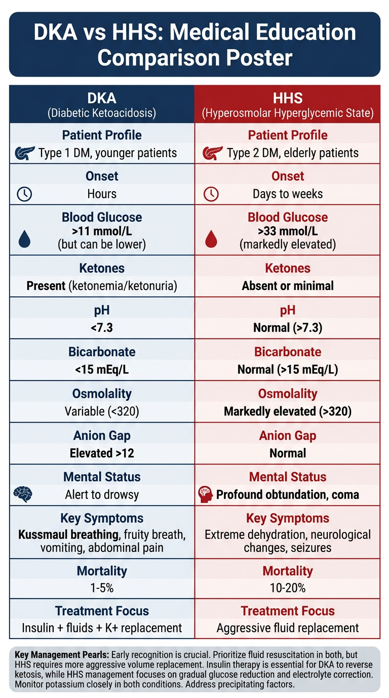
Generate a comparison poster (DKA vs Hyperosmolar Hyperglycemic State)

Generating Image

Medical education comparison poster: DKA (Diabetic Ketoacidosis) vs HHS (Hyperosmolar Hyperglycemic State). Clean clinical reference poster with two clearly divided columns, one for DKA and one for HHS. Include these comparison categories as rows: Patient Profile, Onset, Blood Glucose, Ketones, pH, Bicarbonate, Osmolality, Anion Gap, Mental Status, Key Symptoms, Mortality, Treatment Focus. DKA column data: Type 1 DM, younger patients; Hours; >11 mmol/L (but can be lower); Present (ketonemia/ketonuria); <7.3; <15 mEq/L; Variable (<320); Elevated >12; Alert to drowsy; Kussmaul breathing, fruity breath, vomiting, abdominal pain; 1-5%; Insulin + fluids + K+ replacement. HHS column data: Type 2 DM, elderly patients; Days to weeks; >33 mmol/L (markedly elevated); Absent or minimal; Normal (>7.3); Normal (>15 mEq/L); Markedly elevated (>320); Normal; Profound obtundation, coma; Extreme dehydration, neurological changes, seizures; 10-20%; Aggressive fluid replacement. Use a professional medical poster style with a navy blue and red color scheme for the two columns, clear section headers, bold values for critical differences, and a title banner at the top. Add a footer with key management pearls.

DKA vs HHS Comparison Poster
Here's the full comparison at a glance:
FeatureDKAHHS
PatientType 1 DM, youngerType 2 DM, elderly
OnsetHoursDays to weeks
Blood Glucose>11 mmol/L (variable)>33 mmol/L (markedly high)
KetonesPresent +++Absent or trace
pH<7.3 (acidosis)>7.3 (normal)
Bicarbonate<15 mEq/L>15 mEq/L
OsmolalityVariable (<320)>320 mOsm/kg
Anion GapElevated (>12)Normal
Mental StatusAlert to drowsyProfound obtundation / coma
Key SymptomsKussmaul breathing, fruity breath, vomiting, abdominal painExtreme dehydration, focal neuro deficits, seizures
Mortality1-5%10-20% (higher - older patients, delayed presentation)
Treatment FocusInsulin + fluids + K+ replacementAggressive fluid replacement first
Key clinical pearls:
  • DKA: The problem is lack of insulin - correct the ketoacidosis with insulin. Potassium management is critical (K+ <3.3 = hold insulin; replace first).
  • HHS: The problem is profound dehydration (fluid deficit 8-12 L) - correct with fluids first. Insulin plays a secondary role. Transition slowly to avoid cerebral edema.
  • Overlap: A mixed DKA/HHS picture can occur, especially in Type 2 DM patients under severe physiologic stress.

Generate a step-by-step insulin dosing and potassium replacement protocol

Reading File
Finding Sources
Finding Sources
Reading File
Reading File
I now have all the precise ADA dosing data needed. Generating the protocol poster:
Generating Image

Medical clinical protocol flowchart poster titled "DKA: Insulin Dosing & Potassium Replacement Protocol" for hospital emergency use. Dark navy background with bright white and yellow text. Professional, clean, clinical style. The poster has the following clearly structured flow: TOP SECTION - "STEP 1: CHECK POTASSIUM FIRST (before starting insulin)" with a large decision diamond shape: - K+ LESS THAN 3.3 mEq/L → RED box: "HOLD INSULIN. Give IV KCl 20-40 mEq/hr. Recheck K+ before starting insulin" - K+ 3.3 to 5.5 mEq/L → GREEN box: "START INSULIN. Give 20-40 mEq KCl per liter of IV fluid concurrently" - K+ GREATER THAN 5.5 mEq/L → ORANGE box: "START INSULIN. Hold potassium supplementation. Recheck K+ every 2 hours" MIDDLE SECTION - "STEP 2: INSULIN INFUSION PROTOCOL" - Starting dose: Regular insulin IV infusion 0.1 units/kg/hour (NO IV bolus) - Target glucose drop: 2.8 to 3.9 mmol/L (50-70 mg/dL) per hour - If glucose NOT falling by 50-70 mg/dL in first hour → double the infusion rate - When glucose reaches 11-14 mmol/L (200-250 mg/dL) → ADD dextrose to IV fluid (D5W or D5 0.45% NaCl) to maintain glucose 8-11 mmol/L - KEEP insulin infusion running (do not stop) until ketoacidosis resolves LOWER SECTION - "STEP 3: RESOLUTION CRITERIA (when to transition off IV insulin)" Three criteria in green checkboxes: Blood glucose less than 11 mmol/L AND Bicarbonate greater than 15 mEq/L AND pH greater than 7.3 Then transition arrow: "Overlap IV insulin with subcutaneous insulin by 1-2 hours before stopping infusion" BOTTOM - "MONITORING FREQUENCY" table: - Glucose: every 1 hour - Electrolytes (K+, Na+, bicarb): every 2 hours for first 4 hours, then every 4 hours - ABG/VBG: every 2-4 hours - Urine output: hourly via catheter (target more than 0.5 mL/kg/hr) Footer warning box in red: "CAUTION: Serum K+ does NOT reflect total body K+. All DKA patients are total-body K+ depleted regardless of initial serum level. Aggressive K+ replacement is mandatory." Use clear arrows, boxes, and color coding. Make it readable as a wall-mounted clinical reference poster.

This is a shared conversation. Sign in to Orris to start your own chat.