
What do you mean by sympathetic nervous system
The sympathetic nervous system (SNS) is one of the two main divisions of the autonomic nervous system (the other being the parasympathetic). It prepares the body for "fight or flight" responses - increasing heart rate, dilating airways, redirecting blood to muscles, and inhibiting digestion. It uses norepinephrine as its main neurotransmitter at effector organs, with preganglionic fibers releasing acetylcholine. The SNS originates from the thoracolumbar region (T1-L2) of the spinal cord.
Searching sources...
Reading File
Finding Sources
Searching the Web
sympathetic nervous system function anatomy overview
Reading File
Finding Sources
Reading File
Loading Image
Here is a thorough explanation of the Sympathetic Nervous System (SNS), drawing from Costanzo Physiology 7th Edition and other medical textbooks.
The Sympathetic Nervous System
What Is It?
The sympathetic nervous system is one of the two major divisions of the autonomic nervous system (ANS) - the other being the parasympathetic nervous system. The autonomic nervous system controls involuntary (automatic) body functions. The SNS specifically mobilizes the body for activity, most famously the "fight or flight" response to stress or danger.
"The overall function of the sympathetic nervous system is to mobilize the body for activity. In the extreme, if a person is exposed to a stressful situation, the sympathetic nervous system is activated with a response known as 'fight or flight,' which includes increased arterial pressure, increased blood flow to active muscles, increased metabolic rate, increased blood glucose concentration, and increased mental activity and alertness."
- Costanzo Physiology 7th Edition
Importantly, the SNS is not only active in emergencies - it also continuously regulates the heart, blood vessels, gastrointestinal tract, bronchi, sweat glands, and more under everyday conditions.
Anatomy & Organization
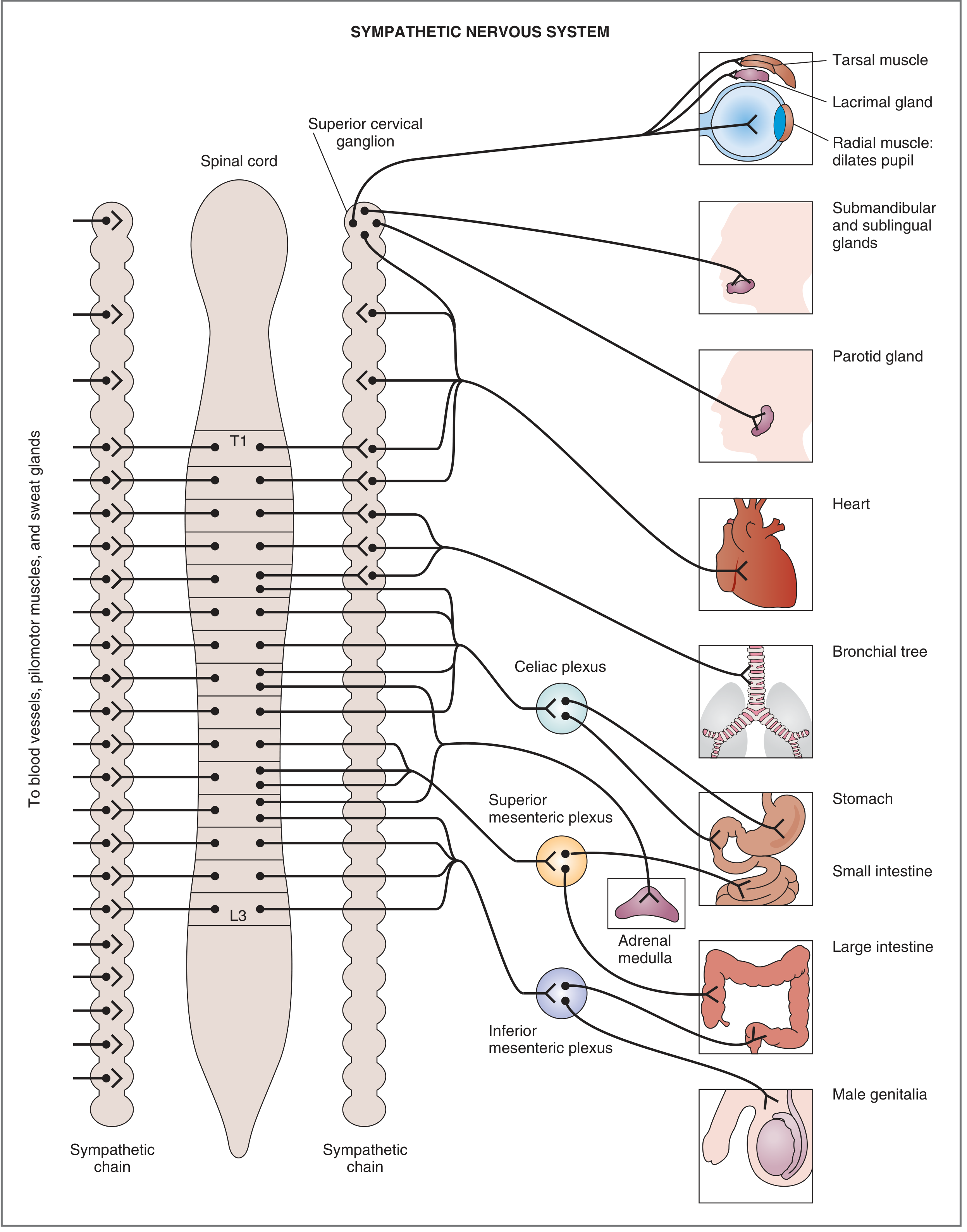
Here is the full innervation diagram from the textbook:
1. Origin (Thoracolumbar Division)
- Preganglionic neurons originate in the thoracic and lumbar spinal cord - specifically T1 to L3.
- This is why the SNS is called the "thoracolumbar" division.
- Upper thoracic segments serve the heart and thoracic organs; lumbar segments serve pelvic organs.
2. Two-Neuron Chain
The SNS uses a two-neuron pathway from the spinal cord to the target organ:
| Neuron | Location | Key Feature |
|---|---|---|
| Preganglionic neuron | Spinal cord (T1-L3) | Short axon; releases acetylcholine (ACh) |
| Postganglionic neuron | Sympathetic ganglia | Long axon; releases norepinephrine (NE) |
3. Ganglia (Relay Stations)
There are two types of sympathetic ganglia where preganglionic and postganglionic neurons synapse:
- Paravertebral ganglia (Sympathetic Chain) - located on either side of the spinal cord. Examples: superior cervical ganglion (for eyes and salivary glands).
- Prevertebral (collateral) ganglia - located closer to the target organs. Examples:
- Celiac ganglion - stomach, small intestine
- Superior mesenteric ganglion - small and large intestine
- Inferior mesenteric ganglion - lower large intestine, bladder, genitalia
4. The Adrenal Medulla - A Special Case
The adrenal medulla is essentially a modified sympathetic ganglion. Its preganglionic fibers (from T5-T9) bypass the sympathetic chain and stimulate the adrenal medulla to release epinephrine (adrenaline) and norepinephrine directly into the bloodstream - acting as hormones rather than local neurotransmitters. This amplifies the fight-or-flight response body-wide.
Neurotransmitters & Receptors
| Step | Neurotransmitter | Receptor Type |
|---|---|---|
| Preganglionic → postganglionic synapse | Acetylcholine (ACh) | Nicotinic (N₂) receptors |
| Postganglionic → effector organ | Norepinephrine (NE) | Adrenergic receptors (α₁, α₂, β₁, β₂) |
| Exception: Sweat glands | Acetylcholine | Muscarinic receptors |
As noted in Costanzo Physiology: "Postganglionic neurons of the sympathetic division are adrenergic in all of the effector organs, except in the thermoregulatory sweat glands (where they are cholinergic)."
Key Effects on Organ Systems
When the SNS is activated, it produces the following effects (all geared toward preparing the body for action):
| Organ / System | Sympathetic Effect |
|---|---|
| Heart | Increased heart rate (chronotropy) and force of contraction (inotropy) |
| Blood vessels | Vasoconstriction (skin, gut) / Vasodilation (skeletal muscle) |
| Lungs (bronchi) | Bronchodilation - opens airways for more oxygen |
| Eyes | Pupil dilation (mydriasis) via radial muscle |
| Liver | Increased glucose release (glycogenolysis) |
| Digestive tract | Decreased motility and secretion - digestion pauses |
| Bladder | Relaxation of detrusor muscle - urine retention |
| Sweat glands | Increased sweating (thermoregulatory) |
| Adrenal medulla | Secretion of epinephrine and norepinephrine |
| Pilomotor muscles | Hair stands up ("goosebumps") |
SNS vs. Parasympathetic Nervous System
| Feature | Sympathetic | Parasympathetic |
|---|---|---|
| Nickname | "Fight or flight" | "Rest and digest" |
| Origin | T1-L3 (thoracolumbar) | Craniosacral (CN III, VII, IX, X + S2-S4) |
| Preganglionic axon | Short | Long |
| Postganglionic axon | Long | Short |
| Main neurotransmitter (post) | Norepinephrine | Acetylcholine |
| Heart rate | Increases | Decreases |
| Digestion | Inhibits | Promotes |
Summary
The sympathetic nervous system is your body's "accelerator" - it gets you ready to respond to challenges by increasing heart rate, redirecting blood flow, releasing energy stores, dilating airways, and sharpening alertness. It works through a two-neuron pathway (preganglionic releasing ACh, postganglionic releasing norepinephrine), with ganglia located near the spinal cord. Its effects are widespread, diffuse, and designed for whole-body mobilization.
- Costanzo Physiology 7th Edition, p. 56-57
- Goldman-Cecil Medicine; Katzung's Basic & Clinical Pharmacology 16th Ed.
How does the parasympathetic nervous system differ from the sympathetic system?
The parasympathetic nervous system (PNS) is the "rest and digest" counterpart to the sympathetic "fight or flight" system. Key differences: it originates from craniosacral regions (brainstem + S2-S4), uses acetylcholine at both synapses, has long preganglionic and short postganglionic fibers, and its ganglia are located near or within target organs. It slows the heart, stimulates digestion, and conserves energy.
Searching sources...
Finding Sources
Searching the Web
parasympathetic vs sympathetic nervous system differences comparison
Reading File
Finding Sources
Reading File
Finding Sources
Loading Image
Reading File
Excellent content from Costanzo Physiology 7th Edition. Here is a complete, detailed comparison.
Parasympathetic vs. Sympathetic Nervous System
Both are divisions of the autonomic nervous system (ANS) and work together to maintain homeostasis - but they differ fundamentally in origin, anatomy, neurotransmitters, and effects. As Costanzo Physiology states: "The overall function of the parasympathetic nervous system is restorative, to conserve energy," in direct contrast to the sympathetic system's role in mobilizing the body.
Side-by-Side Anatomical Comparison
| Feature | Sympathetic | Parasympathetic |
|---|---|---|
| Nickname | "Fight or flight" | "Rest and digest" |
| Origin | Thoracolumbar - T1 to L3 | Craniosacral - brainstem (CN III, VII, IX, X) + sacral cord (S2-S4) |
| Preganglionic axon length | Short | Long |
| Postganglionic axon length | Long | Short |
| Ganglia location | Near the spinal cord (paravertebral chain or prevertebral ganglia) | Near or inside the target organ (e.g., ciliary, otic, pterygopalatine) |
| Divergence | High - 1 preganglionic → many postganglionic neurons (diffuse, widespread effects) | Low - 1 preganglionic → few postganglionic neurons (discrete, localized effects) |
Parasympathetic Innervation Diagram (Costanzo Physiology)
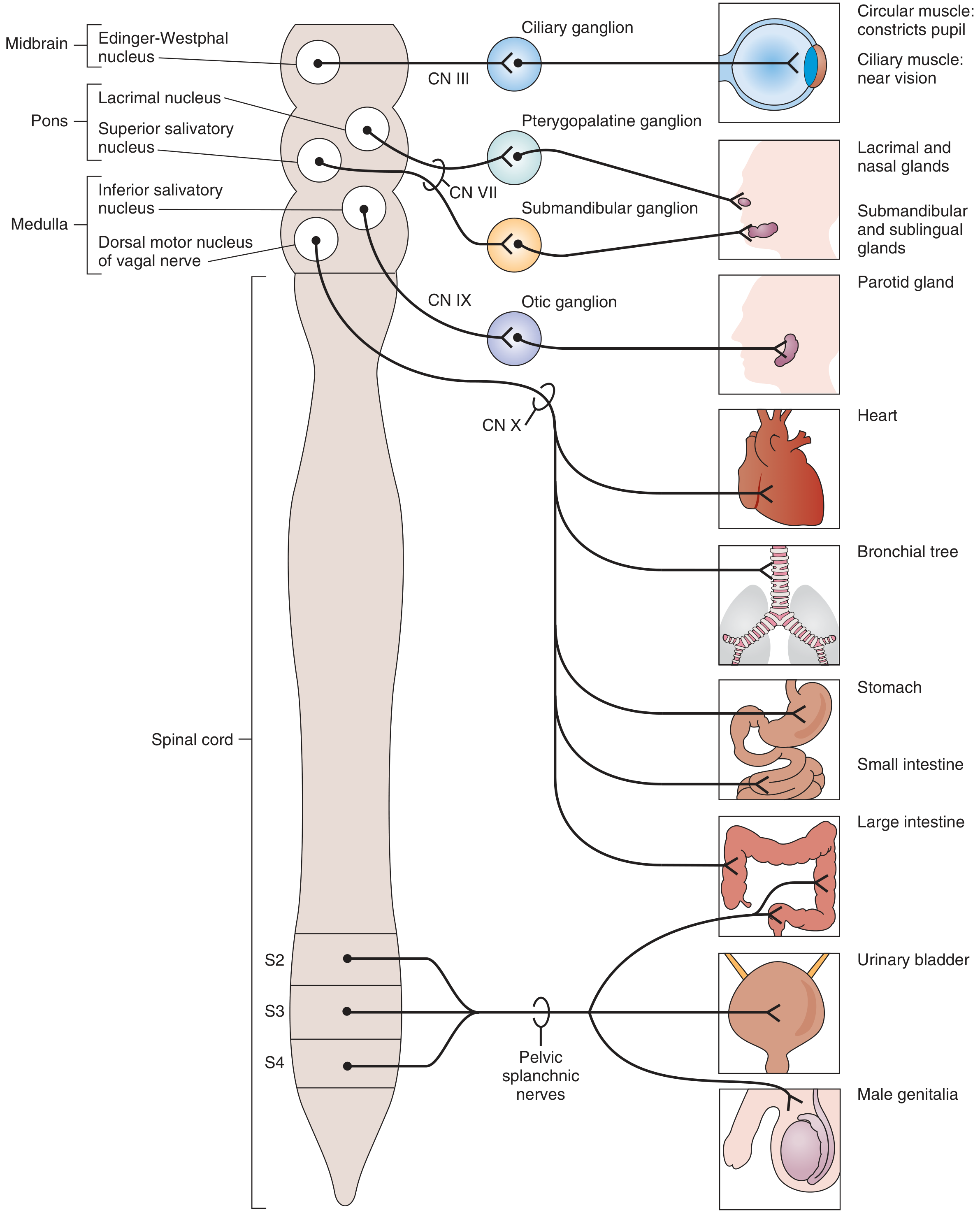
Notice how the parasympathetic system originates from the brainstem (midbrain, pons, medulla) via cranial nerves, and from sacral segments S2-S4 via pelvic splanchnic nerves - compared to the thoracolumbar origin of the sympathetic system seen in the previous response.
Neurotransmitter Comparison
This is the most important pharmacological difference:
| Step | Sympathetic | Parasympathetic |
|---|---|---|
| Preganglionic → ganglion | ACh → Nicotinic (N₂) receptors | ACh → Nicotinic (N₂) receptors |
| Postganglionic → effector organ | Norepinephrine (NE) → Adrenergic receptors (α₁, α₂, β₁, β₂) | Acetylcholine (ACh) → Muscarinic receptors |
| Exception | Sweat glands: ACh → Muscarinic | None |
| Co-transmitters | NE + ATP + Neuropeptide Y | ACh + VIP + Nitric Oxide (NO) |
Key point: Both systems use ACh at the preganglionic synapse (acting on nicotinic receptors). The difference is entirely at the postganglionic effector synapse - adrenergic (NE) for sympathetic vs. cholinergic (ACh/muscarinic) for parasympathetic.
Effects on Organ Systems
| Organ | Sympathetic Effect | Parasympathetic Effect |
|---|---|---|
| Heart rate | Increases (↑) via β₁ | Decreases (↓) via muscarinic |
| Heart contractility | Increases (↑) via β₁ | Decreases slightly |
| Blood pressure | Increases (vasoconstriction via α₁) | Decreases |
| Bronchi (airways) | Dilation (↑ airflow) via β₂ | Constriction (↓ airflow) |
| GI tract motility | Decreases (slows digestion) | Increases (promotes digestion) |
| GI sphincters | Contracts (closes) via α₁ | Relaxes (opens) |
| Salivary glands | Thick, mucous secretion | Thin, watery, copious secretion |
| Pupil (eye) | Dilation - mydriasis (radial muscle) via α₁ | Constriction - miosis (circular muscle) |
| Lens (eye) | Far vision (relaxes ciliary muscle) | Near vision - accommodation (contracts ciliary muscle) via CN III |
| Urinary bladder (detrusor) | Relaxes (fills) via β₂ | Contracts (empties) |
| Bladder sphincter | Contracts (retains urine) via α₁ | Relaxes (allows urination) |
| Male genitalia | Ejaculation | Erection |
| Sweat glands | Increases sweating (cholinergic exception) | No significant innervation |
| Adrenal medulla | Secretes epinephrine + NE | No significant innervation |
Reciprocal (Coordinated) Functions
Most organs receive both sympathetic and parasympathetic innervation that works reciprocally. Costanzo Physiology gives a clear example with the heart:
"An increase in sympathetic activity increases heart rate, and an increase in parasympathetic activity decreases heart rate... If there is a decrease in blood pressure, vasomotor centers in the brain stem respond by producing, simultaneously, an increase in sympathetic activity to the SA node and a decrease in parasympathetic activity. Each of these actions has the effect of increasing heart rate - the sympathetic and parasympathetic actions do not compete but work synergistically."
Another example is the bladder: during filling, sympathetic activity dominates (detrusor relaxes, sphincter contracts); during urination, parasympathetic activity dominates (detrusor contracts, sphincter relaxes).
Key Cranial Nerves of the Parasympathetic System
| Cranial Nerve | Nucleus | Ganglion | Target |
|---|---|---|---|
| CN III (Oculomotor) | Edinger-Westphal (midbrain) | Ciliary ganglion | Pupil constriction, near vision |
| CN VII (Facial) | Lacrimal + Superior salivatory (pons) | Pterygopalatine, Submandibular | Lacrimal, nasal, submandibular, sublingual glands |
| CN IX (Glossopharyngeal) | Inferior salivatory (medulla) | Otic ganglion | Parotid gland |
| CN X (Vagus) | Dorsal motor nucleus (medulla) | Terminal ganglia in organs | Heart, bronchi, stomach, small intestine, large intestine |
| Pelvic splanchnic (S2-S4) | Sacral cord | Pelvic ganglia | Bladder, lower colon, genitalia |
The vagus nerve (CN X) is by far the most important - it carries ~75% of all parasympathetic fibers.
Summary: The Core Difference
| Sympathetic | Parasympathetic | |
|---|---|---|
| Purpose | Mobilize - spend energy | Restore - conserve energy |
| When active | Stress, exercise, danger | Rest, digestion, recovery |
| Spread | Diffuse, whole-body response | Discrete, localized response |
| Speed | Fast, widespread | Slower, targeted |
| Final neurotransmitter | Norepinephrine | Acetylcholine (muscarinic) |
Together, these two systems continuously balance each other to keep the body in homeostasis - the SNS accelerates, the PNS brakes, and the interplay between them governs virtually every involuntary function.
- Costanzo Physiology 7th Edition, pp. 56-62
- Katzung's Basic and Clinical Pharmacology 16th Edition
- Morgan and Mikhail's Clinical Anesthesiology 7e
This is a shared conversation. Sign in to Orris to start your own chat.