Cerebrospinal fluid renoria
cerebrospinal fluid rhinorrhea diagnosis management
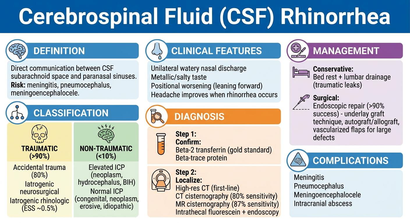
Cerebrospinal Fluid (CSF) Rhinorrhea
Definition
Classification
| Subtype | Notes |
|---|---|
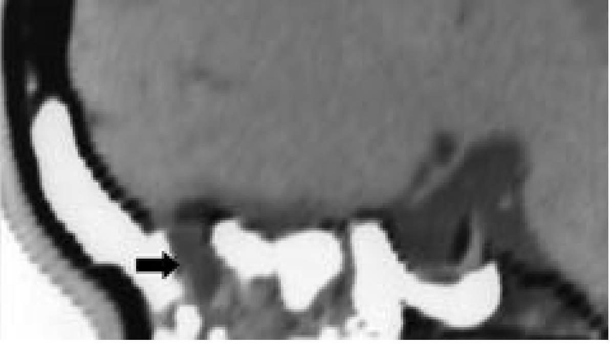
| Accidental trauma (~80% of traumatic) | Most involve the anterior cranial base / cribriform plate; ~80% become evident within 2 days, nearly all within 3 months |
| Iatrogenic - neurosurgical | Craniotomies, transsphenoidal hypophysectomy |
| Iatrogenic - rhinologic | Endoscopic sinus surgery (ESS), septoplasty, other skull base procedures; ESS complication rate ~0.5% |
- Elevated ICP: Intracranial neoplasm, hydrocephalus (noncommunicating/obstructive), benign intracranial hypertension (BIH)
- Normal ICP: Congenital anomaly, skull base neoplasm (nasopharyngeal carcinoma, sinonasal tumors), erosive processes (osteomyelitis, granulomatosis with polyangiitis), idiopathic
Pathophysiology
- CSF pressure is maintained by the balance between choroid plexus secretion (steady rate) and arachnoid villi resorption (primary regulator)
- Disruption of resorption raises ICP
- A long lateral lamella of the cribriform plate (LLCP) increases the risk of spontaneous leak - the combination with elevated ICP (BIH/ESS variant) likely precipitates rhinorrhea in idiopathic cases
Clinical Presentation
- Unilateral watery nasal discharge with a characteristic metallic or salty taste
- Often positional (increases when leaning forward)
- Headache that improves when rhinorrhea occurs and worsens when it stops (CSF pressure-related)
- May be intermittent, making diagnosis challenging
Differential Diagnosis
| Mimicker | Distinguishing Feature |
|---|---|
| Allergic/vasomotor rhinitis | Usually bilateral, seasonal/perennial pattern |
| CSF otorrhea presenting as rhinorrhea | Middle ear effusion + intact tympanic membrane; CSF drains via Eustachian tube |
| Retained saline irrigation fluid | Clears with cessation of irrigations |
| Ruptured sinus retention cyst | Yellow tint on white paper; resolves spontaneously; air-fluid level on imaging |
Diagnosis
Step 1: Confirm CSF Leak
The old "halo test" (ring sign on filter paper) is unreliable and should not be used for diagnosis.
Step 2: Localize the Defect
| Test | Notes |
|---|---|
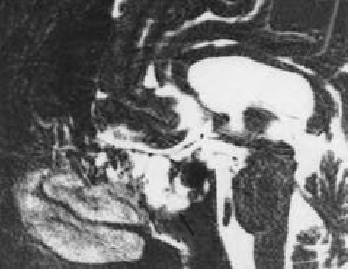
| High-resolution CT (non-contrast) | First-line imaging; identifies bony defects and associated fractures |
| CT cisternography | Intrathecal radiopaque contrast (metrizamide); confirms ~80% of leaks; requires active leak at time of study |
| MR cisternography | Noninvasive; heavily T2-weighted fat-suppressed sequences; sensitivity 87%, specificity 57%, accuracy 78%; best when leak is active |
| Radionuclide cisternography | Intrathecal radioisotope + nasal pledgets; poor spatial resolution; unacceptable false-positive/indeterminate rate - cannot be sole test |
| Intrathecal fluorescein + endoscopy | Confirms AND localizes the defect endoscopically; dilute concentrations mandatory (serious neurologic sequelae reported at higher doses) |
Management
Conservative Management
- Bed rest + lumbar drainage - appropriate first-line for most traumatic CSF leaks
- Most accidental traumatic leaks resolve spontaneously with conservative measures
- Post-surgical CSF leaks may be managed conservatively initially, but most eventually require operative repair
Surgical Repair
- Identify the leak site (with or without fluorescein)
- Close the defect using graft materials:
- Autogenous grafts: fascia lata, free bone, fat ("bath-plug" technique - fat plug held in place by ICP)
- Allograft: acellular dermal allograft (placed on intracranial side - underlay technique)
- Xenogeneic: collagen dural substitutes
- Cover with free mucosal graft
- Secure with surgical sealant + resorbable/nonresorbable packing
- Layer A: Dura
- Layer B: Fascial autograft or acellular dermal allograft (intracranial underlay)
- Layer C: Bone or cartilage autograft (rigid support)
- Layer D: Second fascial layer
- Layer E: Mucosal free autograft
- Layer F: Surgical sealant
Note: Current evidence does not confirm improved outcomes when rigid support or multilayered reconstruction is used for larger defects specifically.
Management by Etiology
| Etiology | Approach |
|---|---|
| Accidental trauma | Conservative (lumbar drain + bed rest) first; operate if fails |
| Intraoperative CSF leak (recognized at surgery) | Repair during that procedure |
| Post-surgical CSF rhinorrhea | Conservative initially; most require operative repair |
| Nontraumatic (idiopathic) | Unlikely to resolve spontaneously; after excluding neoplasm, proceed to operative repair |
| Associated BIH/ESS | Treat elevated ICP (acetazolamide, weight loss, CSF diversion if needed) alongside repair |
Complications
- Meningitis - the most feared complication; risk increases with duration of untreated leak
- Pneumocephalus - air entering the intracranial space through the defect
- Meningocele / meningoencephalocele - herniation of meninges ± brain tissue through the defect
- Intracranial abscess
Recent Literature
- Spontaneous rhinorrhea and idiopathic intracranial hypertension (2024, PMID 38393958) - highlights the complex association between idiopathic CSF rhinorrhea and IIH
- CSF Leaks: Diagnosis, Management, and Outcomes (2025, Neurosurg Clin N Am, PMID 39542549) - comprehensive current review
- CSF Rhinorrhea review (2026, Harefuah, PMID 41736622) - most current published review as of early 2026
Can u create image of the above text
Medical summary infographic titled "Cerebrospinal Fluid (CSF) Rhinorrhea" with clean clinical layout on a white background. Include the following sections clearly organized with icons and colored headers: 1. DEFINITION - "Direct communication between CSF subarachnoid space and paranasal sinuses. Risk: meningitis, pneumocephalus, meningoencephalocele." 2. CLASSIFICATION (two columns): Left - TRAUMATIC (>90%): Accidental trauma (80%), Iatrogenic neurosurgical, Iatrogenic rhinologic (ESS ~0.5%) Right - NON-TRAUMATIC (<10%): Elevated ICP (neoplasm, hydrocephalus, BIH), Normal ICP (congenital, neoplasm, erosive, idiopathic) 3. CLINICAL FEATURES: Unilateral watery nasal discharge, Metallic/salty taste, Positional worsening (leaning forward), Headache improves when rhinorrhea occurs 4. DIAGNOSIS (two steps): Step 1 - Confirm: Beta-2 transferrin (gold standard), Beta-trace protein Step 2 - Localize: High-res CT (first-line), CT cisternography (80% sensitivity), MR cisternography (87% sensitivity), Intrathecal fluorescein + endoscopy 5. MANAGEMENT: Conservative: Bed rest + lumbar drainage (traumatic leaks) Surgical: Endoscopic repair (>90% success) - underlay graft technique, autograft/allograft, vascularized flaps for large defects 6. COMPLICATIONS: Meningitis, Pneumocephalus, Meningoencephalocele, Intracranial abscess Use color coding: blue for headers, light yellow for traumatic, light green for nontraumatic, orange for diagnosis, purple for management. Professional medical poster style.
- Definition and clinical significance
- Classification - traumatic vs. nontraumatic with subtypes
- Clinical features - the hallmark presentation
- Diagnosis - two-step approach (confirm then localize)
- Management - conservative and surgical options
- Complications to watch for
Histology of skin with diagram
Detailed labeled anatomical cross-section diagram of skin histology showing all layers from surface to deep. Title: "Histology of Skin". Include three main compartments clearly separated: EPIDERMIS (top, pink stratified layer) showing from surface down: - Stratum Corneum (flat dead keratinized cells, outermost) - Stratum Lucidum (clear thin layer, only in thick skin) - Stratum Granulosum (granular cells with keratohyalin granules) - Stratum Spinosum (prickle cells with desmosomes, largest layer) - Stratum Basale / Germinativum (single row of columnar cells on basement membrane, mitotically active, contains melanocytes) DERMIS (middle, dense connective tissue) with two sublayers: - Papillary Dermis (superficial loose connective tissue, dermal papillae projecting upward into epidermis, contains capillaries and Meissner's corpuscles) - Reticular Dermis (deeper dense irregular connective tissue, collagen and elastin fibers, contains hair follicles, sebaceous glands, sweat glands, Pacinian corpuscles, blood vessels, nerve fibers) HYPODERMIS / Subcutaneous Layer (bottom, adipose tissue, lobules of fat cells, larger blood vessels) Also label these structures in the dermis/hypodermis: - Hair follicle with hair shaft - Sebaceous gland (attached to hair follicle) - Eccrine sweat gland (coiled, opens to surface) - Arrector pili muscle - Meissner's corpuscle (in dermal papilla) - Pacinian corpuscle (deep dermis) - Blood vessels (capillaries and arterioles) - Basement membrane (between epidermis and dermis) - Melanocyte (in stratum basale) - Langerhans cell (in stratum spinosum) Use clean anatomical illustration style: pink for epidermis, light pink for dermis, yellow for hypodermis/fat. All structures clearly labeled with neat leader lines. Professional medical textbook quality.
400, message: Got more than 4194304 bytes when reading: b'data: {"candidates": [{"content": {"role": "model","parts": [{"inlineData": {"mimeType": "image/png"...'.
We encountered an error while processing your request. Please try again or reach out to us for help.
Histology of skin with diagram
An error occurred (ValidationException) when calling the ConverseStream operation: Expected toolResult blocks at messages.34.content for the following Ids: tooluse_cUrJvdWtT0Cb2Ao0EtjRbi
We encountered an error while processing your request. Please try again or reach out to us for help.
Y?
An error occurred (ValidationException) when calling the ConverseStream operation: Expected toolResult blocks at messages.34.content for the following Ids: tooluse_cUrJvdWtT0Cb2Ao0EtjRbi
We encountered an error while processing your request. Please try again or reach out to us for help.