
Congenital pulmonary airway malformation
"congenital pulmonary airway malformation"
congenital pulmonary airway malformation CPAM pathology CT scan
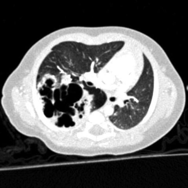
This transverse diagnostic computerized tomography (CT) scan of a pediatric thorax demonstrates a congenital pulmonary airway malformation (CPAM) within the right lung, likely involving the lower lobe. The hallmark feature is a large, multicystic mass consisting of various-sized, air-filled cystic spaces with an irregular 'honeycomb' internal architecture. These cysts are separated by thin septations and surrounded by areas of increased soft tissue density representing pulmonary consolidation or dysplastic parenchyma. The mass exerts a mild mass effect, characterized by the partial displacement of the mediastinum towards the contralateral side. In contrast, the left lung parenchyma appears relatively normal with preserved aeration, visible vascular markings, and no evidence of cystic lesions or structural abnormalities. The image is a classic representation of pediatric thoracic pathology used to teach the radiological presentation of congenital lung malformations, focusing on the differentiation of cystic lung lesions and their impact on adjacent thoracic structures.
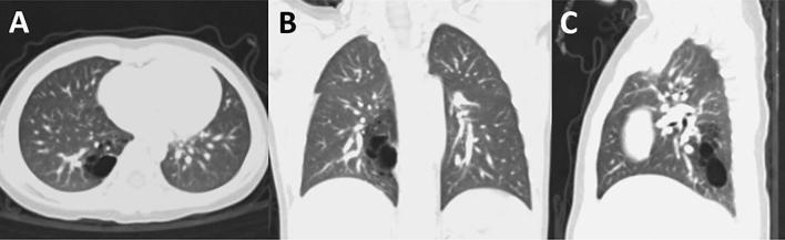
This diagnostic image consists of three chest CT scan views (A: axial, B: coronal, C: sagittal) in a lung window, demonstrating a Congenital Pulmonary Airway Malformation (CPAM). The lesion is characterized by a focal area of well-defined, air-filled cystic spaces with decreased attenuation (radiolucency), localized specifically to the posterior basal segment (S10) of the right lower lobe. The axial view (A) shows the posterior positioning within the right lung field. The coronal (B) and sagittal (C) views further delineate the inferior-superior extent of the malformation near the diaphragmatic surface. The surrounding lung parenchyma and pulmonary vasculature appear normal, with no evidence of significant mediastinal shift, mass effect, or distortion of adjacent anatomical structures. This visual representation is a classic example of a localized Type 1 or Type 2 CPAM, utilized in pediatric radiology and thoracic surgery education to illustrate lung bud developmental anomalies and surgical planning for segmentectomy.
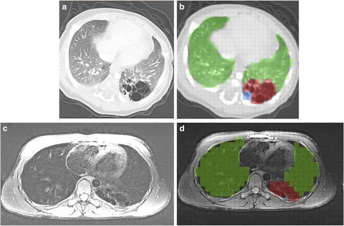
This composite figure demonstrates diagnostic imaging and quantitative analysis of a Congenital Pulmonary Airway Malformation (CPAM) in the left lower lobe. Panel (a) is an axial postnatal CT scan showing a localized, multicystic air-filled lesion with thin internal septations and low attenuation, characteristic of a cystic congenital lung abnormality. Panel (c) provides an axial school-age MRI of the same patient, displaying the lesion's evolution with mixed signal intensity in the left posterior lung field. Panels (b) and (d) illustrate specialized volumetric scoring methods: CLAQ (Congenital Lung Abnormalities Quantification on CT) and CLAM (on MRI), respectively. These panels use color-coded grid overlays to differentiate tissue types: green indicates normal lung parenchyma, red identifies the primary CPAM lesion, and blue highlights lesion-associated abnormalities such as atelectasis. This comparison highlights the utility of multi-modal imaging in the longitudinal follow-up of pediatric congenital lung lesions from infancy to school age.
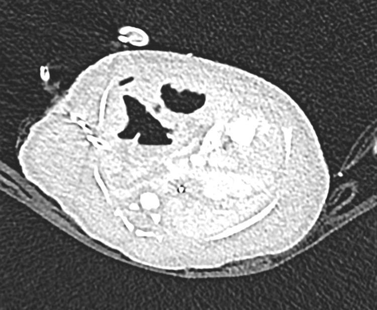
This axial chest computerized tomography (CT) scan of a newborn demonstrates a large congenital cystic airway malformation (CPAM) predominantly occupying the right middle lobe. The lesion is characterized by multiple air-filled cystic spaces of varying sizes and irregular morphology, surrounded by soft tissue density. A significant mass effect is evident, causing a prominent contralateral (leftward) mediastinal shift and compression of the adjacent pulmonary parenchyma. The CT scan also captures the presence of medical hardware, including an external chest drain on the right side and endotracheal/vascular access lines, consistent with neonatal intensive care management. This diagnostic image illustrates a Type 1 CPAM with invasive mucinous features, emphasizing its clinical significance in causing severe neonatal respiratory and cardiovascular instability through space-occupying displacement of vital thoracic structures.
Congenital Pulmonary Airway Malformation (CPAM)
Definition & Pathogenesis
- Prevalence: ~9 per 100,000 total births (estimated incidence 1:25,000 to 1:35,000)
- Pathogenesis: Abnormal branching of primitive bronchioles causing arrested development at various stages of lung morphogenesis - primarily during the pseudoglandular stage but sometimes as late as alveolarization, producing more distal cystic lesions
- Molecular basis: Overexpression of Hoxb5 gene beyond the pseudoglandular stage, as well as overexpression of KGF/Fgf7 (keratinocyte growth factor) and platelet-derived growth factor B (PDGFB), have all been implicated
- Involves only one pulmonary lobe and has a pulmonary blood supply (distinguishing it from bronchopulmonary sequestration)
- In ~40% of cases, CPAM manifests as a hybrid CPAM-BPS with a systemic vascular supply
Classification (Stocker Types 0-4)
| Type | Origin | Characteristics | Frequency |
|---|---|---|---|
| 0 | Tracheal / primary bronchial | Often lethal; incompatible with survival | Rare |
| 1 | Distal bronchus | Most common (50-70%); single large cyst 3-10 cm; significant mass effect | 50-70% |
| 2 | Terminal bronchioles | Evenly spaced small cysts 1-2 cm, often with solid components; associated with extrapulmonary anomalies (renal agenesis, cardiac defects, GI atresia, skeletal abnormalities) | 15-30% |
| 3 | Microcysts (<5 mm) | Cuboidal epithelium resembling fetal lung; appears largely solid on CT; may mimic BPS | Uncommon |
| 4 | Alveolar tissue | Very large peripheral cysts (up to 10 cm); risk of pneumothorax; can be indistinguishable from Type 1 on imaging | Uncommon |
Diagnosis
Antenatal
- Ultrasound: sensitivity 93%, specificity 32% - presents as a solid echodense mass or a mass with large cysts
- Fetal MRI: superior for identifying systemic feeding vessels (sensitivity 71%, specificity 88%) - helps differentiate CPAM from BPS
- Up to one-third of CPAMs identified in early pregnancy resolve spontaneously before birth
- Most cases are now detected prenatally with routine obstetric ultrasound
Postnatal / Adult
- Postnatal CT scan remains the gold standard - lesions persist even when apparently "resolved" on US
- Adults typically present as an incidental finding on imaging, or with recurrent lower respiratory tract infections or pneumothorax
CPAM Volume Ratio (CVR) - Key Prognostic Tool
| CVR | Risk of Hydrops |
|---|---|
| >1.6 (without dominant cyst) | ~80% risk of fetal hydrops |
| <1.6 (without dominant cyst) | <3% risk of hydrops |
| Any dominant cyst (regardless of CVR) | Significant risk |
- Fetuses with CVR >1.6: consider ultrasound surveillance twice weekly
Complications
Fetal/Neonatal
- Fetal hydrops - develops in up to 40% of untreated cases; associated with high mortality
- Pulmonary compression with mediastinal shift
- Polyhydramnios
Pediatric/Adult
- Recurrent pneumonia
- Pneumothorax (especially Type 4)
- Aspergilloma formation in pre-existing cavities → life-threatening hemoptysis
Malignant Transformation
- Type 1 CPAM: associated with mucinous adenocarcinoma (K-ras oncogenic mutations, chromosomal aberrations in chr 2 and 4 in atypical goblet cells identified)
- Type 2 CPAM: associated with rhabdomyosarcoma and pleuropulmonary blastoma
- Type 4 CPAM: associated with pleuropulmonary blastoma
Management
Prenatal Management
- Thoracoamniotic shunting - placement of a shunt into the largest cyst cavity; overall survival ~74%; procedure of choice before 32 weeks
- Percutaneous puncture - minimally invasive but temporary relief
- After 32 weeks: delivery typically recommended; intervention may be considered up to 37 weeks
- Prenatal corticosteroids (standard fetal lung maturation doses): hydrops resolved in ~78% of cases; survival ~85%
- Mechanism unknown - possibly accelerates maturation or involution of the lesion
- Open fetal lobectomy via hysterotomy (21-31 weeks) - reserved for large lesions; 11/22 long-term survivors in one series; hydrops resolves in 1-2 weeks post-surgery with catch-up lung growth
Postnatal / Neonatal Management
- Up to 50% of neonates with CPAM are asymptomatic at birth - can be managed expectantly
- If elective surgical resection is chosen: ideally within 10 months of birth (associated with fewer complications)
- Symptomatic neonates: emergency lobectomy or segmentectomy
Pediatric / Adult Management
- Asymptomatic adults: surveillance with interval imaging; no mandatory resection
- Type 1 or Type 4 CPAM: close follow-up due to slightly increased malignancy risk
- Main indication for surgery in adults: recurrent infections
- Surgical options: lobectomy or segmentectomy (minimally invasive / VATS preferred when feasible)
Imaging Examples
Recent Evidence (2025-2026)
-
Risk of malignant transformation and infections in adults (Pederiva et al., Eur Respir Rev 2025) - [PMID 40174959] - addresses long-term oncologic surveillance needs in adult CPAM
-
Safety of conservative management of asymptomatic CPAMs in children (Thorburn et al., J Pediatr Surg 2026) - [PMID 41643769] - recent 2026 systematic review examining the evidence base for watchful waiting vs. early elective resection in asymptomatic pediatric CPAM; this may update traditional recommendations favoring early surgery
Key Differentials
| Feature | CPAM | Bronchopulmonary Sequestration (BPS) |
|---|---|---|
| Blood supply | Pulmonary | Systemic (aortic branches) |
| Bronchial connection | Yes (communicates) | No (non-communicating) |
| Location | Any lobe | Left > right lower lobe (intralobar) |
| Hybrid lesion possible? | Yes (~40%) | Yes (~40-50% with CPAM) |
| Key imaging clue | Cystic/solid lung mass | Echogenic wedge-shaped mass + "feeding vessel" on Doppler |