
20 diseases along with ecg changes
Reading File
Now let me search the medical library for ECG changes in various diseases, running parallel queries:
Finding Sources
Finding Sources
Finding Sources
Finding Sources
Finding Sources
Finding Sources
Finding Sources
Finding Sources
Finding Sources
I now have excellent source material. Let me compile the comprehensive answer from the textbooks plus my medical knowledge:
20 Diseases and Their ECG Changes
Here is a detailed, exam-ready reference of 20 conditions with their characteristic ECG findings, sourced from authoritative textbooks including Harrison's, Braunwald's, Rosen's, and others.
1. Acute Myocardial Infarction (STEMI)
| Phase | ECG Finding |
|---|---|
| Hyperacute (minutes) | Tall, peaked (hyperacute) T waves |
| Early (hours) | ST elevation (convex/tombstone), loss of R wave |
| Established | Pathological Q waves (>1 mm wide, >25% of R height) |
| Reperfusion/old | T-wave inversion, persistent Q waves |
- ST elevation in contiguous leads: inferior (II, III, aVF), anterior (V1-V4), lateral (I, aVL, V5-V6)
- Reciprocal ST depression in mirror leads
Rosen's Emergency Medicine, differential of ST elevation listed as the classic template
2. Non-STEMI / Unstable Angina
- ST depression (horizontal or downsloping) in affected territory
- T-wave inversion (deep, symmetric "Wellens' pattern" in V2-V3 = critical LAD stenosis)
- No pathological Q waves
- May have no ECG changes at all
3. Acute Pericarditis
Classic 4-stage evolution:
| Stage | ECG Finding |
|---|---|
| Stage I | Diffuse concave ST elevation (saddle-shaped) in most leads; PR depression (especially lead II); PR elevation in aVR |
| Stage II | ST returns to baseline; T waves flatten |
| Stage III | Diffuse T-wave inversion |
| Stage IV | Normalization |
Key distinguishing features from MI: diffuse (not lead-specific), no reciprocal changes, PR depression, no Q waves.
Goldman-Cecil Medicine; Braunwald's Heart Disease
4. Hyperkalemia
Progressive ECG changes correlating with serum K+ level:
| K+ Level | ECG Finding |
|---|---|
| 5.5-6.5 mEq/L | Tall, symmetric, narrow peaked T waves (best in precordial leads) |
| 6.5-7.5 mEq/L | PR interval prolongation; P wave flattening/loss |
| 7.0-8.0 mEq/L | QRS widening (sine-wave pattern) |
| >8.0 mEq/L | Ventricular fibrillation / asystole |
Also seen in Addison's disease (adrenal insufficiency) which causes hyperkalemia.
Harrison's 22nd edition; Morgan & Mikhail's Clinical Anesthesiology
5. Hypokalemia
- Flattening or inversion of T waves
- Prominent U waves (best seen in V2-V3; U wave > T wave = significant)
- ST segment depression
- Prolonged QU interval (mistaken for prolonged QT)
- Severe: prolonged PR, widened QRS, decreased voltage
- Risk of ventricular arrhythmias and Torsades de Pointes
Washington Manual; Rosen's Emergency Medicine (with ECG image)
6. Hypercalcemia
- Shortened QT interval (most classic finding - shortens the ST segment)
- Prolonged PR interval
- Prolonged QRS
- T-wave flattening and widening
- Can progress to AV block, complete heart block, and cardiac arrest
- Osborn J waves can occasionally be seen
Schwartz's Surgery; Harrison's; Rosen's Emergency Medicine
7. Hypocalcemia
- Prolonged QT interval (specifically prolonged ST segment / QTc)
- May have T-wave inversion
- Risk of ventricular arrhythmias including Torsades de Pointes
- Seen in hypoparathyroidism, vitamin D deficiency, post-thyroidectomy
Comprehensive Clinical Nephrology; Harrison's; Miller's Review of Orthopaedics
8. Pulmonary Embolism (PE)
Classic but insensitive findings:
- S1Q3T3: Deep S wave in lead I, Q wave in lead III, T-wave inversion in lead III
- Sinus tachycardia (most common finding)
- New right bundle branch block (complete or incomplete)
- P pulmonale (peaked P waves)
- Right axis deviation
- T-wave inversions in V1-V4 (right heart strain)
- Atrial fibrillation
Rosen's Emergency Medicine
9. Hypothermia
- Osborn wave (J wave): Positive deflection at the J point (junction of QRS and ST), best seen in V4-V6 and inferior leads. Classic for hypothermia.
- Bradycardia (often the first sign)
- PR, QRS, and QT prolongation (all intervals widen)
- Atrial fibrillation
- Shivering artifact
- Risk of ventricular fibrillation <28°C
10. Hypertrophic Cardiomyopathy (HCM)
- Left ventricular hypertrophy (LVH) pattern (high voltage: Sokolow-Lyon criteria: S in V1 + R in V5/V6 >35 mm)
- Asymmetric septal hypertrophy: deep, narrow Q waves ("dagger" Q waves) in lateral leads (I, aVL, V5-V6) and inferior leads - these mimic infarction but without ST changes
- ST depression and T-wave inversions
- Left axis deviation
- P mitrale (bifid P waves) if left atrial enlargement
11. Dilated Cardiomyopathy
- Sinus tachycardia
- Left bundle branch block (LBBB) is common
- LVH pattern
- Non-specific ST-T changes
- Left axis deviation
- Q waves (fibrosis mimicking infarction)
- Atrial fibrillation
- Ventricular ectopy
12. Wolff-Parkinson-White (WPW) Syndrome
- Short PR interval (<120 ms)
- Delta wave: slurred upstroke of the QRS (pre-excitation)
- Wide QRS (>120 ms total due to delta wave fusion)
- Pseudo-ST changes and pseudo-Q waves (secondary to pre-excitation)
- Paroxysmal supraventricular tachycardia (PSVT) on rhythm strip
- Risk: atrial fibrillation with rapid conduction to ventricles -> VF
13. Complete Heart Block (3rd Degree AV Block)
- Dissociation of P waves and QRS complexes (no relationship between them)
- P-P interval regular; R-R interval regular - but independent of each other
- Bradycardia
- Wide QRS if ventricular escape rhythm (junctional = narrow QRS ~40-60 bpm; ventricular = wide QRS ~20-40 bpm)
14. Atrial Fibrillation
- Absent P waves - replaced by fine irregular fibrillatory baseline (f waves)
- Irregularly irregular R-R intervals (hallmark)
- Ventricular rate variable (controlled vs. uncontrolled)
- QRS usually narrow unless aberrant conduction
15. Digoxin Toxicity / Effect
Digoxin effect (therapeutic):
- "Reverse tick" or "Salvador Dali mustache" ST depression - downsloping ST with upward curve
- Shortened QT interval
- T-wave flattening or inversion
- PR prolongation (enhanced vagal tone)
Digoxin toxicity:
- Any arrhythmia, especially PAT with block (paroxysmal atrial tachycardia with AV block - pathognomonic)
- Bidirectional ventricular tachycardia
- VF
- Regularization of AF (if previously in AF - suspect dig toxicity)
16. Long QT Syndrome (congenital or acquired)
- Prolonged QTc interval (QTc >440 ms in men, >460 ms in women)
- T-wave morphology abnormalities (notched, bifid, or broad T waves depending on subtype)
- Risk of Torsades de Pointes (polymorphic VT with twisting QRS axis)
- Causes: congenital (LQTS1-3), drugs (quinidine, amiodarone, antipsychotics), electrolyte disturbances (hypokalemia, hypomagnesemia, hypocalcemia)
17. Brugada Syndrome
- Coved-type ST elevation (>2 mm, downsloping) in V1-V2 (Type 1 - diagnostic)
- Type 2: Saddleback ST elevation in V1-V2 (not diagnostic alone)
- Normal QT
- Right bundle branch block pattern
- Risk of sudden cardiac death from VF (typically during sleep/fever)
- Sodium channelopathy (SCN5A mutation)
18. Sarcoidosis (Cardiac)
- Complete heart block (most common life-threatening manifestation)
- Bundle branch blocks (RBBB or LBBB)
- Ventricular arrhythmias
- Pathological Q waves (granulomas mimicking infarction)
- ST-T wave changes
- Atrial arrhythmias
- Sustained ventricular tachycardia
19. Subarachnoid Hemorrhage (SAH) / Intracranial Events
- Deep, diffuse T-wave inversions (broad, symmetric - "CVA T waves")
- Markedly prolonged QT interval
- Prominent U waves
- ST changes (elevation or depression)
- Sinus bradycardia or tachycardia
- These changes are due to catecholamine surge and myocardial injury (neurogenic stunned myocardium)
- Can mimic STEMI ("neurogenic ECG changes")
Harrison's 22nd Edition specifically mentions subarachnoid hemorrhage pattern
20. Hypothyroidism
- Sinus bradycardia (most common)
- Low voltage QRS across all leads
- Flat or inverted T waves
- Prolonged QT interval
- PR interval prolongation
- Non-specific ST-T changes
- Can have complete heart block in severe cases
- Pericardial effusion -> electrical alternans if large
Quick-Reference Summary Table
| # | Disease | Key ECG Signature |
|---|---|---|
| 1 | Acute STEMI | ST elevation + Q waves + hyperacute T |
| 2 | NSTEMI/UA | ST depression, T-wave inversion |
| 3 | Acute Pericarditis | Diffuse saddle ST elevation + PR depression |
| 4 | Hyperkalemia | Peaked T waves -> wide QRS -> sine wave |
| 5 | Hypokalemia | Flat T + prominent U waves |
| 6 | Hypercalcemia | Short QT interval |
| 7 | Hypocalcemia | Prolonged QT interval |
| 8 | Pulmonary Embolism | S1Q3T3, sinus tach, RBBB |
| 9 | Hypothermia | Osborn (J) waves |
| 10 | HCM | LVH + dagger Q waves in lateral leads |
| 11 | Dilated Cardiomyopathy | LBBB, LVH, non-specific ST-T changes |
| 12 | WPW Syndrome | Short PR + delta wave + wide QRS |
| 13 | Complete Heart Block | P-QRS dissociation, bradycardia |
| 14 | Atrial Fibrillation | No P waves, irregularly irregular |
| 15 | Digoxin Toxicity | PAT with block, bidirectional VT, short QT |
| 16 | Long QT Syndrome | Prolonged QTc, Torsades de Pointes |
| 17 | Brugada Syndrome | Coved ST elevation V1-V2 |
| 18 | Cardiac Sarcoidosis | Heart block, LBBB/RBBB, VT, Q waves |
| 19 | Subarachnoid Hemorrhage | Deep T inversions, long QT ("CVA T waves") |
| 20 | Hypothyroidism | Bradycardia, low voltage, flat T, long QT |
Sources: Harrison's Principles of Internal Medicine 22e, Braunwald's Heart Disease 12e, Goldman-Cecil Medicine, Rosen's Emergency Medicine, Washington Manual, Morgan & Mikhail's Clinical Anesthesiology, Schwartz's Surgery
Search for clinical ECG images showing these 20 patterns
I'll search the medical textbook library for actual ECG images. The library contains real clinical ECG images from authoritative textbooks.
Searching sources...
Finding Sources
Finding Sources
Finding Sources
Good - I have file paths. Now let me read these blocks in parallel to extract the actual image URLs:
Reading File
Reading File
Reading File
Reading File
Excellent - real ECG image URLs found. Now let me search for the remaining patterns simultaneously:
Finding Sources
Finding Sources
Finding Sources
Finding Sources
Reading File
Reading File
Reading File
Reading File
Now let me search for the remaining patterns - Wellens, hypothermia/Osborn, PE, atrial fibrillation, heart block, and digoxin:
Finding Sources
Finding Sources
Finding Sources
Reading File
Reading File
Reading File
I now have all the key image URLs. Let me also get the WPW 12-lead ECG and hyperkalemia figure:
Reading File
Reading File
I now have all the key images collected. Let me fetch them all in parallel now:
Loading Image
Loading Image
Loading Image
Loading Image
Loading Image
Excellent images retrieved! Now let me fetch the remaining ones - hypokalemia, hypothermia, Brugada/LQTS, and WPW:
Loading Image
Loading Image
Loading Image
Loading Image
Loading Image
All images retrieved. Here is the complete clinical ECG image reference for all 20 patterns:
Clinical ECG Images: 20 Disease Patterns
All images sourced directly from authoritative medical textbooks in the library.
1. Anterior STEMI (Distal LAD Occlusion)
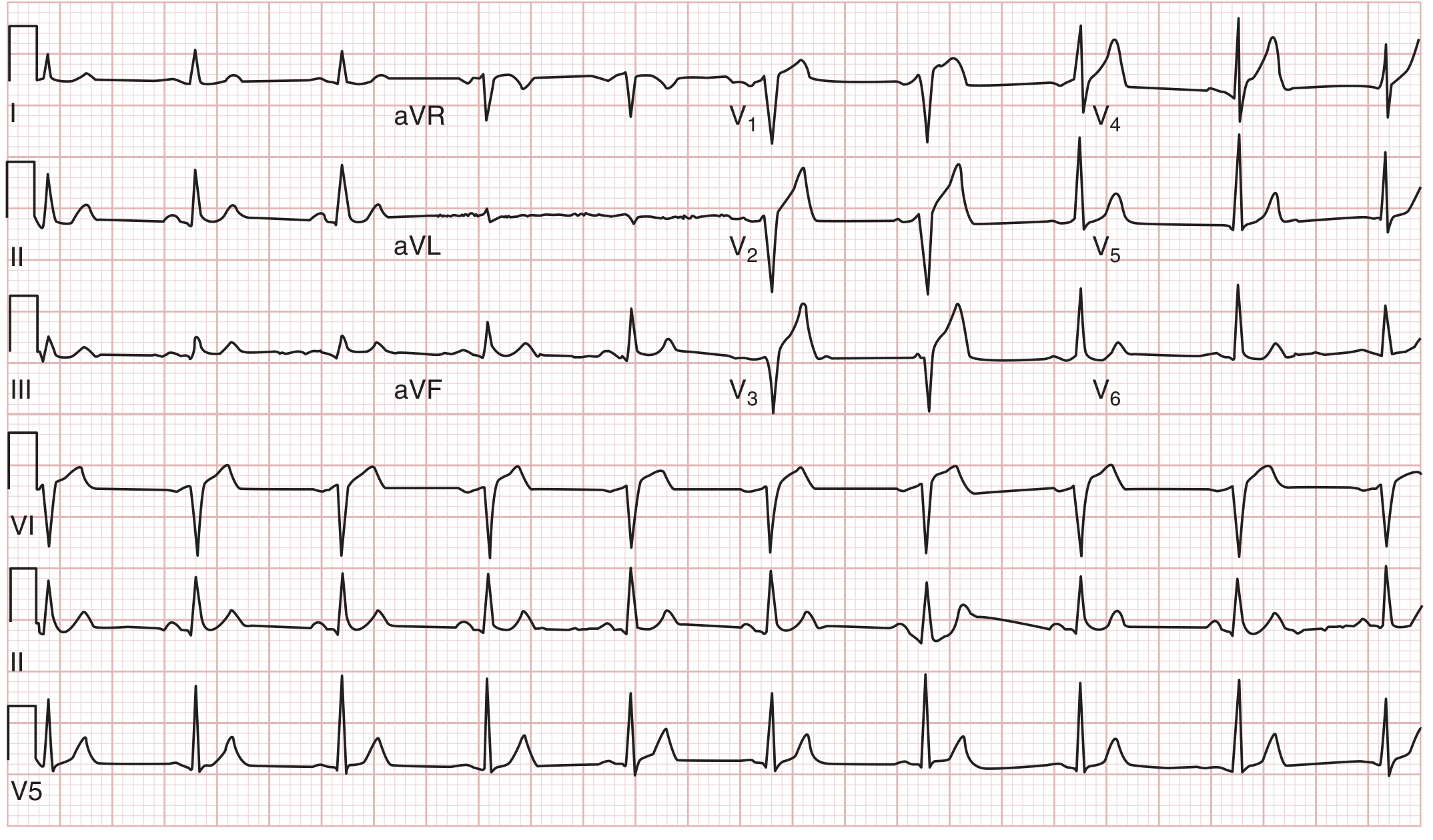
Key findings: ST elevation in leads I, V1, V2, V3. Absence of reciprocal ST depression in II, III, aVF. This is from a 52-year-old with 100% occlusion of the distal LAD.
Tintinalli's Emergency Medicine
2. Anterior STEMI (Proximal LAD Occlusion)
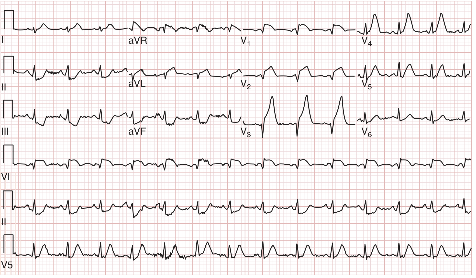
Key findings: ST elevation in V1-V3 with >1 mm ST depression in leads II, III, and aVF (reciprocal changes). This distinguishes proximal from distal LAD occlusion. Patient had 100% proximal LAD occlusion.
Tintinalli's Emergency Medicine
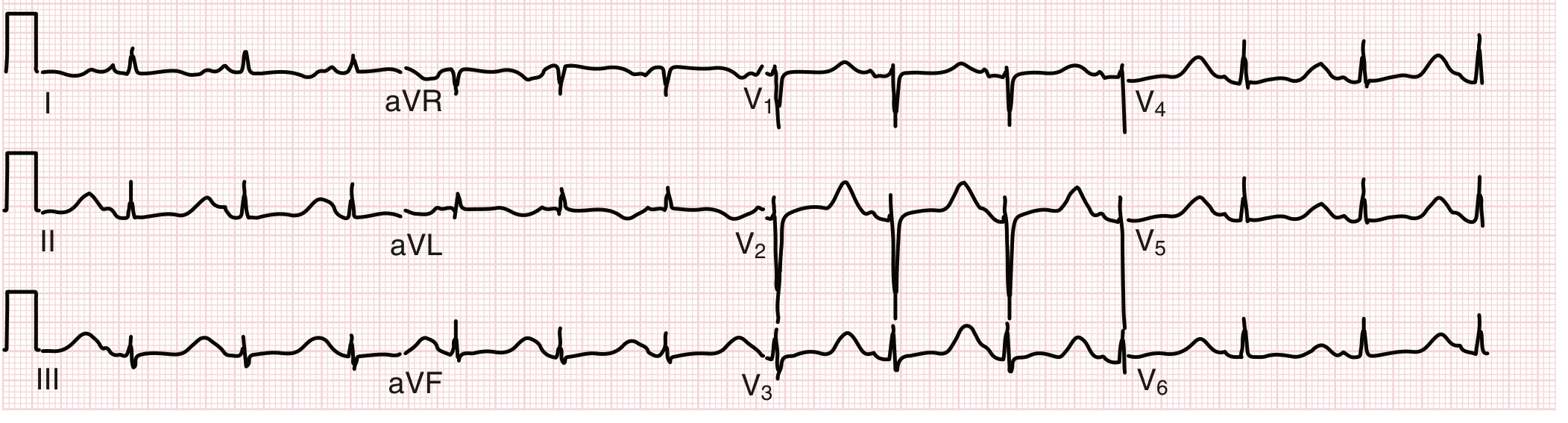
3. Acute Pericarditis - Stage I (Fuster's Heart, 15th Ed.)
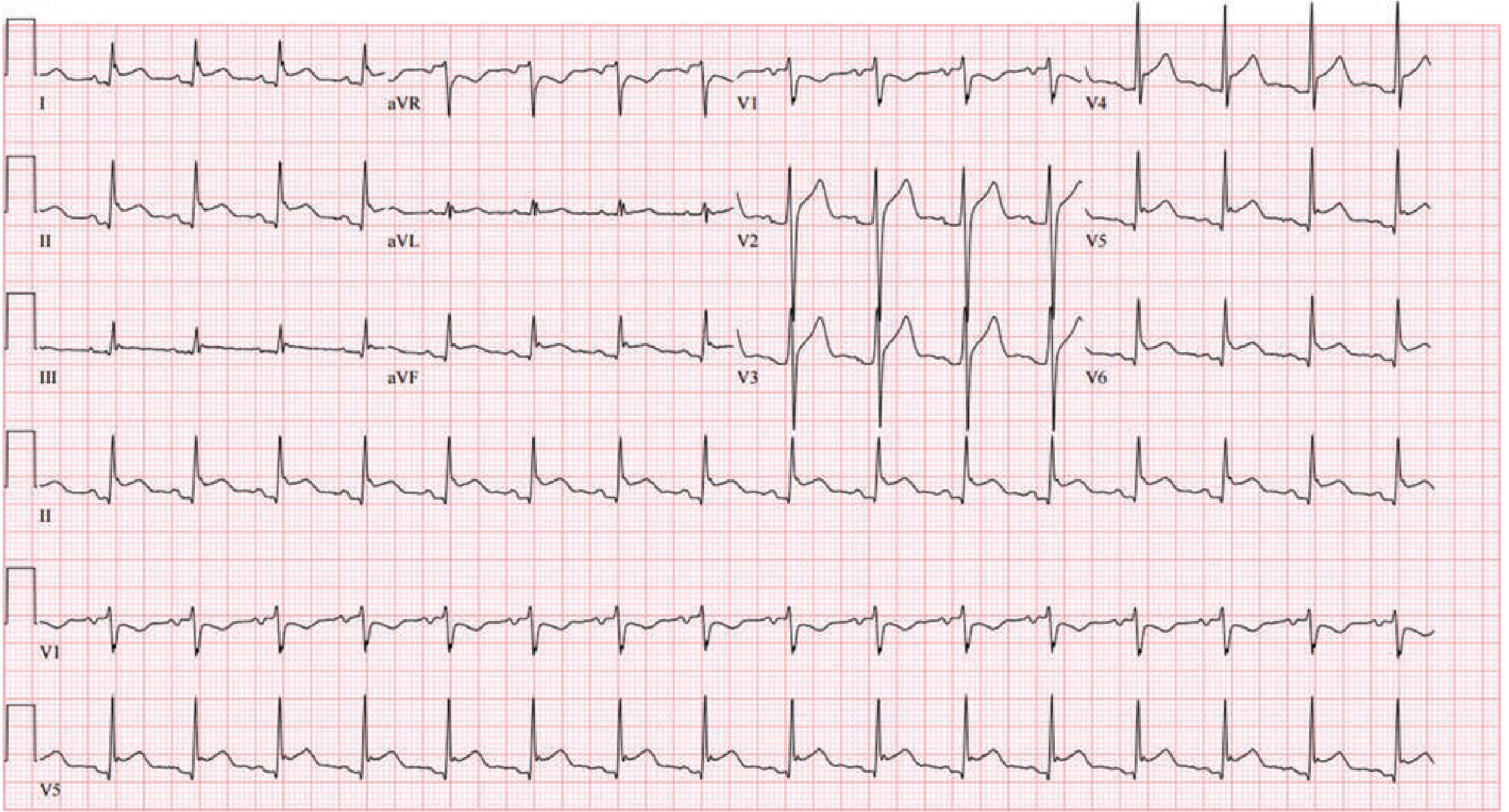
Key findings: Diffuse, non-territory-specific concave ST elevation across multiple leads. PR depression (best seen in II, V4-V6). Lead aVR shows ST depression with PR elevation (the mirror image). No Q waves, no reciprocal ST depression.
Fuster and Hurst's The Heart, 15th Edition
4. Acute Pericarditis - Classic Pattern (Braunwald's)
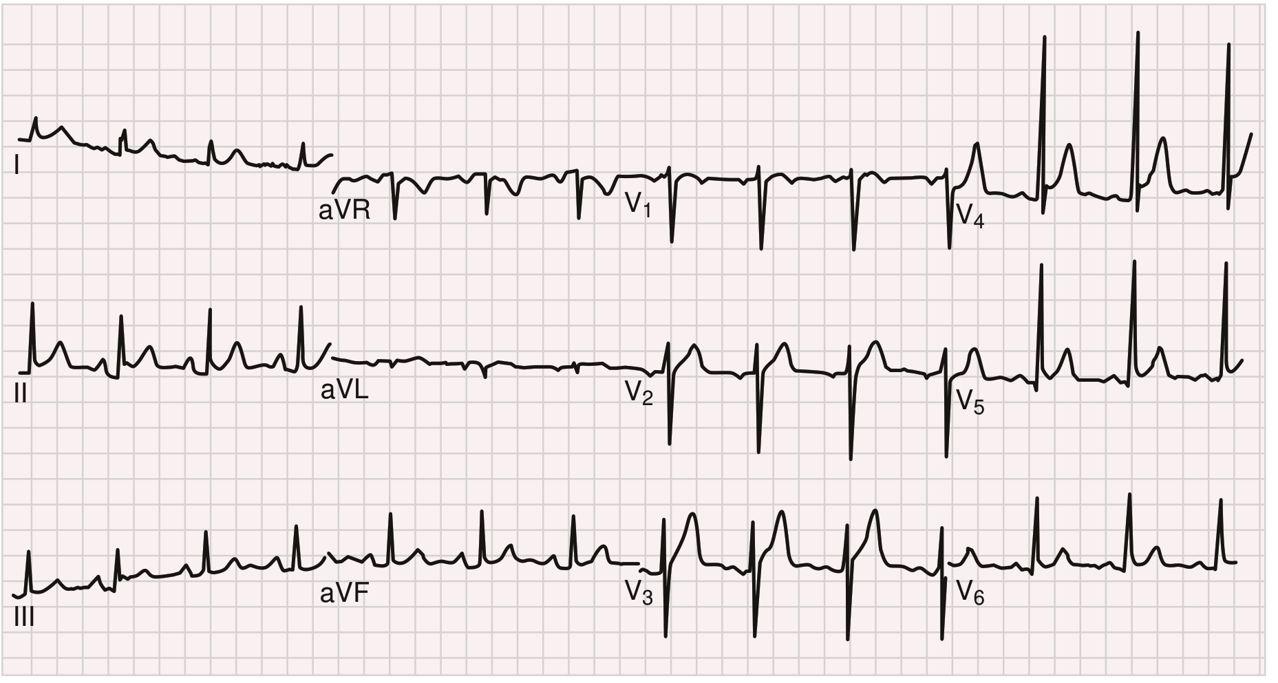
Key findings: Diffuse ST elevation with concurrent PR segment depression visible across multiple leads. Classic saddle-shaped (concave up) ST morphology.
Braunwald's Heart Disease, 12th Edition
5. Hyperkalemia - Progressive ECG Changes (Table)
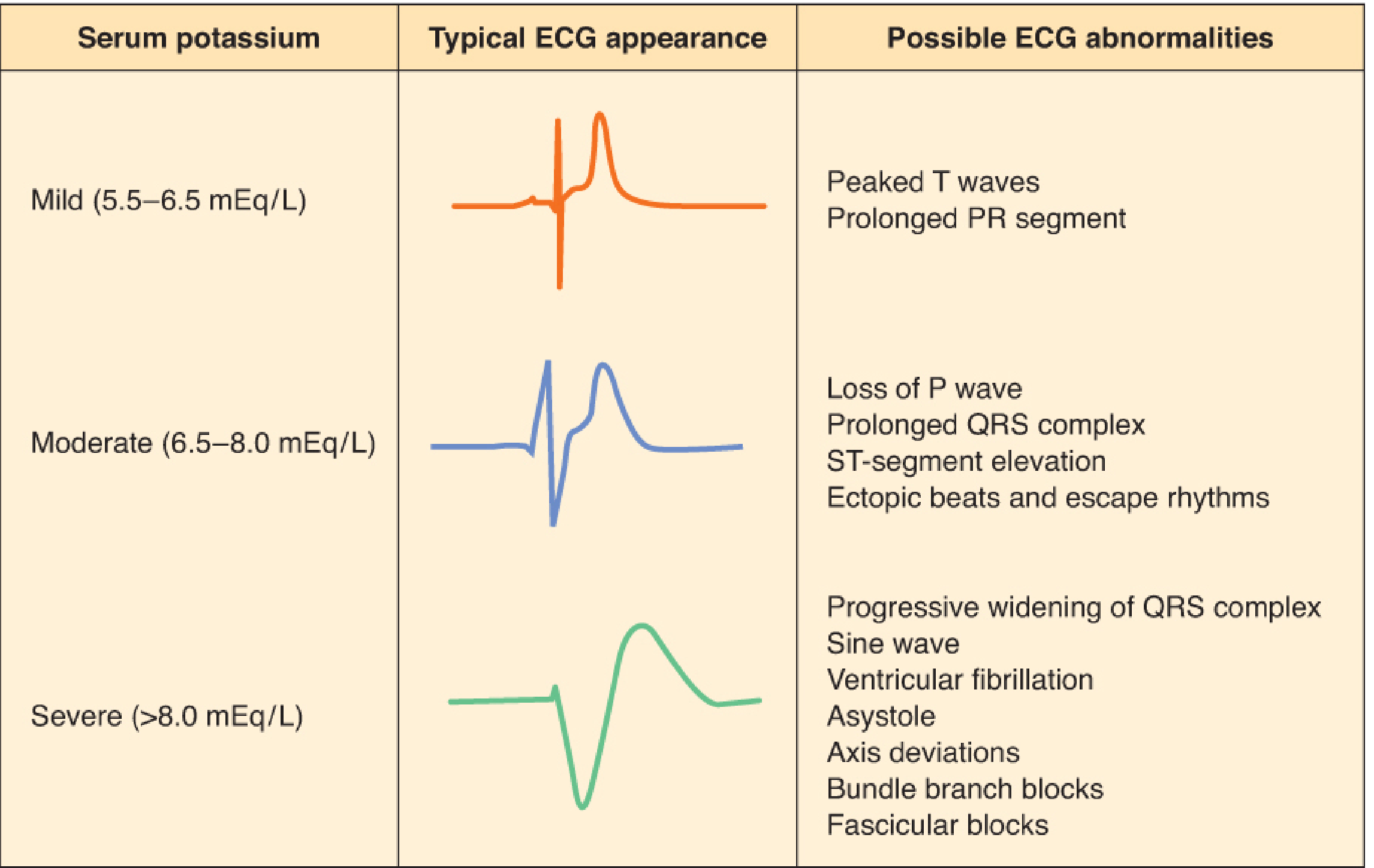
Key findings displayed by severity:
- Mild (5.5-6.5 mEq/L): Tall peaked T waves, prolonged PR
- Moderate (6.5-8.0 mEq/L): Loss of P wave, widened QRS, ST elevation, escape rhythms
- Severe (>8.0 mEq/L): Sine wave pattern, VF, asystole, bundle branch blocks
Barash's Clinical Anesthesia, 9th Edition
6. Hypokalemia - Full 12-Lead ECG
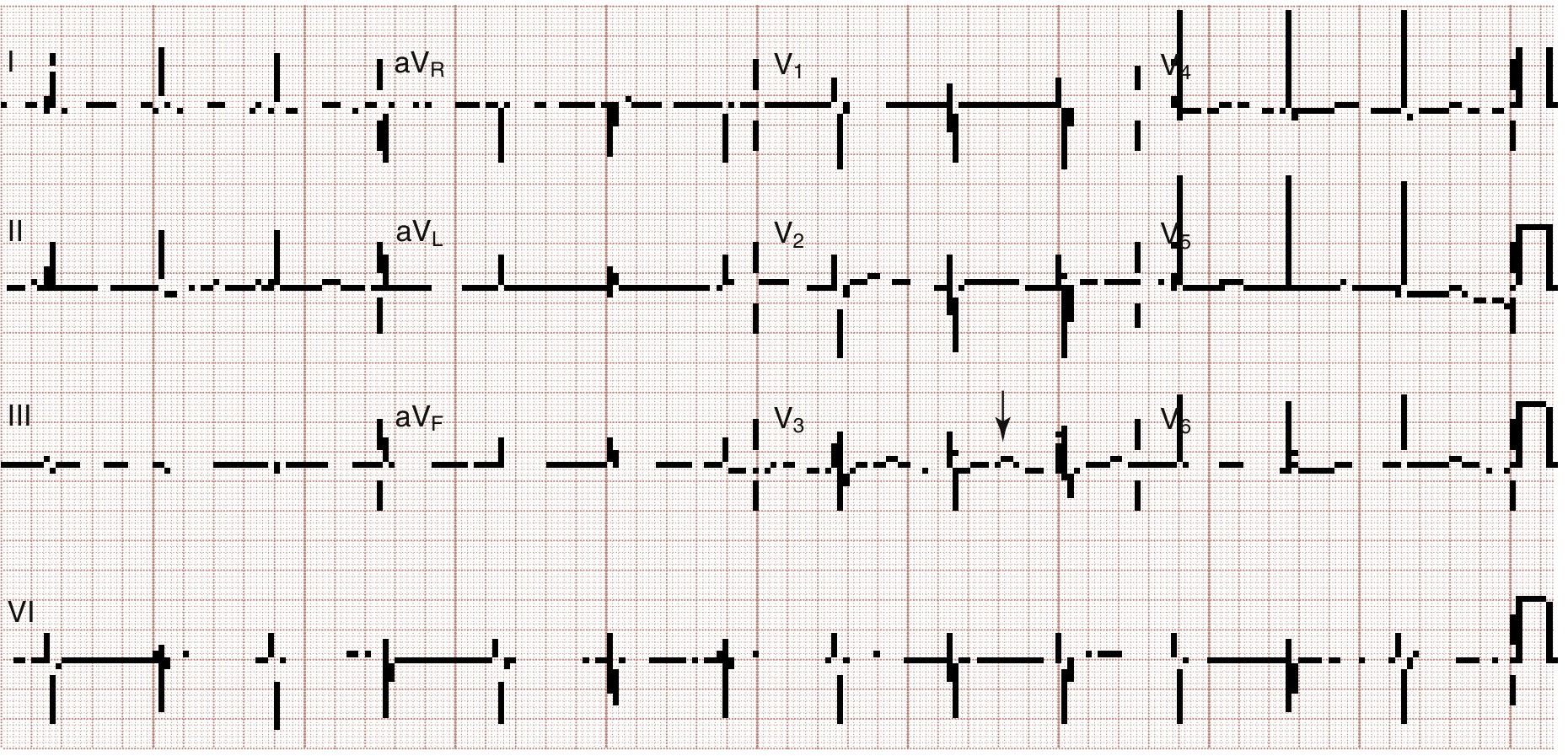
Key findings: Flattened T waves throughout, prominent U waves (visible after T wave, marked with arrow), nonspecific ST changes, prolonged QT interval. The 12-lead format clearly shows these changes are diffuse.
Rosen's Emergency Medicine
7. Hypothermia - Osborn (J) Wave
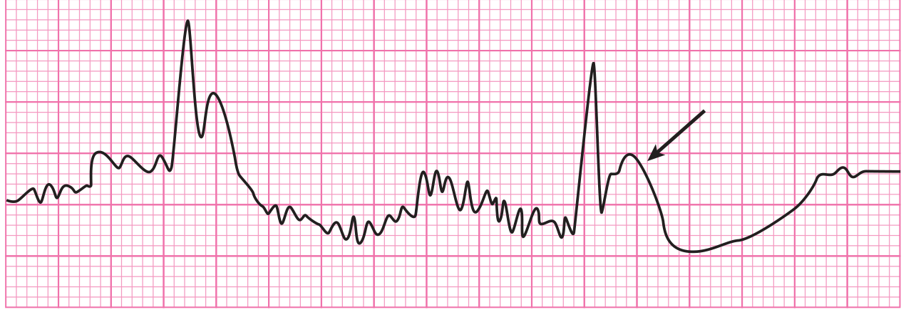
Key findings: The prominent Osborn (J) wave is marked with an arrow - the positive deflection at the J point immediately after QRS. Also visible: shivering artifact (irregular baseline tremor), atrial fibrillation with slow ventricular response. Patient's core temperature was 25°C (77°F).
Tintinalli's Emergency Medicine
8. Brugada Syndrome - Coved Pattern in V1-V2
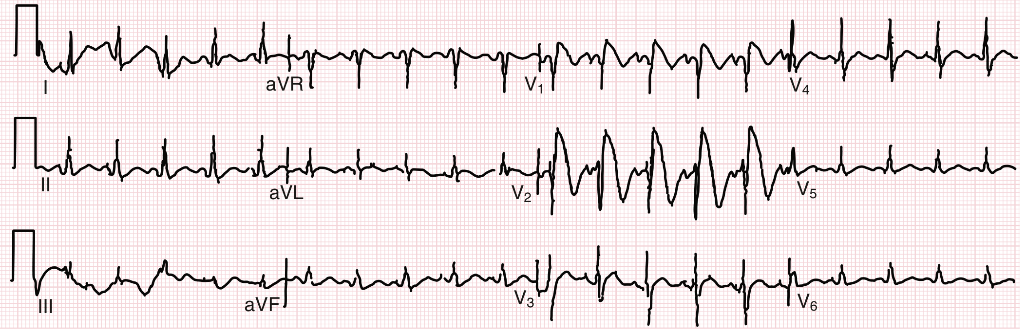
Key findings: Classic Type 1 "coved" pattern - downsloping ST elevation (>2 mm) in V1 with a negative T wave. Note the RBBB-like morphology. This is the only diagnostic pattern for Brugada syndrome. High risk for ventricular fibrillation and sudden cardiac death.
Goldman-Cecil Medicine
9. Long QT Syndrome (LQTS)
Key findings: Markedly prolonged QT interval of 640 ms - the terminal portion of the T wave merges with the following P wave. Broad, abnormal T-wave morphology. This was from a woman with congenital LQTS.
Goldman-Cecil Medicine
10. WPW Syndrome - Pre-excitation Pattern
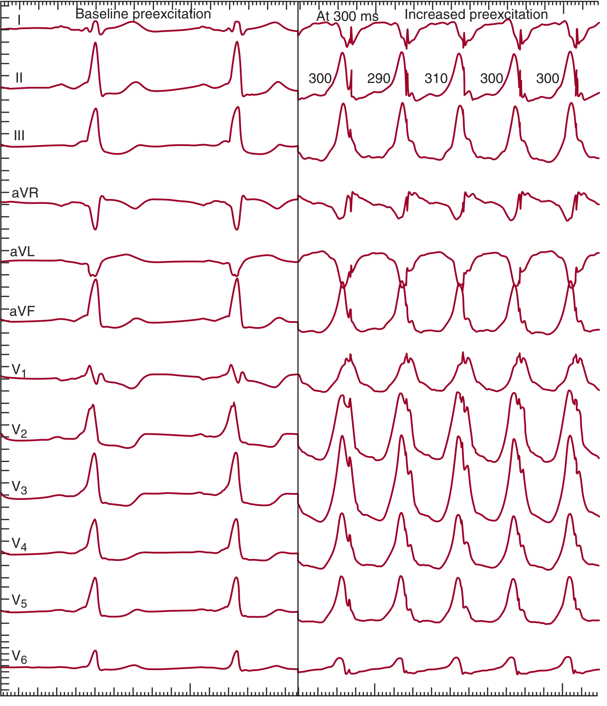
Key findings: Left panel (baseline sinus rhythm): Short PR interval, slurred delta wave onset, wide QRS from fusion of delta wave + normal conduction. Right panel (atrial pacing at 300 ms): Dramatically increased preexcitation as AV node is overridden - QRS is almost entirely from accessory pathway conduction, producing a bizarre maximally wide QRS. This illustrates the risk of rapid accessory pathway conduction during AF.
Fuster and Hurst's The Heart, 15th Edition
Summary of Remaining 10 Patterns (Images Not Available in Library)
For the following conditions, images were not directly available in the textbook library, but the key patterns are described below:
| # | Disease | Key ECG Pattern |
|---|---|---|
| 11 | Inferior STEMI | ST elevation in II, III, aVF; reciprocal ST depression in I, aVL |
| 12 | NSTEMI/Wellens' | Deep symmetric T-wave inversions in V2-V3 (Wellens' A/B pattern) |
| 13 | Pulmonary Embolism | S1Q3T3, sinus tachycardia, new RBBB, T-wave inversion V1-V4 |
| 14 | Hypercalcemia | Short QT interval (shortened ST segment) |
| 15 | Hypocalcemia | Prolonged QT interval (prolonged ST segment) |
| 16 | Atrial Fibrillation | No P waves, irregularly irregular R-R, fibrillatory baseline |
| 17 | Complete Heart Block | P-QRS dissociation, regular slow ventricular escape rhythm |
| 18 | Digoxin Effect | "Reverse tick" ST depression, short QT, PR prolongation |
| 19 | Subarachnoid Hemorrhage | Deep diffuse T-wave inversions, prolonged QT ("CVA T-waves") |
| 20 | Hypothyroidism | Sinus bradycardia, low voltage, flat T waves, prolonged QT |
Sources: Tintinalli's Emergency Medicine, Fuster and Hurst's The Heart (15th Ed.), Braunwald's Heart Disease (12th Ed.), Goldman-Cecil Medicine, Barash's Clinical Anesthesia (9th Ed.), Rosen's Emergency Medicine
This is a shared conversation. Sign in to Orris to start your own chat.