
Neural tube and derivatives of neural crest
Reading File
Now let me search the medical library for neural tube and neural crest content in parallel.
Finding Sources
Finding Sources
Excellent! Langman's Medical Embryology and The Developing Human are the best sources. Let me read the key sections in parallel.
Reading File
Reading File
Finding Sources
Great - now let me get the neural crest derivatives table from Langman's and also the THIEME Atlas section, plus the neural tube derivatives from Histology textbook.
Reading File
Reading File
Reading File
Excellent content gathered. Now let me fetch the key figures from Langman's showing neural crest migration and formation.
Loading Image
Loading Image
Reading File
I now have comprehensive information. Let me compile a thorough, well-structured answer.
Neural Tube and Neural Crest: Development and Derivatives
1. Neurulation - Formation of the Neural Tube
The neural tube is the embryonic precursor to the entire central nervous system. Its formation (neurulation) proceeds as follows:
- Neural plate induction (Week 3): The notochord signals the overlying ectoderm via BMP inhibitors (chordin, noggin, follistatin) to thicken into the neural plate - a slipper-shaped neuroectodermal thickening.
- Neural groove formation: The lateral edges of the neural plate elevate to form neural folds; the midline depresses into a neural groove.
- Neural tube closure (Weeks 3-4): The neural folds elevate, appose, and fuse dorsally, converting the groove into a hollow tube. Fusion begins in the cervical region and proceeds in both directions (rostral and caudal).
- Neuropores: The tube remains temporarily open at both ends - the anterior neuropore closes around day 25 (Week 4); the posterior neuropore closes around day 28.
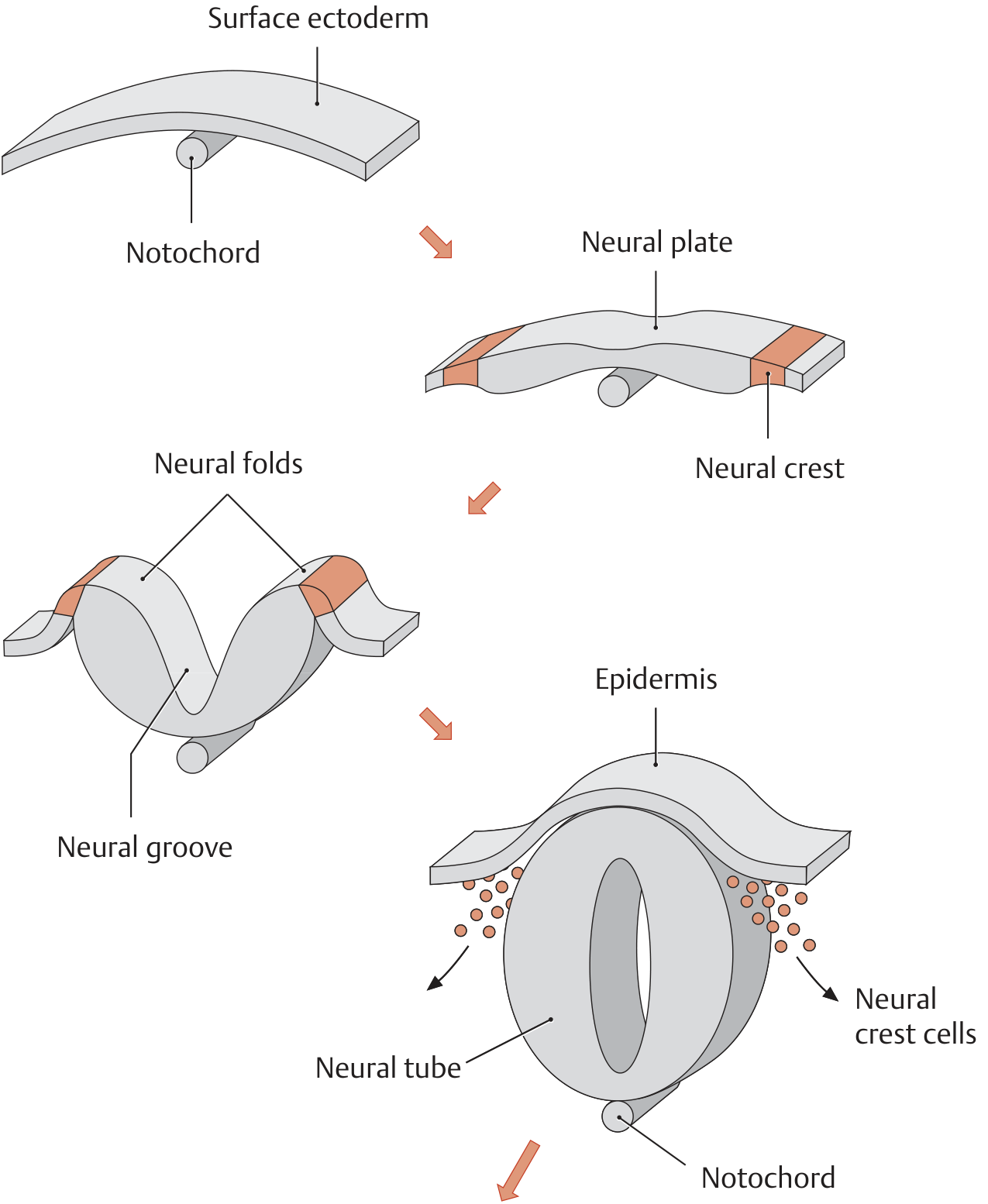
Neural plate → neural folds → neural groove → closed neural tube, with neural crest cells dissociating at closure.
2. Derivatives of the Neural Tube
The neural tube gives rise to all central nervous system structures:
| Region of Neural Tube | Derivative |
|---|---|
| Prosencephalon (forebrain) | Telencephalon (cerebral hemispheres, basal ganglia) + Diencephalon (thalamus, hypothalamus, retina) |
| Mesencephalon (midbrain) | Midbrain structures |
| Rhombencephalon (hindbrain) | Metencephalon (pons, cerebellum) + Myelencephalon (medulla oblongata) |
| Caudal neural tube | Spinal cord |
| Lumen of tube | Ventricular system and central canal |
Beyond the CNS itself, the neural tube also produces:
- Ependymal cells (lining the ventricular system and central canal)
- Oligodendrocytes and astrocytes (CNS glia - from neural tube proper)
- Pineal gland
- Posterior pituitary (neurohypophysis)
- Sensory epithelium of the eye (retina), ear, and nose
"Neuroectoderm gives rise to the neural tube and its derivatives, including components of the CNS, ependymia, pineal body, posterior lobe of the pituitary gland (neurohypophysis), and the sensory epithelium of the eye, ear, and nose."
- Histology: A Text and Atlas (Pawlina)
3. Neural Crest Formation
As the neural folds fuse to close the neural tube, cells at the lateral border (crest) of the neuroectoderm:
- Lose epithelial adhesion to neighboring cells
- Undergo epithelial-to-mesenchymal transition (EMT)
- Detach from the neuroectoderm
- Migrate actively into the underlying mesoderm
The process is regulated by:
- Wnt/β-catenin signaling activating GBX2
- BMPs (intermediate concentrations at the neural plate/surface ectoderm border induce NCC)
- Transcription factors: FOXD3, SNAIL2, SOX9, SOX10
- Guidance molecules: ephrins guide specific migration streams
Neural crest cells are sometimes called "the fourth germ layer" because of their extraordinary pluripotency and contribution to so many organ systems. They are implicated in at least one-third of all birth defects.
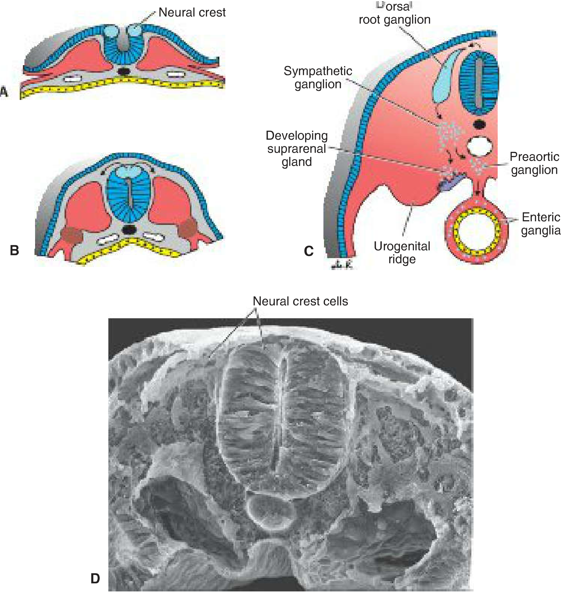
A,B: Crest cells form at tips of neural folds. C: After migration, crest cells contribute to dorsal root ganglia, sympathetic chain ganglia, adrenal medulla, and other tissues. D: Scanning EM showing migrating crest cells.
4. Migration Pathways (Trunk Neural Crest)
Neural crest cells in the trunk travel along three main routes:
| Pathway | Destination |
|---|---|
| ① Dorsolateral | Through the dermis → enter epidermis → become melanocytes (melanoblasts) |
| ② Ventrolateral | Through anterior half of each somite → dorsal root (spinal) ganglia (sensory neurons) |
| ③ Ventral | Paravertebral sympathetic ganglia, adrenal medulla (chromaffin cells), enteric ganglia, preaortic ganglia |
Cranial neural crest cells leave the neural tube before closure in the cephalic region and follow additional pathways into the face, pharyngeal arches, and cranium.
5. Derivatives of the Neural Crest (Complete Table)
Nervous System
| Derivative | Notes |
|---|---|
| Spinal (dorsal root) ganglia | Sensory neurons |
| Sympathetic chain and preaortic ganglia | Autonomic |
| Parasympathetic ganglia of GI tract (enteric nervous system) | Meissner's and Auerbach's plexuses |
| Ganglia of cranial nerves V, VII, IX, X | Partly from neural crest |
| Schwann cells | Peripheral nerve myelin |
| Glial cells (satellite cells) | Peripheral glial support |
| Meninges (arachnoid + pia mater of forebrain) | CNS coverings |
Endocrine / Secretory
| Derivative | Notes |
|---|---|
| Adrenal medulla | Chromaffin cells (catecholamines) |
| C cells (parafollicular cells) of thyroid | Calcitonin secretion |
| APUD cells (diffuse neuroendocrine system) | Enteroendocrine cells |
Craniofacial / Skeletal (Cranial Neural Crest)
| Derivative | Notes |
|---|---|
| Connective tissue and bones of face and skull | Facial bones, hyoid bone, portions of thyroid cartilage |
| Cartilage of pharyngeal arches | Meckel's cartilage, etc. |
| Dermis of face and neck | |
| Muscles of facial expression | |
| Odontoblasts | Form dentine of teeth |
| Corneal endothelium | |
| Smooth muscle of blood vessels (face and forebrain) |
Cardiac
| Derivative | Notes |
|---|---|
| Conotruncal (aorticopulmonary) septum | Separates aorta from pulmonary artery |
Pigment
| Derivative | Notes |
|---|---|
| Melanocytes | Throughout skin, hair follicles, uveal tract |
6. Neural Tube Defects (Clinical Correlates)
Failure of neural tube closure causes neural tube defects (NTDs):
- Anencephaly: failure of anterior neuropore to close → absent forebrain (lethal)
- Spina bifida: failure of posterior closure (most common at lumbosacral level)
- Spina bifida occulta: defect in vertebral arch only, cord intact
- Meningocele: meninges herniate through defect
- Myelomeningocele: cord + meninges herniate (most severe)
- Risk factor: folate deficiency; prevented by periconceptional folic acid supplementation
- Screening: elevated maternal serum AFP; confirmed by ultrasound/amniocentesis
7. Diseases of Neural Crest Derivatives
| Neural Crest Cell Type | Disease |
|---|---|
| Parasympathetic visceral ganglia | Neuroblastoma (malignant childhood tumor) |
| Enteric nervous system | Hirschsprung disease (aganglionic colon) |
| Schwann cells / satellite cells | Neurofibromatosis (von Recklinghausen disease) |
| Melanocytes | Malignant melanoma; albinism |
| Adrenal medulla | Pheochromocytoma |
| C cells of thyroid | Medullary thyroid carcinoma |
| Endocrine cells of lung/heart | Carcinoids |
| Conotruncal migration failure | Conotruncal heart defects (DiGeorge-type) |
The broad syndrome of neurocristopathies encompasses all conditions arising from defective neural crest migration, differentiation, or proliferation.
Sources: Langman's Medical Embryology, Table 6.1; The Developing Human (Moore), 10th ed.; THIEME Atlas of Anatomy; Histology: A Text and Atlas (Pawlina), pp. 308-309.
This is a shared conversation. Sign in to Orris to start your own chat.